Raymond de Vieussens (1641–1715): connoisseur of cardiologic anatomy and pathological forms thereof
- Affiliations
-
- 1Department of Anatomy, All India Institute of Medical Sciences, Patna, India
- KMID: 2523571
- DOI: http://doi.org/10.5115/acb.21.108
Abstract
- Raymond de Vieussens was an exemplary anatomist who made seminal contributions in the field of cardiology. During initial part of his academic career, he adopted human dissection based experiments as medium of his research. This was in accordance with prevailing trend among anatomists during 17th century. He discovered the presence of tiny venous tributaries communicating between cardiac veins and chambers of heart (ducti carnosi/venae cordis minimae). He reported the existence of a collateral circulatory pathway between right and left coronary arterial systems (Vieussens arterial ring). He was the first to note the valve at the junction of great cardiac vein and coronary sinus (valve of Vieussens) and the prominent oval margin of the fossa ovalis (Vieussens Annulus). All his findings were associated with considerable clinical significance as evidenced in literature that followed. Vieussens accurately demonstrated the three-layered orientation of myocardium and gave a precise description of coronary arteries and their branches. At the onset of 18th century, buoyed by royal patronage from King Louis XIV of France, the second half of Vieussens illustrious career was defined by pathologic anatomy (autopsy based) and anatomo clinical correlations. This was a new trend initiated by Vieussens in anatomical research and was later adopted as a signature method by anatomists of 18th century. As a true connoisseur of cardiologic anatomy, Vieussens accurately charted the anatomo clinical correlations of cardiac tamponade, mitral stenosis and aortic regurgitation. His contributions were pivotal elements in metamorphosis of cardiology as a robust discipline of medicine in modern times.
Keyword
Figure
-
Fig. 1 A portrait of Raymond de Vieussens. Image in public domain and free from copyright issues. Source of image: Wikimedia Commons.

Fig. 2 Illustrations showing the structure of tricuspid valve as observed from a superior view. The valve is shown after opening the walls of right atrium and also after opening the walls of right ventricle. In the right ventricle the tricuspid valve is shown along with the three papillary muscles which are attached to the valve cusps via chordae tendinae. The illustration was prepared by Lason (artist) in Montpellier at the time of dissection by Vieussens. Later the drawings were converted to engravings by Simmoneau (engraver) in Paris. Image in public domain and free from copyright issues. Source of image: Wikimedia Commons.
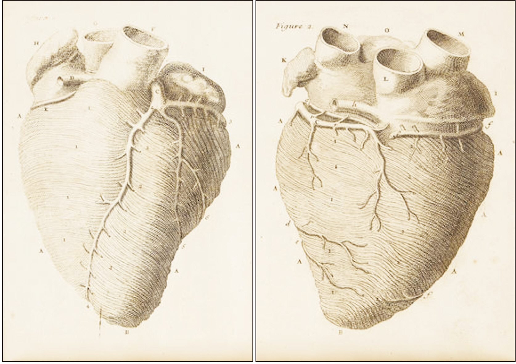
Fig. 3 Illustrations showing the middle layer of myocardium which has fibres in horizontal direction in the right ventricle and oblique direction in the left ventricle. Remarkably the distribution of the coronary arteries have been kept intact on the surface of the heart by Lason (artist) who prepared the drawing for Vieussens. Image in public domain and free from copyright issues. Source of image: Wikimedia Commons.
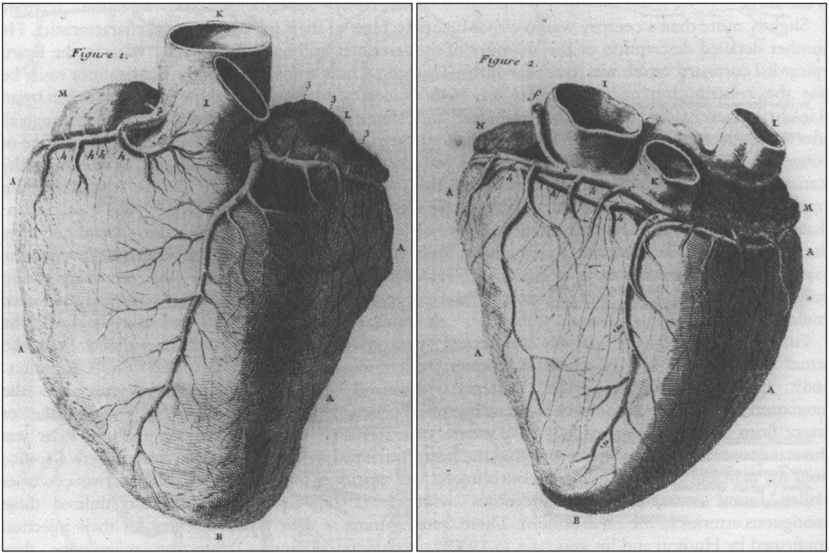
Fig. 4 Illustrations showing distribution of coronary arteries on the surface of heart. On the anterior surface of heart course of right coronary artery and those of left anterior descending artery as well as circumflex artery (both branches of left coronary artery) are demonstrated. On the inferior surface course of posterior descending artery (branch of right coronary artery) and circumflex artery (branch of left coronary artery) are depicted. Image in public domain and free from copyright issues. Source of image: Wikimedia Commons.

Fig. 5 Illustration showing the interior of right ventricle in a case of mitral stenosis. The internal features of the right ventricle are abnormally enlarged and its walls (myocardium) are inordinately thickened. The anatomical features observed in right ventricle of the heart are a consequence of mitral stenosis which is the pathological condition involving the heart. Image in public domain and free from copyright issues. Source of image: Wikimedia Commons.
Reference
-
References
1. Kellett CE. 1942; The life and work of Raymond de Vieussens. Ann Med Hist. 4:31–54. PMID: 33943704. PMCID: PMC7942442.2. Amalric P. 1983; Birth of neuro-ophthalmology in Montpellier in the 17th century through the works of Raymond Vieussens and Guilhem Briggs. Bull Soc Ophtalmol Fr. 83:83–8. French. PMID: 6347423.3. Podolsky E. 1952; Raymond Vieussens and the affairs of the heart. Med Womans J. 59:29–31. passim. PMID: 14910015.4. Loukas M, Clarke P, Tubbs RS, Kapos T. 2007; Raymond de Vieussens. Anat Sci Int. 82:233–6. DOI: 10.1111/j.1447-073X.2007.00192.x. PMID: 18062153.
Article5. Dulieu L. 1967; Raymond Vieussens. Monspel Hippocrates. 10:9–26. PMID: 33944162. PMCID: PMC7945240.6. Mounier-Kuhn P, Guerrier Y. 1982; Vieussens et l'anatomie de l'oreille. Vieussens and the anatomy of the ear. Acta Otorhinolaryngol Belg. 36:1029–38. French.7. Bonnel F, Lavabre-Bertrand T, Bonnel C. 2019; The teaching of anatomy in Montpellier University during VIII centuries (1220-2020). Surg Radiol Anat. 41:1119–28. DOI: 10.1007/s00276-019-02289-6. PMID: 31363840.
Article8. Ghosh SK. 2015; Human cadaveric dissection: a historical account from ancient Greece to the modern era. Anat Cell Biol. 48:153–69. DOI: 10.5115/acb.2015.48.3.153. PMID: 26417475. PMCID: PMC4582158.
Article9. Ghosh SK, Kumar A. 2018; Marcello Malpighi (1628-1694): Pioneer of microscopic anatomy and exponent of the scientific revolution of the 17th Century. Eur J Anat. 22:433–9.10. Ghosh SK, Sharma S, Biswas S, Chakraborty S. 2014; Adriaan van den Spiegel (1578-1625): anatomist, physician, and botanist. Clin Anat. 27:952–7. DOI: 10.1002/ca.22414. PMID: 24811238.
Article11. Vergani F, Morris CM, Mitchell P, Duffau H. 2012; Raymond de Vieussens and his contribution to the study of white matter anatomy: historical vignette. J Neurosurg. 117:1070–5. DOI: 10.3171/2012.8.JNS12387. PMID: 22998052.12. Ghosh SK, Narayan RK. 2020; Anatomy of nervous system and emergence of neuroscience: a chronological journey across centuries. Morphologie. 104:267–79. DOI: 10.1016/j.morpho.2020.05.005. PMID: 32534997.
Article13. Vieussens R. 1685. Neurographia universalis. Apud Joannem Certe;Lyons: p. 252. Latin.14. Harvey W. 1628. Exercitatio anatomica de motu cordis et sanguinis in animalibus. Fitzeri;Frankfurt: p. 236. Latin.15. Vieussens R. 1705. Novum vasorum corporis humani systema. Apud Paulum Marret;Amsterdam: p. 144. Latin.16. Snodgrass BT. 2012; Vessels described by Thebesius and Pratt are distinct from those described by Vieussens and Wearn. Am J Cardiol. 110:160. DOI: 10.1016/j.amjcard.2012.04.005. PMID: 22704295.
Article17. Mazurak M, Kusa J. 2019; Adam Christian Thebesius' channels into the human heart: the Thebesian veins and the Thebesian valve. Tex Heart Inst J. 46:175–8. DOI: 10.14503/THIJ-18-6604. PMID: 31708698. PMCID: PMC6827465.
Article18. Standring S. 2020. Gray's anatomy: the anatomical basis of clinical practice. 42nd ed. Elsevier;London: p. 1606.19. Vieussens R. 1706. Nouvelles decouvertes sur le coeur. Chez Laurent D'Houry;Paris: p. 43. French.20. Doğan N, Dursun A, Özkan H. 2019; Vieussens' arterial ring: a rare coronary variant anatomy. Diagn Interv Radiol. 25:109–13. DOI: 10.5152/dir.2019.17449. PMID: 30860074. PMCID: PMC6411272.
Article21. Corcoran SJ, Lawrence C, McGuire MA. 1999; The valve of Vieussens: an important cause of difficulty in advancing catheters into the cardiac veins. J Cardiovasc Electrophysiol. 10:804–8. DOI: 10.1111/j.1540-8167.1999.tb00260.x. PMID: 10376917.
Article22. Caliot P, Bousquet V, Cabanie P, Midy D. 1984; The nerve loops crossing below the subclavian artery and their anatomical variations. Anat Clin. 6:209–13. DOI: 10.1007/BF01784315. PMID: 6518114.
Article23. Vieussens R. 1715. Traité nouveau de la structure et des causes du mouvement naturel du coeur. Jean Guillemette;Toulouse: p. 102. French.24. Pearlman ES, Weber KT, Janicki JS, Pietra GG, Fishman AP. 1982; Muscle fiber orientation and connective tissue content in the hypertrophied human heart. Lab Invest. 46:158–64. PMID: 6460896.25. Major RH. 1932; Raymond Vieussens and his treatise on the heart. Ann Med Hist. 4:147–54. PMID: 33944169. PMCID: PMC7945248.26. Spodick DH. 1970; Medical history of the pericardium. The hairy hearts of hoary heroes. Am J Cardiol. 26:447–54. DOI: 10.1016/0002-9149(70)90701-0. PMID: 4920772.27. Buess H. 1951; Theophil Bonet (1620-1689) und die grundsätzliche Bedeutung seines Sepulchretum in der Geschichte der pathologischen Anatomie. Theophil Bonet and the significance of his Sepulchretum in the history of pathologic anatomy. Gesnerus. 8:32–52. German. DOI: 10.1163/22977953-0080102005. PMID: 14860449.
Article28. Kellett CE. 1959; Raymond de Vieussens on mitral stenosis. Br Heart J. 21:440–4. DOI: 10.1136/hrt.21.3.440. PMID: 14405255. PMCID: PMC1017600.
Article29. Suvarna JC. 2008; Watson's water hammer pulse. J Postgrad Med. 54:163–5. DOI: 10.4103/0022-3859.40791. PMID: 18480541.
Article30. Reichert P. 1978; A history of the development of cardiology as a medical specialty. Clin Cardiol. 1:5–15. DOI: 10.1002/clc.4960010102. PMID: 389510.
Article31. Ghosh SK, Raheja S, Tuli A. 2014; Obstructive Thebesian valve: anatomical study and implications for invasive cardiologic procedures. Anat Sci Int. 89:85–94. DOI: 10.1007/s12565-013-0203-0. PMID: 24043316.
Article32. Gumpangseth T, Lekawanvijit S, Mahakkanukrauh P. 2020; Histological assessment of the human heart valves and its relationship with age. Anat Cell Biol. 53:261–71. DOI: 10.5115/acb.20.093. PMID: 32727956. PMCID: PMC7527117.
Article33. Ghosh SK. 2017; Thomas Bartholin (1616-1680): Danish anatomist and his cardinal contributions towards the discovery of the lymphatic system. Eur J Anat. 21:261–8.34. Ghosh SK. 2017; Giovanni Battista Morgagni (1682-1771): father of pathologic anatomy and pioneer of modern medicine. Anat Sci Int. 92:305–12. DOI: 10.1007/s12565-016-0373-7. PMID: 27629485.
Article35. Chitwood WR Jr. 1977; John and William Hunter on aneurysms. Arch Surg. 112:829–36. DOI: 10.1001/archsurg.1977.01370070043005. PMID: 327974.
Article
- Full Text Links
-
- Actions
-
Cited
- CITED
-
- Close
- Share
-
- Similar articles
-
- An Unusual Form of Coronary Artery Fistula: A Small Aneurysm of Vieussens' Arterial Ring Communicating with the Pulmonary Artery
- The rich heritage of anatomical texts during Renaissance and thereafter: a lead up to Henry Gray's masterpiece
- Analysis of Bony Landmarks and Muscles Classification for Visual Artists
- Rencontres de la recherche et de l'innovation
- Inappropriate Authorship and Kinship in Research Evaluation
