Hematochezia in Patient with Rectal Tumor: Consideration of Various Diagnostic Possibilities
- Affiliations
-
- 1Department of Internal Medicine, Hallym University College of Medicine, Chuncheon, Korea
- KMID: 2522720
- DOI: http://doi.org/10.5946/ce.2021.243
Figure
-
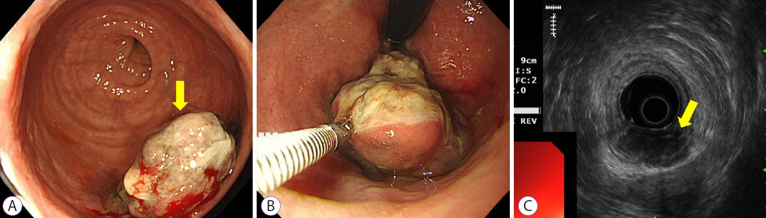
Fig. 1. (A, B) Colonoscopy demonstrating 3 cm mass covered with exudates near anal canal. (C) Endoscopic ultrasonography demonstrating heterogenous hypoechoic lesion in the submucosal layer.

Fig. 2. Histopathological findings of the biopsy specimens. Small malignant oval-shaped cells with high nuclear-to-cytoplasmic ratio with poor differentiation (normal glandular structure was not observed) (hematoxylin and eosin stain ×200).
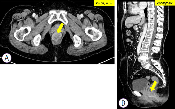
Fig. 3. (A, B) Contrast-enhanced computed tomography demonstrating 3.5 cm enhancing lesion involving distal rectum and anorectal junction.
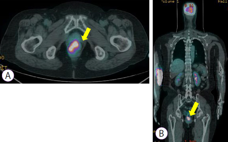
Fig. 4. (A, B) Positron emission tomography demonstrating 3-3.5 cm mass with increased FDG uptake (SUVmax=10.79) in the distal rectum.
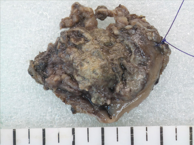
Fig. 5. Surgical specimen demonstrates an ill demarcated ulcerative and fungating mass, measuring 3.3×2.7 cm extending to the pericolic soft tissue.
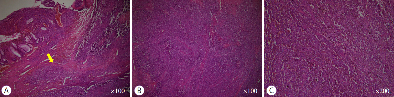
Fig. 6. Histopathological findings of the surgically resected specimen. (A, B) Round cells with a high nuclear-to-cytoplasmic ratio without macronucleoli which invaded whole mucosal layer (hematoxylin and eosin stain ×100). (C) Diffuse brown colored pigmentation (hematoxylin and eosin stain ×200)

Fig. 7. Immunohistochemistry findings of the surgically resected specimen. (A, B) The tumor cells are diffusely positive for HMB-45 and S-100 (hematoxylin and eosin stain ×100).
Reference
-
1. Xu X, Ge T, Wang G. Primary anorectal malignant melanoma: a case report. Medicine (Baltimore). 2020; 99:e19028.2. Chang AE, Karnell LH, Menck HR. The National Cancer Data Base report on cutaneous and noncutaneous melanoma: a summary of 84,836 cases from the past decade. The American College of Surgeons Commission on Cancer and the American Cancer Society. Cancer. 1998; 83:1664–1678.3. Sahoo MR, Gowda MS, Kaladagi RM. Primary amelanotic melanoma of the rectum mimicking adenocarcinoma. Am J Case Rep. 2013; 14:280–283.
Article4. Maqbool A, Lintner R, Bokhari A, Habib T, Rahman I, Rao BK. Anorectal melanoma--3 case reports and a review of the literature. Cutis. 2004; 73:409–413.5. Stefanou A, Nalamati SPM. Anorectal melanoma. Clin Colon Rectal Surg. 2011; 24:171–6.
Article6. Kim KW, Ha HK, Kim AY, et al. Primary malignant melanoma of the rectum: CT findings in eight patients. Radiology. 2004; 232:181–186.
Article7. Malik A, Hull TL, Milsom J. Long-term survivor of anorectal melanoma: report of a case. Dis Colon Rectum. 2002; 45:1412–1415. discussion 1415-1417.8. Tokuhara K, Nakatani K, Tanimura H, Yoshioka K, Kiyohara T, Kon M. A first reported case of metastatic anorectal amelanotic melanoma with a marked response to anti-PD-1 antibody nivolumab: a case report. Int J Surg Case Rep. 2017; 31:188–192.
Article9. Nafees R, Khan H, Ahmed S, Ahmed Samo K, Siraj Memon A. Primary rectal amelanotic malignant melanoma: a rare case report. Cureus. 2020; 12:e8115.
Article
- Full Text Links
-
- Actions
-
Cited
- CITED
-
- Close
- Share
-
- Similar articles
-
- Clinical Observation on Patients with Hematochezia
- Update of Korean Standard Classification of Diseases for Rectal Carcinoid and Its Clinical Implication
- Role of Colonoscopy in Patients with Hematochezia
- Imaging Diagnosis of Locally Advanced Rectal Cancer: Tumor Staging before and after Preoperative Chemoradiotherapy
- A Case of Rectal Schwannoma Presenting with Hematochezia
