Systematic Review of Reciprocal Changes after Spinal Reconstruction Surgery : Do Not Miss the Forest for the Trees
- Affiliations
-
- 1Department of Neurosurgery, Spine Center, Seoul National University Bundang Hospital, Seoul National University College of Medicine, Seongnam, Korea
- KMID: 2521974
- DOI: http://doi.org/10.3340/jkns.2020.0234
Abstract
- The purpose of this review was to synthesize the research on global spinal alignment and reciprocal changes following cervical or thoracolumbar reconstruction surgery. We carried out a search of PubMed, EMBASE, and Cochrane Library for studies through May 2020, and ultimately included 11 articles. The optimal goal of a truly balanced spine is to maintain the head over the femoral heads. When spinal imbalance occurs, the human body reacts through various compensatory mechanisms to maintain the head over the pelvis and to retain a horizontal gaze. Historically, deformity correction has focused on correcting scoliosis and preventing scoliotic curve progression. Following substantial correction of a spinal deformity, reciprocal changes take place in the flexible segments proximal and distal to the area of correction. Restoration of lumbar lordosis following surgery to correct a thoracolumbar deformity induces reciprocal changes in T1 slope, cervical lordosis, pelvic shift, and lower extremity parameters. Patients with cervical kyphosis exhibit different patterns of reciprocal changes depending on whether they have head-balanced or trunk-balanced kyphosis. These reciprocal changes should be considered to in order to prevent secondary spine disorders. We emphasize the importance of evaluating the global spinal alignment to assess postoperative changes.
Figure
-
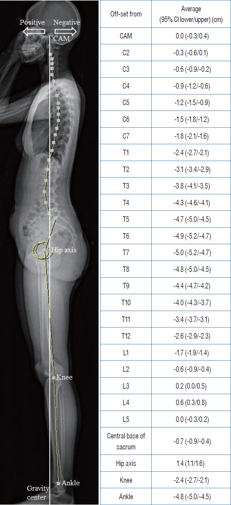
Fig. 1. Normative offset distance between bony landmarks and the gravity line [17]. Positive means anterior to the gravity line and negative means posterior to the gravity line. CAM : center of acoustic meatus, CI : confidence interval.
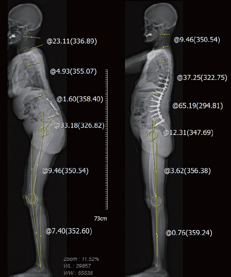
Fig. 2. Whole body image of thoracolumbar malalignment (left) exhibiting cervical hyperlordosis, posterior pelvic shift and knee flexion. Postoperative whole body image of the same patient (right) exhibiting restoration of lumbar lordosis (from 1.6° to 65.2°) with reciprocal changes in thoracic kyphosis (from -4.9° to 37.2°) cervical lordosis (from 23.1° to 9.5°), pelvic tilt (from 33.1° to 12.3°), knee flexion (from 9.5° to 3.6°) and ankle dorsiflexion (from 7.4° to 0.7°).
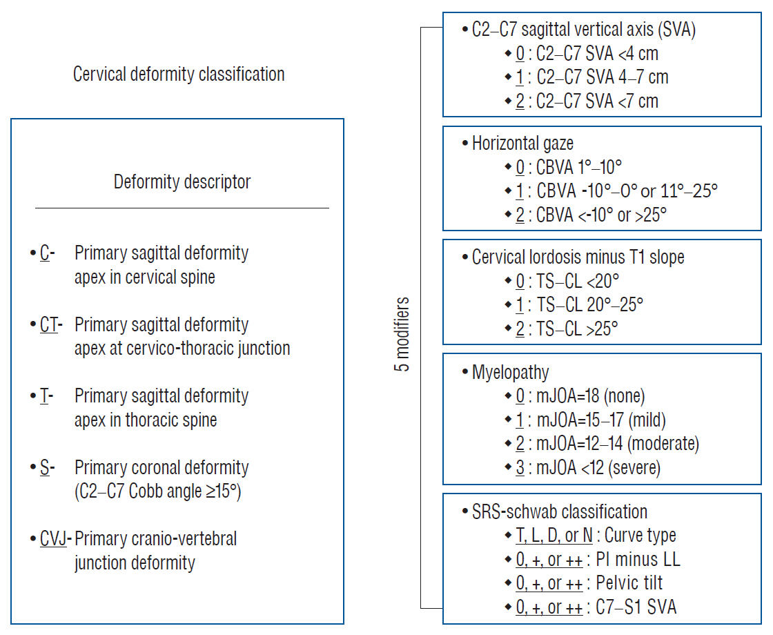
Fig. 3. Revised cervical spinal deformity classification system [18], which consists of a deformity descriptor and five modifiers. T : thoracic major, CBVA : chin-brow to vertical angle, TS : T1 slope, CL : cervical lordosis, mJOA : modified Japanese Orthopaedic Association, SRS : Scoliosis Research Society, L : lumbar major, D : double, N : no scoliosis, LL : lumbar lordosis.
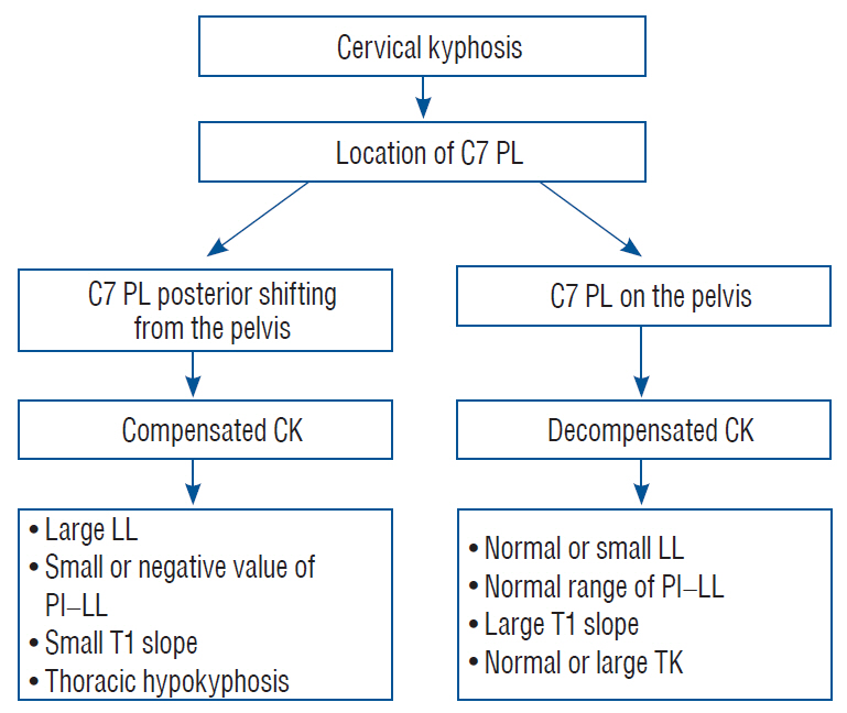
Fig. 4. Compensation mechanisms in patients with symptomatic primary cervical kyphosis. PL : plumb line, CK : cervical kyphosis, LL : lumbar lordosis, PI : pelvic incidence, TK : thoracic kyphosis.
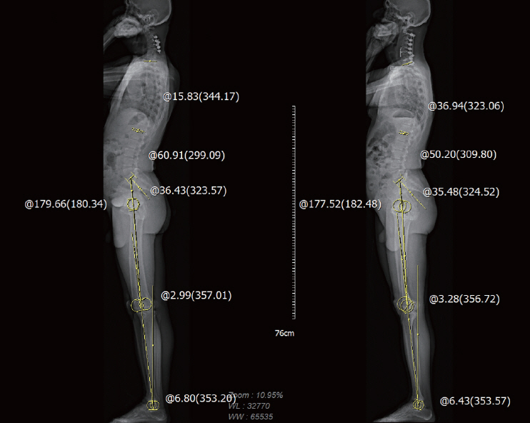
Fig. 5. Whole body images of a head-balanced (compensated) patient. While T1 slope, thoracic kyphosis, and lumbar lordosis changed, spinopelvic- and lower extremity parameters did not change following cervical kyphosis correction.
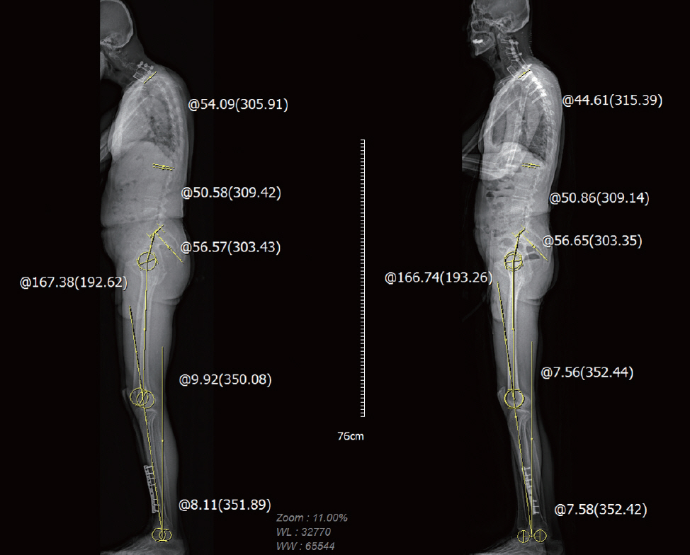
Fig. 6. Whole body images of a trunk-balanced (decompensated) patient. While T1 slope and thoracic kyphosis decreased, lumbar lordosis, spinopelvic- and lower extremity parameters did not change following cervical kyphosis correction.
Cited by 2 articles
-
Pediatric Spine Trauma
Sungjae An, Seung-Jae Hyun
J Korean Neurosurg Soc. 2022;65(3):361-369. doi: 10.3340/jkns.2021.0282.The Sagittal Balance of Cervical Spine : Comprehensive Review of Recent Update
Sang Hoon Lee, Tae Hwan Kim, Seok Woo Kim, Hyun Take Rim, Heui Seung Lee, Ji Hee Kim, In Bok Chang, Joon Ho Song, Yong Kil Hong, Jae Keun Oh
J Korean Neurosurg Soc. 2023;66(6):611-617. doi: 10.3340/jkns.2023.0146.
Reference
-
References
1. Ames CP, Smith JS, Eastlack R, Blaskiewicz DJ, Shaffrey CI, Schwab F, et al. Reliability assessment of a novel cervical spine deformity classification system. J Neurosurg Spine. 23:673–683. 2015.
Article2. Ames CP, Smith JS, Scheer JK, Bess S, Bederman SS, Deviren V, et al. Impact of spinopelvic alignment on decision making in deformity surgery in adults: a review. J Neurosurg Spine. 16:547–564. 2012.
Article3. Barrey C, Roussouly P, Le Huec JC, D’Acunzi G, Perrin G. Compensatory mechanisms contributing to keep the sagittal balance of the spine. Eur Spine J 22 Suppl. 6:S834–S841. 2013.
Article4. Barrey C, Roussouly P, Perrin G, Le Huec JC. Sagittal balance disorders in severe degenerative spine. Can we identify the compensatory mechanisms? Eur Spine J 20 Suppl. 5:626–633. 2011.5. Blondel B, Lafage V, Schwab F, Farcy JP, Bollini G, Jouve JL. Reciprocal sagittal alignment changes after posterior fusion in the setting of adolescent idiopathic scoliosis. Eur Spine J. 21:1964–1971. 2012.
Article6. Choi HY, Hyun SJ, Kim KJ, Jahng TA, Kim HJ. Radiographic and clinical outcomes following pedicle subtraction osteotomy : minimum 2-year follow-up data. J Korean Neurosurg Soc. 63:99–107. 2020.
Article7. Day LM, Ramchandran S, Jalai CM, Diebo BG, Liabaud B, Lafage R, et al. Thoracolumbar realignment surgery results in simultaneous reciprocal changes in lower extremities and cervical spine. Spine (Phila Pa 1976). 42:799–807. 2017.
Article8. Deschênes S, Charron G, Beaudoin G, Labelle H, Dubois J, Miron MC, et al. Diagnostic imaging of spinal deformities: reducing patients radiation dose with a new slot-scanning X-ray imager. Spine (Phila Pa 1976). 35:989–994. 2010.9. Diebo BG, Ferrero E, Lafage R, Challier V, Liabaud B, Liu S, et al. Recruitment of compensatory mechanisms in sagittal spinal malalignment is age and regional deformity dependent: a full-standing axis analysis of key radiographical parameters. Spine (Phila Pa 1976). 40:642–649. 2015.
Article10. Dru AB, Lockney DT, Vaziri S, Decker M, Polifka AJ, Fox WC, et al. Cervical spine deformity correction techniques. Neurospine. 16:470–482. 2019.
Article11. Dubousset J. Three-dimensional analysis of the scoliotic deformity. In : Weinstein SL, editor. The pediatric spine: principles and practice, ed 1. New York: Raven Press;1994. p. 479–496.12. Ferrero E, Liabaud B, Challier V, Lafage R, Diebo BG, Vira S, et al. Role of pelvic translation and lower-extremity compensation to maintain gravity line position in spinal deformity. J Neurosurg Spine. 24:436–446. 2016.
Article13. Glaser DA, Doan J, Newton PO. Comparison of 3-dimensional spinal reconstruction accuracy: biplanar radiographs with EOS versus computed tomography. Spine (Phila Pa 1976). 37:1391–1397. 2012.14. Glassman SD, Berven S, Bridwell K, Horton W, Dimar JR. Correlation of radiographic parameters and clinical symptoms in adult scoliosis. Spine (Phila Pa 1976). 30:682–688. 2005.
Article15. Glassman SD, Bridwell K, Dimar JR, Horton W, Berven S, Schwab F. The impact of positive sagittal balance in adult spinal deformity. Spine (Phila Pa 1976). 30:2024–2029. 2005.
Article16. Ha Y, Schwab F, Lafage V, Mundis G, Shaffrey C, Smith J, et al. Reciprocal changes in cervical spine alignment after corrective thoracolumbar deformity surgery. Eur Spine J. 23:552–559. 2014.
Article17. Hasegawa K, Okamoto M, Hatsushikano S, Shimoda H, Ono M, Homma T, et al. Standing sagittal alignment of the whole axial skeleton with reference to the gravity line in humans. J Anat. 230:619–630. 2017.
Article18. Hyun SJ, Han S, Kim KJ, Jahng TA, Kim HJ. Assessment of T1 slope minus cervical lordosis and C2-7 sagittal vertical axis criteria of a cervical spine deformity classification system using long-term follow-up data after multilevel posterior cervical fusion surgery. Oper Neurosurg (Hagerstown). 16:20–26. 2019.
Article19. Hyun SJ, Jung JM. Spinal deformity surgery : it becomes an essential part of neurosurgery. J Korean Neurosurg Soc. 61:661–668. 2018.
Article20. Iyer S, Nemani VM, Nguyen J, Elysee J, Burapachaisri A, Ames CP, et al. Impact of cervical sagittal alignment parameters on neck disability. Spine (Phila Pa 1976). 41:371–377. 2016.
Article21. Kang J, Hosogane N, Ames C, Schwab F, Hart R, Burton D, et al. Diversity in surgical decision strategies for adult spine deformity treatment: the effects of neurosurgery or orthopedic training background and surgical experience. Neurospine. 15:353–361. 2018.
Article22. Knott PT, Mardjetko SM, Techy F. The use of the T1 sagittal angle in predicting overall sagittal balance of the spine. Spine J. 10:994–998. 2010.
Article23. Kuntz C 4th, Levin LS, Ondra SL, Shaffrey CI, Morgan CJ. Neutral upright sagittal spinal alignment from the occiput to the pelvis in asymptomatic adults: a review and resynthesis of the literature. J Neurosurg Spine. 6:104–112. 2007.
Article24. Lafage R, Challier V, Liabaud B, Vira S, Ferrero E, Diebo BG, et al. Natural head posture in the setting of sagittal spinal deformity: validation of chin-brow vertical angle, slope of line of sight, and McGregor’s slope with health-related quality of life. Neurosurgery. 79:108–115. 2016.
Article25. Lafage V, Schwab F, Skalli W, Hawkinson N, Gagey PM, Ondra S, et al. Standing balance and sagittal plane spinal deformity: analysis of spinopelvic and gravity line parameters. Spine (Phila Pa 1976). 33:1572–1578. 2008.26. Lee DH, Ha JK, Chung JH, Hwang CJ, Lee CS, Cho JH. A retrospective study to reveal the effect of surgical correction of cervical kyphosis on thoraco-lumbo-pelvic sagittal alignment. Eur Spine J. 25:2286–2293. 2016.
Article27. Lee SH, Kim KT, Seo EM, Suk KS, Kwack YH, Son ES. The influence of thoracic inlet alignment on the craniocervical sagittal balance in asymptomatic adults. J Spinal Disord Tech. 25:E41–E47. 2012.
Article28. Lee SH, Son DW, Lee JS, Kim DH, Sung SK, Lee SW, et al. Differences in cervical sagittal alignment changes in patients undergoing laminoplasty and anterior cervical discectomy and fusion. Neurospine. 15:91–100. 2018.
Article29. Makhni MC, Shillingford JN, Laratta JL, Hyun SJ, Kim YJ. Restoration of sagittal balance in spinal deformity surgery. J Korean Neurosurg Soc. 61:167–179. 2018.
Article30. McClendon J Jr, Graham RB, Sugrue PA, Smith TR, Thompson SE, Koski TR. Cranial center of mass compared to C7 plumb line alignment in adult spinal deformity. World Neurosurg. 91:199–204. 2016.
Article31. Melhem E, Assi A, El Rachkidi R, Ghanem I. EOS(®) biplanar X-ray imaging: concept, developments, benefits, and limitations. J Child Orthop. 10:1–14. 2016.
Article32. Miyazaki M, Hymanson HJ, Morishita Y, He W, Zhang H, Wu G, et al. Kinematic analysis of the relationship between sagittal alignment and disc degeneration in the cervical spine. Spine (Phila Pa 1976). 33:E870–E876. 2008.
Article33. Mizutani J, Strom R, Abumi K, Endo K, Ishii K, Yagi M, et al. How cervical reconstruction surgery affects global spinal alignment. Neurosurgery. 84:898–907. 2019.
Article34. Mizutani J, Verma K, Endo K, Ishii K, Abumi K, Yagi M, et al. Global spinal alignment in cervical kyphotic deformity: the importance of head position and thoracolumbar alignment in the compensatory mechanism. Neurosurgery. 82:686–694. 2018.
Article35. Obeid I, Boniello A, Boissiere L, Bourghli A, Pointillart V, Gille O, et al. Cervical spine alignment following lumbar pedicle subtraction osteotomy for sagittal imbalance. Eur Spine J. 24:1191–1198. 2015.
Article36. Park JH, Hyun SJ, Kim KJ, Jahng TA. Comparative study between pedicle subtraction osteotomy (PSO) and closing-opening wedge osteotomy (fish-mouth PSO) for sagittal plane deformity correction. Spine (Phila Pa 1976). 42:E899–E905. 2017.
Article37. Roussouly P, Pinheiro-Franco JL. Sagittal parameters of the spine: biomechanical approach. Eur Spine J 20 Suppl. 5:578–585. 2011.
Article38. Scheer JK, Ames CP, Deviren V. Assessment and treatment of cervical deformity. Neurosurg Clin N Am. 24:249–274. 2013.
Article39. Scheer JK, Tang JA, Smith JS, Acosta FL Jr, Protopsaltis TS, Blondel B, et al. Cervical spine alignment, sagittal deformity, and clinical implications: a review. J Neurosurg Spine. 19:141–159. 2013.40. Sharp J, Bouton D, Anabell L, Degan T, Sienko S, Welborn MC. Image distortion in biplanar slot scanning: part 2 technology-specific factors. J Pediatr Orthop. 40:587–591. 2020.
Article41. Smith JS, Shaffrey CI, Lafage V, Blondel B, Schwab F, Hostin R, et al. Spontaneous improvement of cervical alignment after correction of global sagittal balance following pedicle subtraction osteotomy. J Neurosurg Spine. 17:300–307. 2012.
Article42. Suk KS, Kim KT, Lee SH, Kim JM. Significance of chin-brow vertical angle in correction of kyphotic deformity of ankylosing spondylitis patients. Spine (Phila Pa 1976). 28:2001–2005. 2003.
Article43. Tang JA, Scheer JK, Smith JS, Deviren V, Bess S, Hart RA, et al. The impact of standing regional cervical sagittal alignment on outcomes in posterior cervical fusion surgery. Neurosurgery. 71:662–669. discussion 669. 2012.
Article44. Vrtovec T, Janssen MM, Pernuš F, Castelein RM, Viergever MA. Analysis of pelvic incidence from 3-dimensional images of a normal population. Spine (Phila Pa 1976). 37:E479–E485. 2012.
Article45. Wui SH, Hyun SJ, Kang B, Kim KJ, Jahng TA, Kim HJ. Bicortical screw purchase at upper instrumented vertebra (UIV) can cause UIV fracture after adult spinal deformity surgery: a finite element analysis study. Neurospine. 17:377–383. 2020.
Article46. Wybier M, Bossard P. Musculoskeletal imaging in progress: the EOS imaging system. Joint Bone Spine. 80:238–243. 2013.
Article
- Full Text Links
-
- Actions
-
Cited
- CITED
-
- Close
- Share
-
- Similar articles
-
- Let's Not Miss the Forest for the Trees
- Minimally Invasive Spinal Surgery for Adult Spinal Deformity
- Minimally Invasive Spine Surgery: Techniques, Technologies, and Indications
- Systematic Review of Forest Therapy Program for Adult Patients with Diseases
- Surgical Impact on Global Sagittal Alignment and Health-Related Quality of Life Following Cervical Kyphosis Correction Surgery: Systematic Review
