Korean J Orthod.
2021 Sep;51(5):301-303. 10.4041/kjod.2021.51.5.301.
READER’S FORUM
- Affiliations
-
- 1Division of Orthodontics, Department of Dentistry, St. Vincent’s Hospital, College of Medicine, The Catholic University of Korea, Seoul, Korea
- KMID: 2520369
- DOI: http://doi.org/10.4041/kjod.2021.51.5.301
Abstract
- No abstract available
Figure
-
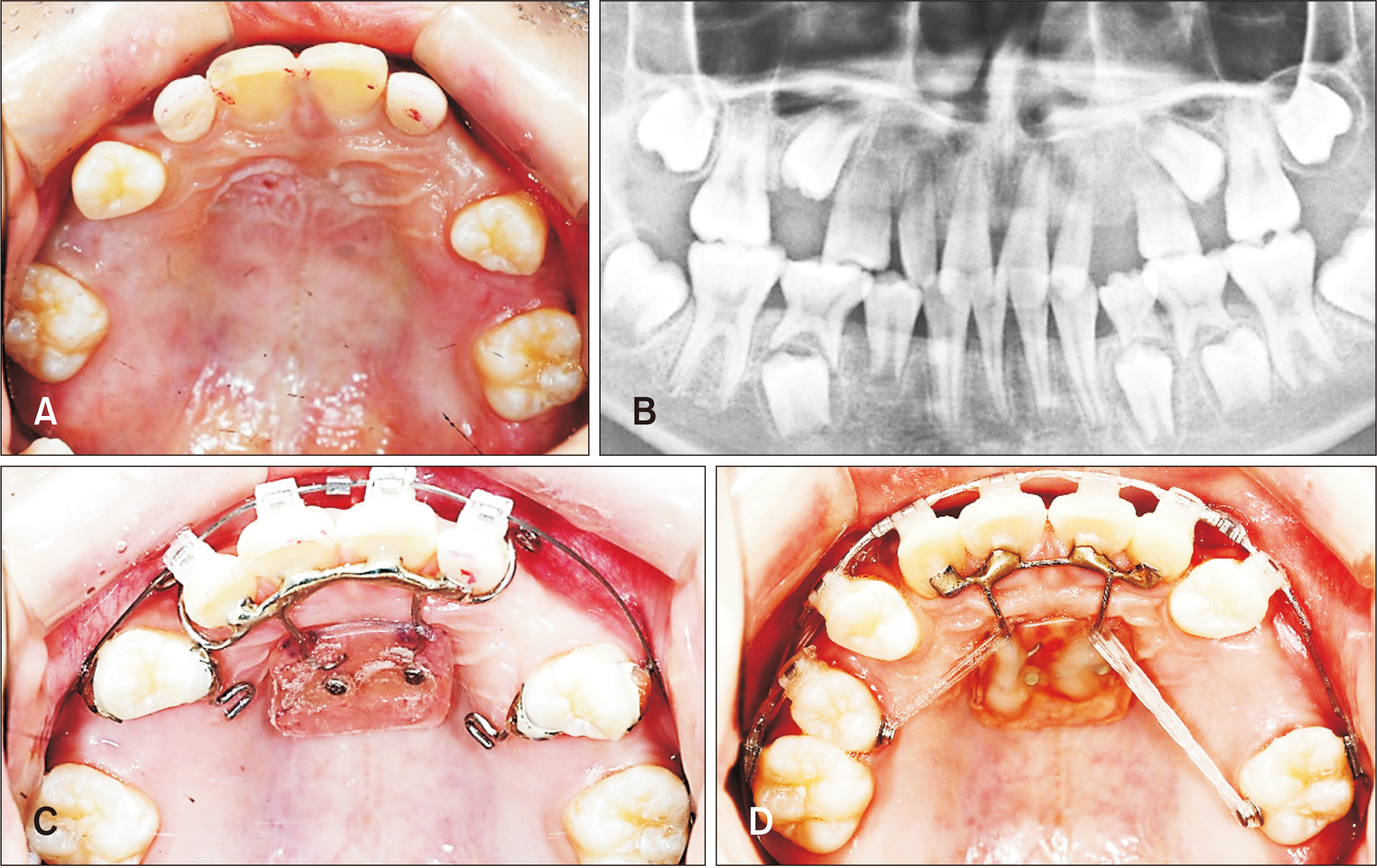
Figure 1 A ten-year-old male patient who requires whole posterior dentition protraction without anterior retraction. A, Pretreatment occlusal photograph. B, Pretreatment panoramic radiograph. C, Two 1.6 × 8 mm self-drilling type miniscrews (Proto type of Bio-Action Screw; Jin Biomed Co., Bucheon, Korea) were placed in the anterior palate area about 3 mm lateral to the midpalatal suture. D, After eight months of treatment occlusal photograph.
Reference
-
References
1. Bjoerk A, Krebs A, Solow B. 1964; A method for epidemiological registration of malocclusion. Acta Odontol Scand. 22:27–41. DOI: 10.3109/00016356408993963. PMID: 14158468.2. Kim SH, Yoon HG, Choi YS, Hwang EH, Kook YA, Nelson G. 2009; Evaluation of interdental space of the maxillary posterior area for orthodontic mini-implants with cone-beam computed tomography. Am J Orthod Dentofacial Orthop. 135:635–41. DOI: 10.1016/j.ajodo.2007.06.013. PMID: 19409346.
Article3. Lee JA, Ahn HW, Oh SH, Park KH, Kim SH, Nelson G. 2021; Evaluation of interradicular space, soft tissue, and hard tissue of the posterior palatal alveolar process for orthodontic mini-implant, using cone-beam computed tomography. Am J Orthod Dentofacial Orthop. 159:460–9. DOI: 10.1016/j.ajodo.2020.01.019. PMID: 33526299.
Article4. Kim SH, Park KH, Noh MK, Chung KR, Neson G. 2018; The biocreative strategy. Part 5: labial and lingual space closure in extraction treatment. J Clin Orthod. 52:528–49. PMID: 30346932.
