Results in Operative Treatment of Open Calcaneal Fracture
- Affiliations
-
- 1Department of Orthopaedic Surgery, College of Medicine, Chosun University, Gwangju, Korea
- KMID: 2520104
- DOI: http://doi.org/10.14193/jkfas.2021.25.3.133
Abstract
- Purpose
This paper reports the surgical treatment results of open calcaneal fractures performed at the author’s clinics focusing on open calcaneal fractures to help understand the appropriate treatment and realistic outcomes.
Materials and Methods
This study was conducted on 22 cases out of 30 patients who visited the hospital from February 2009 to December 2019 and were followed up for more than one year. In open fractures, the fracture was classified using the Gustilo-Anderson classification and was evaluated using the soft tissue status at the time of visit. Intra-articular calcaneal fractures were classified using Sanders classification. The radiological parameters were measured for the Böhler angle, Gissane angle, calcaneal length, height, and width before and after surgery, and at the last follow-up. The clinical outcomes were evaluated using the American Orthopaedic Foot and Ankle Society (AOFAS) ankle-hindfoot scale and investigated complications. In addition, statistical analysis of the incidence and associated factors of posttraumatic arthritis was conducted.
Results
In all cases, the surgical treatment was performed by minimally invasive surgery. The AOFAS ankle-hindfoot scale conducted for a clinical evaluation of the final follow-up was averaged 72.5 points. In the classification of open fractures, the Gustilo-Anderson classification type IIIA was the most common, and the Sanders type III was the most common. Of the 22 cases after surgery, 15 cases had complications, 11 cases had posttraumatic arthritis, eight cases had an infection, and 4 cases had both complications. Only the Sanders classification showed a statistically significant correlation with the incidence of posttraumatic osteoarthritis (p-value 0.032).
Conclusion
In treating open calcaneal fractures, internal fixation by a minimally invasive approach showed relatively satisfactory results. However, follow-up research will be needed, including the results of a long-term follow-up through a large number of cases and comparative studies with other surgical methods.
Keyword
Figure
-

Figure. 1 (A~D) Postoperative radiographs. The definitive fixation using Steinmann pin and 4.0-, 6.5-cannulated screws was performed. Postoperative X-ray (A: Ankle lateral view, B: Calcaneal axial view, C: Brodens view), Böhler angle, and Gissane angle have recovered. (D) Computed tomography after 6 months of surgery (semicoronal view) shows well reducted articular surface.
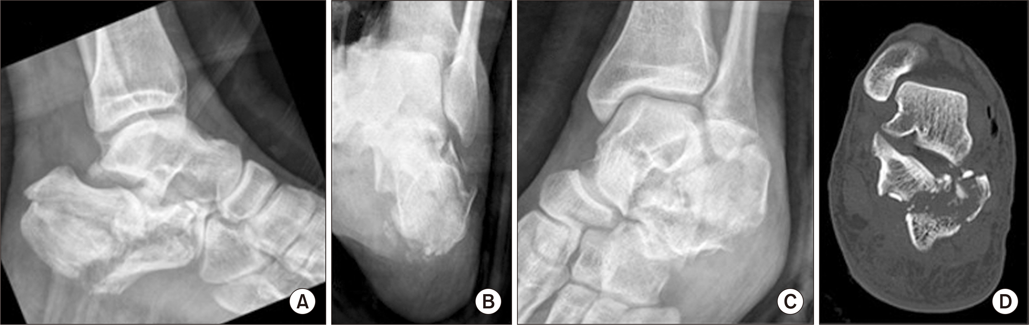
Figure. 2 Preoperative radiographs. Preoperative X-ray (A: ankle lateral view, B: calcaneal axial view, C: Brodens view), It is joint depressive calcaneal fracture. The posterior joint surface is depressive and lateral wall is bulging on calcaneal axial view. (D) Preoperative computed tomography (semicoronal view) shows joint depressive type. Sander type IIIAB calcaneal fracture.
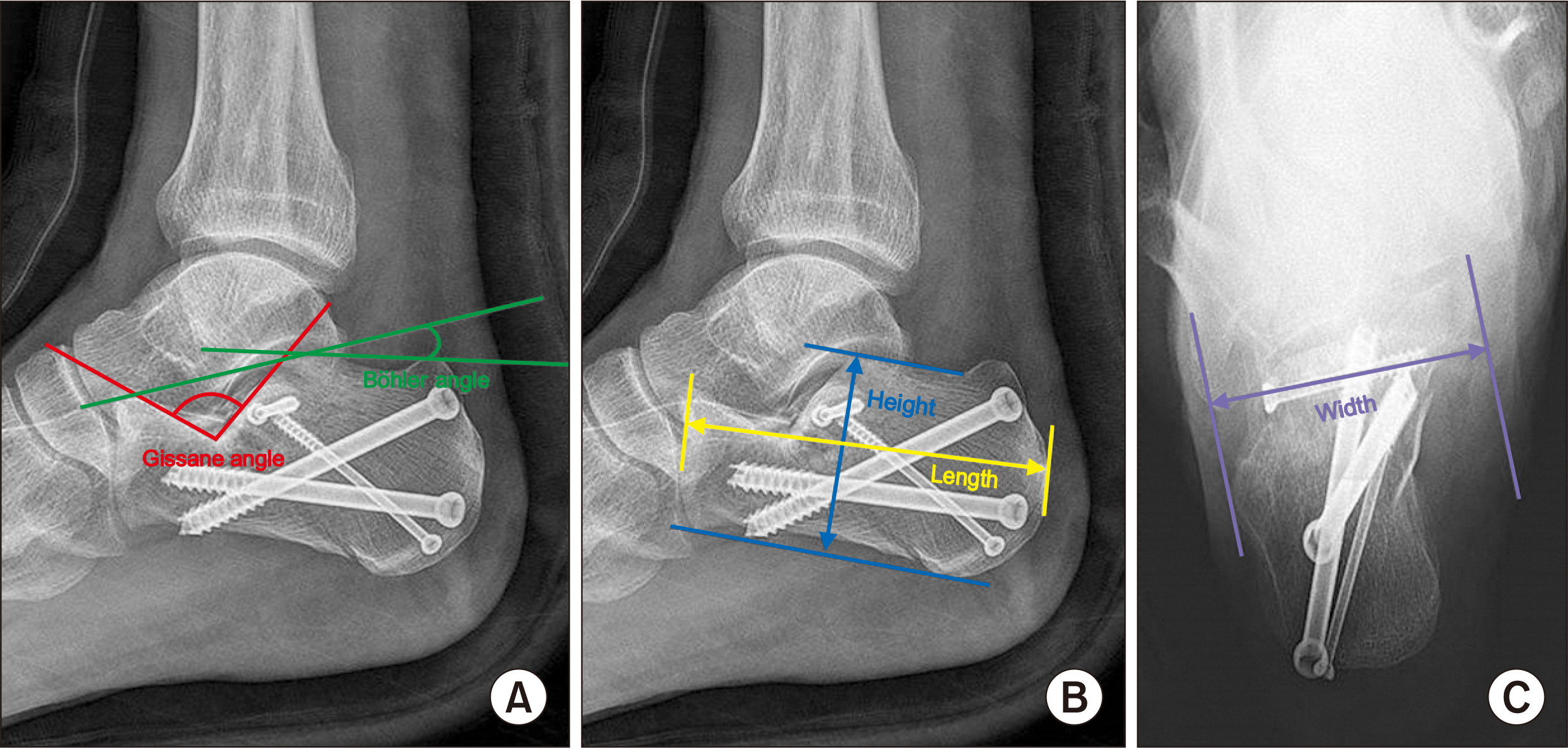
Figure. 3 Radiologic assessment. (A) Ankle lateral X-ray. Gissane angle, Böhler angle, and Gissane angle are formed by the downward and upward slopes of the calcaneal superior surface. Böhler angle is composed of a line drawn from the highest point of the anterior process of the calcaneus to the highest point of the posterior facet and a line drawn tangential to the superior edge of the tuberosity. (B) Ankle lateral X-ray: calcaneal height, calcaneal length. The calcaneal height and calcaneal length were measured at the highest and lowest length and the longest length from anterior to posterior in the lateral X-ray. (C) Calcaneal axial view. Calcaneal width calcaneal width measures the widest width including sustentaculum tali on calcaneal axial view.
Reference
-
1. Sanders R, Fortin P, DiPasquale T, Walling A. 1993; Operative treatment in 120 displaced intraarticular calcaneal fractures. Results using a prognostic computed tomography scan classification. Clin Orthop Relat Res. (290):87–95. DOI: 10.1097/00003086-199305000-00012.2. Park H, Shin SJ, Kim SR, Nam KW, Choi SW, Seo KB, et al. 2011; Bilateral open transcalcaneal fracture with talonavicular dislocation: a case report. J Korean Fract Soc. 24:87–91. doi: 10.12671/jkfs.2011.24.1.87. DOI: 10.12671/jkfs.2011.24.1.87.3. Ebraheim NA, Savolaine ER, Paley K, Jackson WT. 1993; Comminuted fracture of the calcaneus associated with subluxation of the talus. Foot Ankle. 14:380–4. doi: 10.1177/107110079301400702. DOI: 10.1177/107110079301400702. PMID: 8406256.
Article4. Sohn HM, Lee JY, Ha SH, Jo SH. 2007; The comparison of radiographic parameters and clinical results after operative treatment of displaced intraarticular calcaneal fractures. J Korean Fract Soc. 20:227–32. doi: 10.12671/jkfs.2007.20.3.227. DOI: 10.12671/jkfs.2007.20.3.227.
Article5. Folk JW, Starr AJ, Early JS. 1999; Early wound complications of operative treatment of calcaneus fractures: analysis of 190 fractures. J Orthop Trauma. 13:369–72. doi: 10.1097/00005131-199906000-00008. DOI: 10.1097/00005131-199906000-00008. PMID: 10406705.
Article6. Heier KA, Infante AF, Walling AK, Sanders RW. 2000; The natural history and treatment of open calcaneal fractures. J Orthop Trauma. 14:141–2. DOI: 10.1097/00005131-200002000-00056.
Article7. Siebert CH, Hansen M, Wolter D. 1998; Follow-up evaluation of open intra-articular fractures of the calcaneus. Arch Orthop Trauma Surg. 117:442–7. doi: 10.1007/s004020050289. DOI: 10.1007/s004020050289. PMID: 9801778.
Article8. Aldridge JM 3rd, Easley M, Nunley JA. 2004; Open calcaneal fractures: results of operative treatment. J Orthop Trauma. 18:7–11. doi: 10.1097/00005131-200401000-00002. DOI: 10.1097/00005131-200401000-00002. PMID: 14676550.9. Heier KA, Infante AF, Walling AK, Sanders RW. 2003; Open fractures of the calcaneus: soft-tissue injury determines outcome. J Bone Joint Surg Am. 85:2276–82. doi: 10.2106/00004623-200312000-00002. DOI: 10.2106/00004623-200312000-00002.
Article10. Bèzes H, Massart P, Delvaux D, Fourquet JP, Tazi F. 1993; The operative treatment of intraarticular calcaneal fractures. Indications, technique, and results in 257 cases. Clin Orthop Relat Res. (290):55–9. DOI: 10.1097/00003086-199305000-00008.11. Letournel E. 1993; Open treatment of acute calcaneal fractures. Clin Orthop Relat Res. (290):60–7. DOI: 10.1097/00003086-199305000-00009.
Article12. Paley D, Hall H. 1993; Intra-articular fractures of the calcaneus. A critical analysis of results and prognostic factors. J Bone Joint Surg Am. 75:342–54. doi: 10.2106/00004623-199303000-00005. DOI: 10.2106/00004623-199303000-00005. PMID: 8444912.
Article13. Zwipp H, Tscherne H, Thermann H, Weber T. 1993; Osteosynthesis of displaced intraarticular fractures of the calcaneus. Results in 123 cases. Clin Orthop Relat Res. (290):76–86. DOI: 10.1097/00003086-199305000-00011.14. Gustilo RB, Anderson JT. 1976; Prevention of infection in the treatment of one thousand and twenty-five open fractures of long bones: retrospective and prospective analyses. J Bone Joint Surg Am. 58:453–8. doi: 10.2106/00004623-197658040-00004. DOI: 10.2106/00004623-197658040-00004.15. Tornetta P 3rd, Ricci WM, Ostrum RF, McQueen MM, McKee MD, Court-Brown CM. 2020. Rockwood and Green’s fractures in adults. 9th ed. Wolters Kluwer;Philadelphia:16. Byun YS, Cho YH, Park JW, Lee JS, Kim JH. 2004; Early postoperative complications of calcaneal fractures following operative treatment by a lateral extensile approach. J Korean Fract Soc. 17:323–7. doi: 10.12671/jkfs.2004.17.4.323. DOI: 10.12671/jkfs.2004.17.4.323.
Article17. Acello AN, Wallace GF, Pachuda NM. 1995; Treatment of open fractures of the foot and ankle: a preliminary report. J Foot Ankle Surg. 34:329–46. doi: 10.1016/S1067-2516(09)80002-1. DOI: 10.1016/S1067-2516(09)80002-1.
Article18. Franklin JL, Johnson KD, Hansen ST Jr Jr. 1984; Immediate internal fixation of open ankle fractures. Report of thirty-eight cases treated with a standard protocol. J Bone Joint Surg Am. 66:1349–56. DOI: 10.2106/00004623-198466090-00004. PMID: 6438107.
Article19. Bray TJ, Endicott M, Capra SE. 1989; Treatment of open ankle fractures. Immediate internal fixation versus closed immobilization and delayed fixation. Clin Orthop Relat Res. (240):47–52. DOI: 10.1097/00003086-198903000-00007.20. Spierings KE, Min M, Nooijen LE, Swords MP, Schepers T. 2019; Managing the open calcaneal fracture: a systematic review. Foot Ankle Surg. 25:707–13. doi: 10.1016/j.fas.2018.10.005. DOI: 10.1016/j.fas.2018.10.005. PMID: 30467055.
Article21. Sanders R, Vaupel ZM, Erdogan M, Downes K. 2014; Operative treatment of displaced intraarticular calcaneal fractures: long-term (10-20 years) results in 108 fractures using a prognostic CT classification. J Orthop Trauma. 28:551–63. doi: 10.1097/BOT.0000000000000169. DOI: 10.1097/BOT.0000000000000169. PMID: 25243849.22. Backes M, Schepers T, Beerekamp MS, Luitse JS, Goslings JC, Schep NW. 2014; Wound infections following open reduction and internal fixation of calcaneal fractures with an extended lateral approach. Int Orthop. 38:767–73. doi: 10.1007/s00264-013-2181-1. DOI: 10.1007/s00264-013-2181-1. PMID: 24281853. PMCID: PMC3971279.
Article23. Lawrence SJ. 2004; Open calcaneal fractures. Orthopedics. 27:737–41. quiz 742–3. doi: 10.3928/0147-7447-20040701-14. DOI: 10.3928/0147-7447-20040701-14. PMID: 15315044.
Article24. Miric A, Patterson BM. 1998; Pathoanatomy of intra-articular fractures of the calcaneus. J Bone Joint Surg Am. 80:207–12. doi: 10.2106/00004623-199802000-00007. DOI: 10.2106/00004623-199802000-00007. PMID: 9486726.
Article25. Rammelt S, Barthel S, Biewener A, Gavlik JM, Zwipp H. 2003; [Calcaneus fractures. Open reduction and internal fixation]. Zentralbl Chir. 128:517–28. German doi: 10.1055/s-2003-40627. DOI: 10.1055/s-2003-40627. PMID: 12865959.26. Gao X, Fan HY, Huang R, Sui YQ, Li F, Yin HL. 2021; Management of open calcaneal fractures with medial wounds by one-stage sequential reduction and frame structure fixation using percutaneous Kirschner wires. Orthop Surg. 13:225–36. doi: 10.1111/os.12902. DOI: 10.1111/os.12902. PMID: 33403804. PMCID: PMC7862139.
Article27. Zhang T, Yan Y, Xie X, Mu W. 2016; Minimally invasive sinus tarsi approach with cannulated screw fixation combined with vacuum-assisted closure for treatment of severe open calcaneal fractures with medial wounds. J Foot Ankle Surg. 55:112–6. doi: 10.1053/j.jfas.2015.07.023. DOI: 10.1053/j.jfas.2015.07.023. PMID: 26372552.28. Rammelt S, Amlang M, Barthel S, Gavlik JM, Zwipp H. 2010; Percutaneous treatment of less severe intraarticular calcaneal fractures. Clin Orthop Relat Res. 468:983–90. doi: 10.1007/s11999-009-0964-x. DOI: 10.1007/s11999-009-0964-x. PMID: 19582524. PMCID: PMC2835587.
Article
- Full Text Links
-
- Actions
-
Cited
- CITED
-
- Close
- Share
-
- Similar articles
-
- Operative Treatment of Intraarticular Calcaneal Fracture: Comparison of Outcomes between Open Reduction and Closed Reduction
- Management of Displaced Intra-articular Calcaneal Fracture
- Operative Treatment of Calcaneal Fracture
- Atraumatic Avulsion Fracture of Calcaneal Tuberosity in a Patient with Peripheral Neuropathy: A Case Report
- Operative Treatment of Avulsion Fractures of the Calcaneal Tuberosity
