A Case of Schwannoma in Subfrontal Area and Nasal Cavity
- Affiliations
-
- 1Department of Otolaryngology-Head and Neck Surgery, Dong-A University College of Medicine, Busan, Korea
- 2Department of Pathology, Dong-A University College of Medicine, Busan, Korea
- KMID: 2518680
- DOI: http://doi.org/10.18787/jr.2021.00354
Abstract
- Schwannoma is a benign solitary neoplasm emerging from the Schwann cells of the peripheral, cranial and autonomic nerves. Approximately 25 to 45% of schwannomas occur in the head and neck region. However, schwannoma in the subfrontal area, nasal cavity or paranasal sinus is very rare and accounts for only 4% of these neoplasms. We experienced a case of schwannoma in the subfrontal area and left nasal cavity in a 74-year-old man who complained of recurrent rhinorrhea. We report this unusual case of schwannoma with a review of the literature.
Figure
-
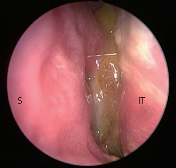
Fig. 1. Preoperative nasal endoscopy shows a yellowish, smooth surfaced mass between the nasal septum and the left inferior turbinate. S: Nasal septum, IT: Inferior turbinate.
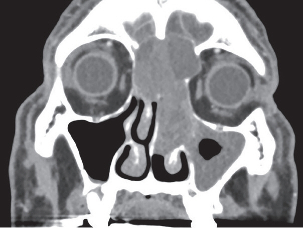
Fig. 2. Preoperative CT scan shows about 2×5.5 cm sized expansile mass in left nasal cavity involving both frontal sinus, anterior cranial fossa, frontal base.

Fig. 3. Brain magnetic resonance images (MRI). Coronal T1-weighted scan (A) and T2-weighted scan (B) show ill-defined inhomogeneous mass of intermediate signal intensity in nasal cavity, frontal sinus, frontal base and left maxillary sinus.
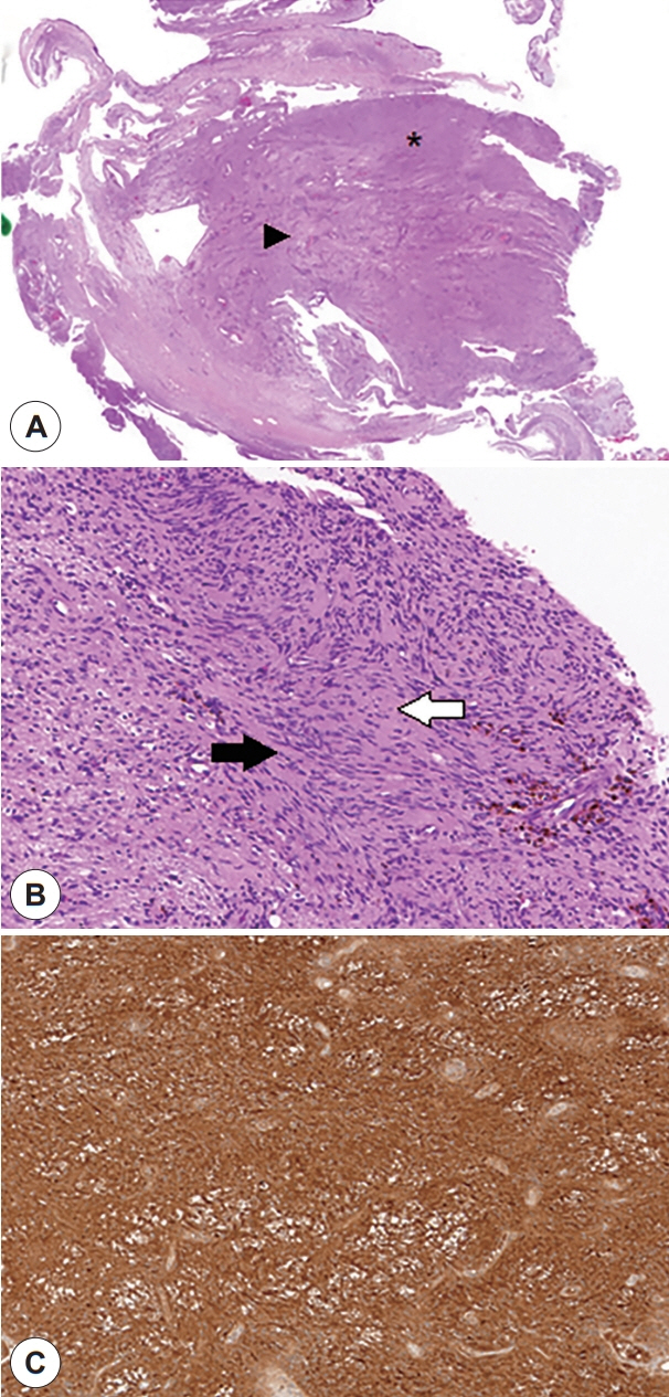
Fig. 4. Histopathologic finding of schwannoma. A well-encapsulated tumor shows areas of dense (astrix) and loose cellular population (arrowhead) (×50) (A). Tumor cells are showing Antoni A areas with classic spindle shaped cells and and elongated palisading nuclei which is called Verocay body (black arrow) and hypocellular Antoni B areas (white arrow) (×200) (B) and immunoreactive for S-100 protein (×200) (C).
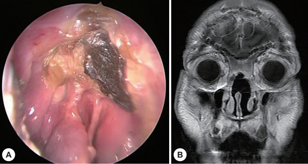
Fig. 5. Post-operative findings. Endoscopic exam shows well healing state of the nasal cavity 2 months after the operation (A). Postoperative MRI shows no sign of recurrence or remnant tumor (B).
Reference
-
References
1. Seo YI, Nam SY, An KH, Kim SY, Lee KS. Extracranial nerve sheath tumors of the head and neck. Korean J Otolaryngol. 1997; 40:908–13.2. Berlucchi M, Piazza C, Blanzuoli L, Battaglia G, Nicolai P. Schwannoma of the nasal septum: a case report with review of the literature. Eur Arch Otorhinolaryngol. 2000; 257(7):402–5.3. Kim SH, Lee JH, Sung SK, Choi CH. Subfrontal Schwannoma Extended Broadly to Nasal Cavity Treated by Gamma Knife Radiosurgery Following Surgical Excision: A Case Report. Brain Tumor Res Treat. 2017; 5(2):116–9.4. Min HJ, Kim KS. Differential Diagnosis Between Nasal Septal Schwannoma and Nasal Septal Neurofibroma. J Craniofac Surg. 2017; 28(7):1780–3.5. Nyte CP, Ryan WD. Schwannoma of the Nasal Ala in a 13 Year-Old Boy. Ear Nose Throat J. 2020; 99(1):42–3.6. Rajagopal S, Kaushik V, Irion K, Herd ME, Bhatnagar RK. Schwannoma of the nasal septum. Br J Radiol. 2006; 79(943):16–8.7. Thompson LD. Olfactory neuroblastoma. Head Neck Pathol. 2009; 3(3):252–9.8. Kim TH, Shin JH, Kwak EK. A case of a schwannoma in the nasal columella. SAGE Open Med Case Rep. 2019; 1(9):7.9. Hirao M, Gushiken T, Imokawa H, Kawai S, Inaba H, Tsukuda M. Solitary neurofibroma of the nasal cavity: resection with endoscopic surgery. J Laryngol Otol. 2001; 115(12):1012–4.10. Choe H, Jun YJ, Cho WS, Kim TH. A case of schwannoma of the nasal cavity mimicking olfactory neuroblastoma. Korean J Otolaryngol. 2007; 50(6):548–51.11. Taha MM, AlBakry A, ElSheikh M, AbdelBary TH. Olfactory Nerve Schwannoma: A Case Report and Review of the Literature. Surg J. 2018; 4(3):164–6.12. Gupta M, Rao N, Kour C, Kaur I. Septal Schwannoma of the Nose: A Rare Case. Turk Arch Otorhinolaryngol. 2017; 55(1):41–3.13. Min HJ, Hong SC, Kim KS. Nasal septal schwannoma: advances in diagonosis and treatment. J Craniofac Surg. 2017; 28(1):97–101.14. Tsai PY, Chan MY, Chen SH, Lin CY, Chen PH. The prognosis and recurrence of head and neck schwannomas: an 8-year retrospective study. Taiwan J Oral Maxillofac Surg. 2011; 22:165–74.
- Full Text Links
-
- Actions
-
Cited
- CITED
-
- Close
- Share
-
- Similar articles
-
- Subfrontal Schwannoma Extended Broadly to Nasal Cavity Treated by Gamma Knife Radiosurgery Following Surgical Excision: A Case Report
- A Case of Schwannoma of the Nasal Cavity Mimicking Olfactory Neuroblastoma
- A Case of Nasal Schwannoma Coexisting with Epidermal Cyst
- A Case of Neurilemmoma in the Nasal Vestibule
- A Case of Huge Fungus Ball in Nasal Cavity Misdiagnosed as Rhinolith on Nasal Septum
