Radiographic Evaluation of the Normal Distal Tibiofibular Syndesmosis in Neutral to Dorsiflexion on Weight-Bearing
- Affiliations
-
- 1Department of Orthopedic Surgery, Seoul Medical Center, Seoul, Korea
- 2Department of Orthopedic Surgery, Nowon Eulji Medical Center, Eulji University, Seoul, Korea
- 3K. T. Lee Orthopaedic Hospital, Seoul, Korea
- KMID: 2513512
- DOI: http://doi.org/10.5763/kjsm.2021.39.1.1
Abstract
- Purpose
Reliable landmarks of ankle syndesmosis change in various positions is important for managing ankle injury. The purpose of our study was to investigate and compare radiographic landmarks of normal ankle in various positions.
Methods
The study involved both ankle radiographs of 30 subjects (15 males, 15 females) without clinical or radiographic abnormality. Tibiofibular clear space (TFCS) and tibiofibular overlap (TFO) were measured on anteroposterior (AP) and mortise radiographs in non-standing (NS) and standing (S) neutral and dorsiflexion 10° (DF10) and 20° (DF20). The radiographic measurements were used to calculate means, standard deviations, and intra- and interobserver reliabilities, and compare TFCS and TFO in various positions and genders.
Results
On the AP view, the mean TFCS in NS, S, DF10, and DF20 positions were 4.00±0.97, 4.00±0.83, 4.35±0.95, and 4.45±0.89 mm and the mean TFO on the same positions were 6.58±2.27, 4.27±1.90, 3.44±1.96, and 2.38±1.91 mm. On the mortise view, the mean TFCS in NS, DF10, and DF20 positions were 3.62±0.88, 4.08±0.86, and 3.88±0.97 mm and the mean TFO on the same positions were 3.57±2.13, 2.31±1.77, and 3.57±2.14 mm. The reliabilities in all positions except TFCS on some positions were excellent. No measurement was significantly different between females and males except TFO in NS on mortise view (p=0.006) and DF10 on AP view (p=0.032).
Conclusion
Increase of TFCS and decrease of TFO on AP view reflects syndesmosis change from NS to DF20 on standing. Clinically, the effect of weight-bearing and reliability of TFO should be considered.
Keyword
Figure
-

Fig. 1 Measurements made on the anteroposterior view of the ankle at 1 cm above the plafond. (A) Measuring tibiofibular clear space and tibiofibular overlap on the anteroposterior view of standard radiograph in standing position, (B) its schematic drawing, and (C) cross-sectional schematic drawing matched with landmarks on panel B. L: lateral border of fibula, A: anterior tibial tubercle, M: medial border of fibula, P: posterior tibial tubercle, I: floor of incisura fibularis. All landmarks on panel C were added with superscript apostrophe to match landmarks on panel B.
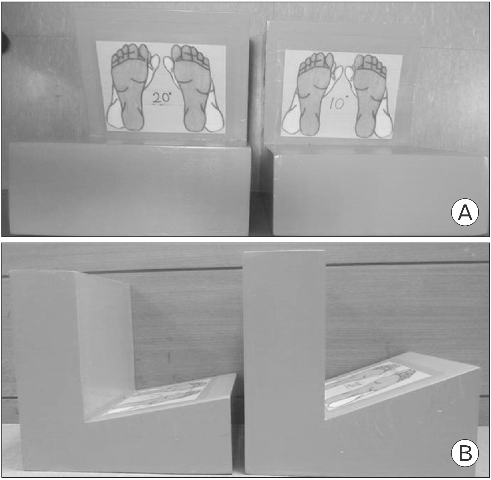
Fig. 2 Footholds for 10° and 20° ankle dorsiflexion. Left foothold is for 10° and right one for 20° in each image. (A) View from above. Blue footprints mean neutral position and green mean 15° internal rotation. (B) Sideview.

Fig. 3 Regression analysis of various ankle position: neutral in non-standing (NS) and standing (S) and dorsiflexion 10° (DF10) and 20° (DF20) in standing position of standard radiograph. (A) Tibiofibular clear space (TFCS) on the anteroposterior (AP) view. Coefficient of determination (linear)=0.042. (B) Tibiofibular overlap (TFO) on the AP view. Coefficient of determination (quadratic)=0.368. (C) TFCS on the mortise view. Coefficient of determination (quadratic)=0.041. (D) TFO on the mortise view. Coefficient of determination (quadratic)=0.081. (E) TFCS on the AP view excluding NS position. Coefficient of determination (quadratic)=0.045.
Reference
-
1. Kellett JJ. 2011; The clinical features of ankle syndesmosis injuries: a general review. Clin J Sport Med. 21:524–9. DOI: 10.1097/JSM.0b013e318234be7d. PMID: 22011797.2. Dubin JC, Comeau D, McClelland RI, Dubin RA, Ferrel E. 2011; Lateral and syndesmotic ankle sprain injuries: a narrative literature review. J Chiropr Med. 10:204–19. DOI: 10.1016/j.jcm.2011.02.001. PMID: 22014912. PMCID: PMC3259913.
Article3. Purvis GD. Displaced, unstable ankle fractures: classification, incidence, and management of a consecutive series. Clin Orthop Relat Res. 1982:91–8. DOI: 10.1097/00003086-198205000-00013. PMID: 6804148.4. Takebe K, Nakagawa A, Minami H, Kanazawa H, Hirohata K. Role of the fibula in weight-bearing. Clin Orthop Relat Res. 1984:289–92. DOI: 10.1097/00003086-198404000-00047. PMID: 6705357.
Article5. Goh JC, Mech AM, Lee EH, Ang EJ, Bayon P, Pho RW. Biomechanical study on the load-bearing characteristics of the fibula and the effects of fibular resection. Clin Orthop Relat Res. 1992:223–8. DOI: 10.1097/00003086-199206000-00028. PMID: 1600659.
Article6. Lambert KL. 1971; The weight-bearing function of the fibula: a strain gauge study. J Bone Joint Surg Am. 53:507–13. DOI: 10.2106/00004623-197153030-00007. PMID: 5580009.7. Van Heest TJ, Lafferty PM. 2014; Injuries to the ankle syndesmosis. J Bone Joint Surg Am. 96:603–13. DOI: 10.2106/JBJS.M.00094. PMID: 24695928.
Article8. Close JR. 1956; Some applications of the functional anatomy of the ankle joint. J Bone Joint Surg Am. 38(A):761–81. DOI: 10.2106/00004623-195638040-00005. PMID: 13331972.
Article9. Rasmussen O. 1985; Stability of the ankle joint: analysis of the function and traumatology of the ankle ligaments. Acta Orthop Scand Suppl. 211:1–75. DOI: 10.3109/17453678509154152. PMID: 3856377.10. Xenos JS, Hopkinson WJ, Mulligan ME, Olson EJ, Popovic NA. 1995; The tibiofibular syndesmosis: evaluation of the ligamentous structures, methods of fixation, and radiographic assessment. J Bone Joint Surg Am. 77:847–56. DOI: 10.2106/00004623-199506000-00005. PMID: 7782357.
Article11. Pettrone FA, Gail M, Pee D, Fitzpatrick T, Van Herpe LB. 1983; Quantitative criteria for prediction of the results after displaced fracture of the ankle. J Bone Joint Surg Am. 65:667–77. DOI: 10.2106/00004623-198365050-00013. PMID: 6406511.
Article12. Takao M, Ochi M, Oae K, Naito K, Uchio Y. 2003; Diagnosis of a tear of the tibiofibular syndesmosis: the role of arthroscopy of the ankle. J Bone Joint Surg Br. 85:324–9. DOI: 10.1302/0301-620X.85B3.13174. PMID: 12729102.13. Shah AS, Kadakia AR, Tan GJ, Karadsheh MS, Wolter TD, Sabb B. 2012; Radiographic evaluation of the normal distal tibiofibular syndesmosis. Foot Ankle Int. 33:870–6. DOI: 10.3113/FAI.2012.0870. PMID: 23050712.
Article14. Amin A, Janney C, Sheu C, Jupiter DC, Panchbhavi VK. 2019; Weight-bearing radiographic analysis of the tibiofibular syndesmosis. Foot Ankle Spec. 12:211–7. DOI: 10.1177/1938640018766631. PMID: 29607668.
Article15. Carrara C, Caravaggi P, Belvedere C, Leardini A. 2020; Radiographic angular measurements of the foot and ankle in weight-bearing: a literature review. Foot Ankle Surg. 26:509–17. DOI: 10.1016/j.fas.2019.07.008. PMID: 31402285.
Article16. Patel S, Malhotra K, Cullen NP, Singh D, Goldberg AJ, Welck MJ. 2019; Defining reference values for the normal tibiofibular syndesmosis in adults using weight-bearing CT. Bone Joint J. 101(B):348–52. DOI: 10.1302/0301-620X.101B3.BJJ-2018-0829.R1. PMID: 30813789.
Article17. Elgafy H, Semaan HB, Blessinger B, Wassef A, Ebraheim NA. 2010; Computed tomography of normal distal tibiofibular syndesmosis. Skeletal Radiol. 39:559–64. DOI: 10.1007/s00256-009-0809-4. PMID: 19830423.
Article18. Gardner MJ, Demetrakopoulos D, Briggs SM, Helfet DL, Lorich DG. 2006; Malreduction of the tibiofibular syndesmosis in ankle fractures. Foot Ankle Int. 27:788–92. DOI: 10.1177/107110070602701005. PMID: 17054878.
Article19. Chen Y, Qiang M, Zhang K, Li H, Dai H. 2015; A reliable radiographic measurement for evaluation of normal distal tibiofibular syndesmosis: a multi-detector computed tomography study in adults. J Foot Ankle Res. 8:32. DOI: 10.1186/s13047-015-0093-6. PMID: 26213578. PMCID: PMC4514948.
Article20. Lamer S, Hebert-Davies J, Dube V, et al. 2019; Effect of a controlled ankle motion walking boot on syndesmotic instability during weightbearing: a cadaveric study. Orthop J Sports Med. 7:2325967119864018. DOI: 10.1177/2325967119864018. PMID: 31457067. PMCID: PMC6702776.
Article21. Ostrum RF, De Meo P, Subramanian R. 1995; A critical analysis of the anterior-posterior radiographic anatomy of the ankle syndesmosis. Foot Ankle Int. 16:128–31. DOI: 10.1177/107110079501600304. PMID: 7599729.
Article22. Weisell RC. 2002; Body mass index as an indicator of obesity. Asia Pac J Clin Nutr. 11:S681–4. DOI: 10.1046/j.1440-6047.11.s8.5.x.
Article23. Fowler JR, Ilyas AM. 2011; The accuracy of digital radiography in orthopaedic applications. Clin Orthop Relat Res. 469:1781–4. DOI: 10.1007/s11999-010-1628-6. PMID: 20972654. PMCID: PMC3094611.
Article24. Bartonicek J. 2003; Anatomy of the tibiofibular syndesmosis and its clinical relevance. Surg Radiol Anat. 25:379–86. DOI: 10.1007/s00276-003-0156-4. PMID: 14504816.25. Beumer A, van Hemert WL, Niesing R, et al. Radiographic measurement of the distal tibiofibular syndesmosis has limited use. Clin Orthop Relat Res. 2004:227–34. DOI: 10.1097/01.blo.0000129152.81015.ad. PMID: 15232454.
Article26. Brage ME, Bennett CR, Whitehurst JB, Getty PJ, Toledano A. 1997; Observer reliability in ankle radiographic measurements. Foot Ankle Int. 18:324–9. DOI: 10.1177/107110079701800602. PMID: 9208288.
Article27. Harper MC, Keller TS. 1989; A radiographic evaluation of the tibiofibular syndesmosis. Foot Ankle. 10:156–60. DOI: 10.1177/107110078901000308. PMID: 2613128.
Article28. Kim JO, Choi HY, Yoo YW. 1995; Measurement of radiologic criteria for evaluation of the syndesmosis in Korean men. J Korean Fract Soc. 8:600–5. DOI: 10.12671/jksf.1995.8.3.600.
Article29. Malhotra K, Welck M, Cullen N, Singh D, Goldberg AJ. 2019; The effects of weight bearing on the distal tibiofibular syndesmosis: a study comparing weight bearing-CT with conventional CT. Foot Ankle Surg. 25:511–6. DOI: 10.1016/j.fas.2018.03.006. PMID: 30321955.
Article30. Anand Prakash A. 2017; Is incisura fibularis a reliable landmark for assessing syndesmotic stability? A systematic review of morphometric studies. Foot Ankle Spec. 10:246–51. DOI: 10.1177/1938640016685152. PMID: 28027658.
Article
- Full Text Links
-
- Actions
-
Cited
- CITED
-
- Close
- Share
-
- Similar articles
-
- Diagnostic Landmarks of Ankle Syndesmosis Separation Measured on Standard Ankle Anterior - posterior Radiographs of Normal Korean Adults
- Heterotopic Ossification of Distal Tibiofibular Syndesmosis after Ankle Fractures
- Injury of Distal Tibio-Fibular Syndesmosis Treated with Trans-Syndesmotic Screw Fixation
- The Effect of a Proximal and Distal Tibiofibular Joint Manipulation on Dorsiflexion and Balance in Individuals with a History of Lateral Ankle Sprain
- Treatement of Diastasis of the Distal Tibiofibular Syndesmosis
