Endocrinol Metab.
2021 Feb;36(1):201-202. 10.3803/EnM.2020.884.
Anaplastic Thyroid Carcinoma with Initial Ultrasonography Features Mimicking Subacute Thyroiditis
- Affiliations
-
- 1Department of Internal Medicine, Asan Medical Center, University of Ulsan College of Medicine, Seoul, Korea
- KMID: 2513303
- DOI: http://doi.org/10.3803/EnM.2020.884
Figure
-
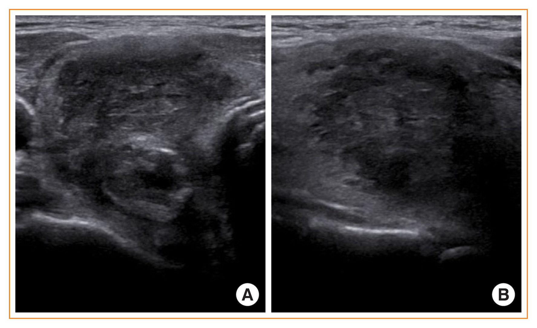
Fig. 1 Initial thyroid ultrasonography results showing a poorly defined and irregularly shaped hypoechogenic lesion. (A) Coronal view. (B) Longitudinal view.
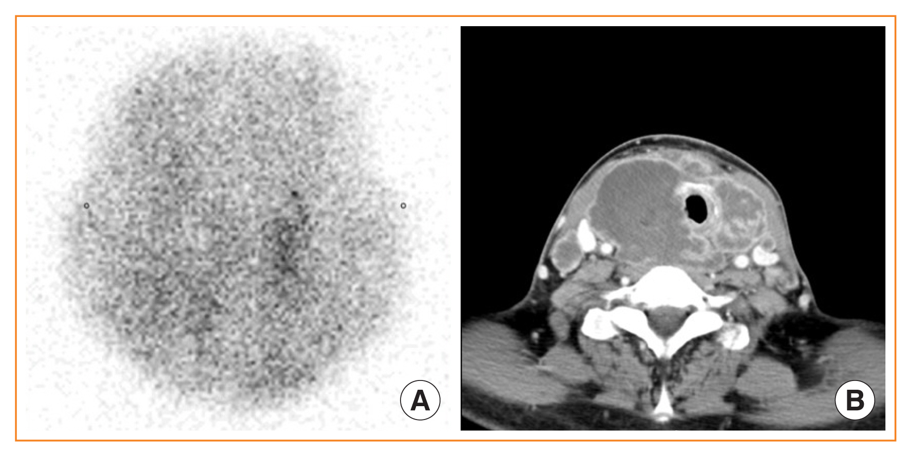
Fig. 2 Initial thyroid scan and neck computed tomography (CT) taken after the diagnosis of anaplastic thyroid carcinoma. (A) Initial thyroid scan showing severely decreased uptake. (B) Neck CT showing a huge necrotic mass in both thyroid lobes compressing the airway.
Reference
-
1. Bennedbaek FN, Hegedus L. The value of ultrasonography in the diagnosis and follow-up of subacute thyroiditis. Thyroid. 1997; 7:45–50.
Article2. Fatourechi V, Aniszewski JP, Fatourechi GZ, Atkinson EJ, Jacobsen SJ. Clinical features and outcome of subacute thyroiditis in an incidence cohort: Olmsted County, Minnesota, study. J Clin Endocrinol Metab. 2003; 88:2100–5.
Article3. Meier DA, Nagle CE. Differential diagnosis of a tender goiter. J Nucl Med. 1996; 37:1745–7.4. Hamburger JI, Miller JM, Kini SR. Lymphoma of the thyroid. Ann Intern Med. 1983; 99:685–93.
Article
- Full Text Links
-
- Actions
-
Cited
- CITED
-
- Close
- Share
-
- Similar articles
-
- Ultrasonographic Findings of Papillary Thyroid Cancer with or without Hashimoto's Thyroiditis
- A Case of Riedel's Thyroiditis Associated with a Benign Nodule
- A Case of Subacute Thyroiditis Associated with Papillary Thyroid Carcinoma and Takayasu's Arteritis
- A Case of Graves' Disease Following Subacute Thyroiditis
- A case of an autonomously functioning thyroid nodule combined with subacute thyroiditis
