Esophageal Stricture Caused by the Ingestion of Undissolved Picosulfate Powder
- Affiliations
-
- 1Department of Internal Medicine, Chungbuk National University Hospital, Cheongju, Korea
- 2Department of Internal Medicine, Chungbuk National University College of Medicine, Cheongju, Korea
- KMID: 2512317
- DOI: http://doi.org/10.5946/ce.2019.206
Abstract
- Picosulfate solution is widely used as a small volume bowel cleansing agent and is considered to be effective and relatively safe. A case of a 75-year-old woman ingested picosulfate powder and drank a small volume of water, subsequently experienced severe burning pain in the chest. Endoscopy was performed and showed a submucosal hemorrhage and exudative ulcers at the mid to lower esophagus. At 2 weeks, her symptoms improved with conservative treatment. However, liquid food dysphagia developed 11 weeks after ingestion. A follow-up endoscopy revealed multiple esophageal strictures, which were treated with a fully covered metal stent and esophageal balloon dilation. Consequently, the esophageal strictures improved after one year. As this case demonstrates, detailed information about picosulfate powder ingestion after dissolving it in more than 200 mL of water should be presented to patients to avoid esophageal injury.
Figure
-
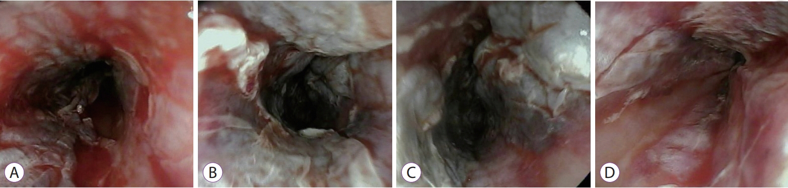
Fig. 1. Endoscopic findings at 1 day after ingestion of picosulfate powder. Endoscopy reveals submucosal hemorrhage, ulcerative lesions, and spotty necrosis along the whole esophagus. Caustic injury is more severe in the mid esophagus (B, C) than the upper (A) or lower (D) parts.
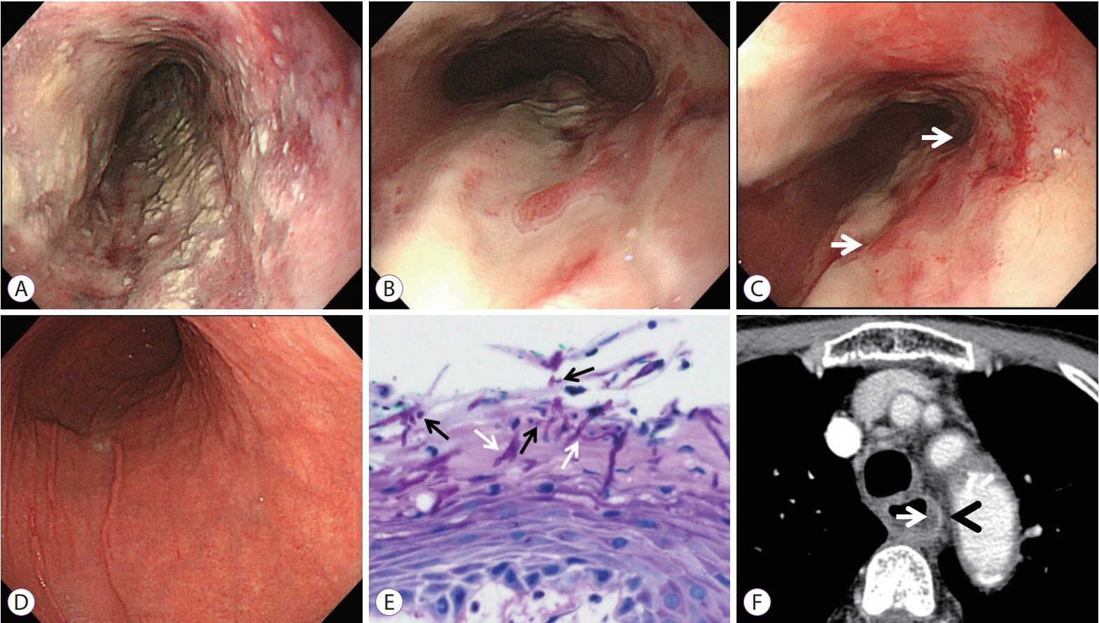
Fig. 2. Image findings at 1 week after ingestion. Endoscopy reveals diffusely scattered yellowish exudates (A), mucosal sloughing (B), and multiple ulcers (C, arrows) throughout the esophagus and linear erythematous lesions in the stomach (D). An endoscopic biopsy (E) shows fungal pseudohyphae (white arrows) and a few yeasts (black arrows) in the esophageal mucosa on PAS stain (original magnification, X400). A neck CT scan (F) reveals esophageal edema (arrow) and increased wall enhancement (arrowhead).
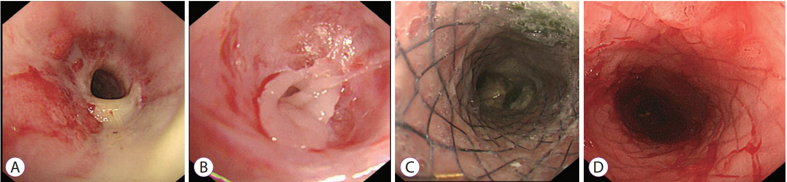
Fig. 3. Endoscopic findings at 2 weeks and 11 weeks after ingestion. Luminal narrowing and esophageal ulcers covered with exudates at 2 weeks (A). The narrowest part of the esophageal stricture (B) caused by healed ulcer scars at 11 weeks after ingestion. Esophageal stricture was treated with fully covered metal stenting for 3 months (C, D).
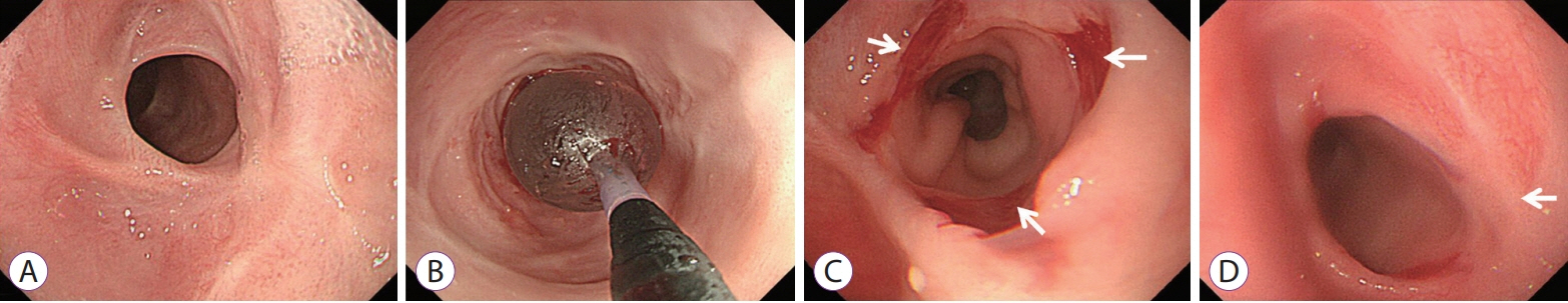
Fig. 4. Endoscopic findings at 8 months and 12 months after ingestion. Restenosis at the mid esophagus after stenting (A) is treated with esophageal balloon dilation (B). Luminal stenosis relief by mucosal tearing (C, arrows) and remaining ulcer scars (D, arrow).
Reference
-
1. Love J, Bernard EJ, Cockeram A, et al. A multicentre, observational study of sodium picosulfate and magnesium citrate as a precolonoscopy bowel preparation. Can J Gastroenterol. 2009; 23:706–710.
Article2. Hoy SM, Scott LJ, Wagstaff AJ. Sodium picosulfate/magnesium citrate: a review of its use as a colorectal cleanser. Drugs. 2009; 69:123–136.3. Suh JP, Choi YS, Lee SH. Education and imaging. Gastroenterology: acute mucosal injury of esophagus and stomach induced by sodium picosulfate/magnesium citrate for bowel preparation. J Gastroenterol Hepatol. 2014; 29:1571.4. Lee DS, Kim HS, Lee SH, Jeon JH, Lee YK. A case of PICOLIGHT powder induced thermal injury of the gastric mucosa. Korean J Helicobacter Up Gastrointest Res. 2014; 14:58–60.
Article5. Seo JY, Kang KJ, Kang HS, et al. Corrosive esophagitis caused by ingestion of picosulfate. Clin Endosc. 2015; 48:66–69.
Article6. Kim GB, Hwang SY, Shin TG, et al. Upper airway obstruction resulting from acute mucosal injury induced by direct ingestion of sodium picosulfate/magnesium citrate powder. Clin Exp Emerg Med. 2016; 3:109–111.
Article7. Ze EY, Choi CH, Kim JW. Acute gastric injury caused by undissolved sodium picosulfate/magnesium citrate powder. Clin Endosc. 2017; 50:87–90.
Article8. Yang DH, Bang BW, Kwon KS, Kim HK, Shin YW. A case of thermal esophageal injury induced by sodium picosulfate with magnesium citrate. Case Rep Gastrointest Med. 2017; 2017:9879843.
Article9. Poley JW, Steyerberg EW, Kuipers EJ, et al. Ingestion of acid and alkaline agents: outcome and prognostic value of early upper endoscopy. Gastrointest Endosc. 2004; 60:372–377.
Article10. Hollenbach M, Tunnemann J, Struck MF, et al. Endoscopic findings and outcome in caustic ingestion of acidic and alkaline agents in adults: a retrospective analysis. Medicine (Baltimore). 2019; 98:e16729.11. Zargar SA, Kochhar R, Mehta S, Mehta SK. The role of fiberoptic endoscopy in the management of corrosive ingestion and modified endoscopic classification of burns. Gastrointest Endosc. 1991; 37:165–169.12. Katz A, Kluger Y. Caustic material ingestion injuries-paradigm shift in diagnosis and treatment. Health Care Curr Rev. 2015; 3:152.
Article13. Ryu HH, Jeung KW, Lee BK, et al. Caustic injury: can CT grading system enable prediction of esophageal stricture? Clin Toxicol (Phila). 2010; 48:137–142.
Article14. Methasate A, Lohsiriwat V. Role of endoscopy in caustic injury of the esophagus. World J Gastrointest Endosc. 2018; 10:274–282.
Article15. Tharavej C, Pungpapong SU, Chanswangphuvana P. Outcome of dilatation and predictors of failed dilatation in patients with acid-induced corrosive esophageal strictures. Surg Endosc. 2018; 32:900–907.
Article16. Chirica M, Bonavina L, Kelly MD, Sarfati E, Cattan P. Caustic ingestion. Lancet. 2017; 389:2041–2052.
Article17. Siersema PD. How to approach a patient with refractory or recurrent benign esophageal stricture. Gastroenterology. 2019; 156:7–10.
Article
- Full Text Links
-
- Actions
-
Cited
- CITED
-
- Close
- Share
-
- Similar articles
-
- Acute Gastric Injury Caused by Undissolved Sodium Picosulfate/Magnesium Citrate Powder
- Corrosive Esophagitis Caused by Ingestion of Picosulfate
- A Case of Upper Airway and Esophageal Injury after Ingestion of Sodium Picosulfate and Magnesium Citrate for Colonoscopy
- A Case of Gastric Stricture Caused by Chemical Fertilizer
- A Case of Refractory Esophageal Stricture Induced by Lye Ingestion and Treated by Temporary Placement of Newly Designed Self-Expanding Metal Stent and Wetting with Mitomycin C
