Anat Cell Biol.
2020 Dec;53(4):516-518. 10.5115/acb.20.095.
Case report of a vertical straight sinus with hydrocephalus and Chiari I malformation
- Affiliations
-
- 1Department of Neurosurgery, Tulane Center for Clinical Neurosciences, Tulane University School of Medicine, New Orleans, LA, USA
- 2Department of Structural & Cellular Biology, Tulane University School of Medicine, New Orleans, LA, USA
- 3Department of Neurosurgery and Ochsner Neuroscience Institute, Ochsner Health System, New Orleans, LA, USA
- 4Department of Anatomical Sciences, St. George’s University, St. George’s, Grenada
- KMID: 2509700
- DOI: http://doi.org/10.5115/acb.20.095
Abstract
- The straight sinus is a division of the dural venous sinuses, found beneath the splenium of the corpus callosum. At the internal occipital protuberance, it comes together with the superior sagittal sinus and transverse sinus to form the torcular Herophili. It functions as a major site of venous drainage for the cerebellum, inferior sagittal sinus, and vein of Galen. Many morphological variations have been reported involving the angulation, positioning, and number of straight sinuses present. Patients with Chiari II and III malformations have been observed to have a high incidence of anatomical variation with their dural venous sinuses, including vertically oriented straight sinuses. Additionally, there is a high rate of hydrocephalus in this patient population. Herein, we report a vertically oriented straight sinus in a child.
Figure
-
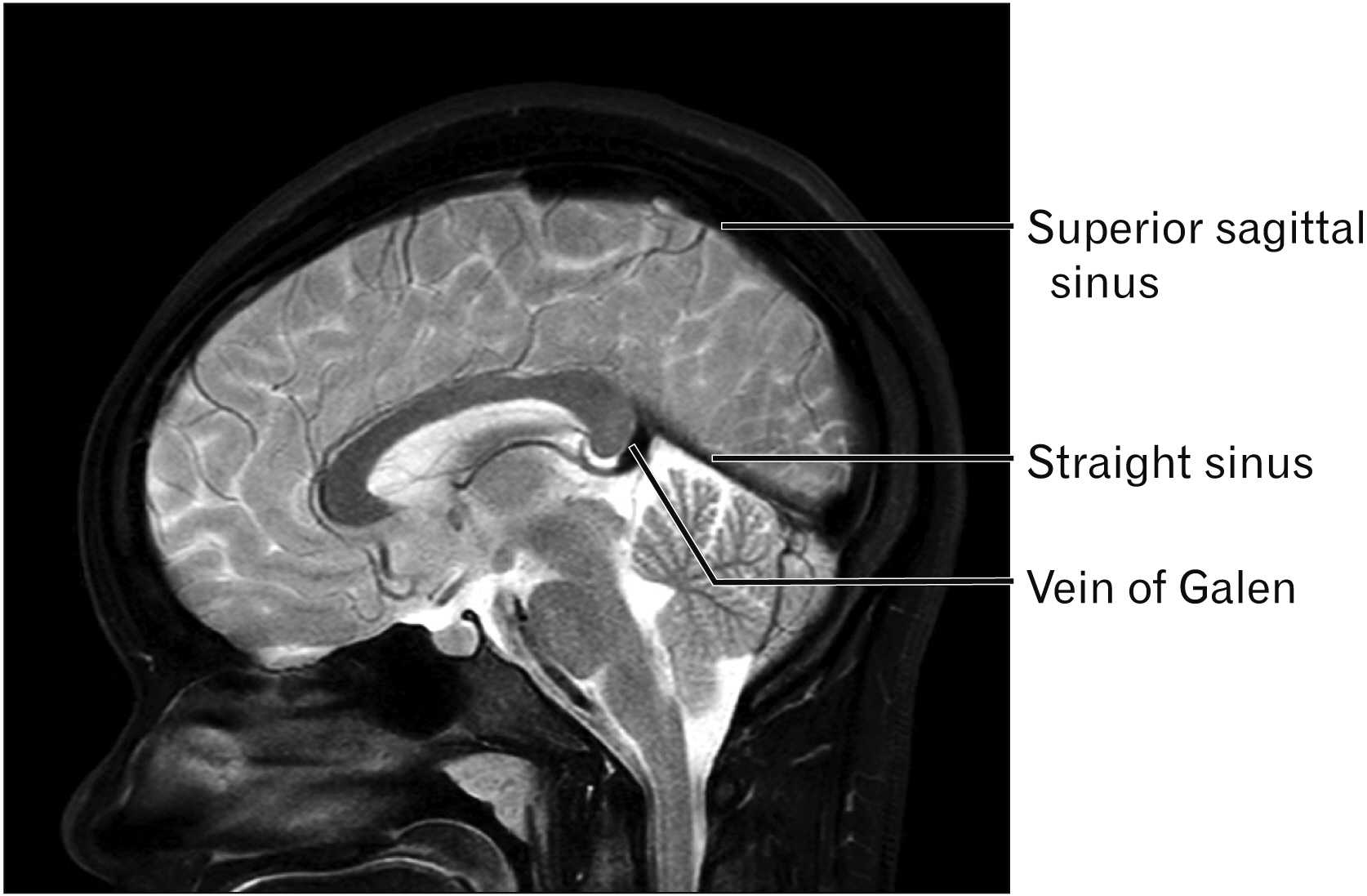
Fig. 1 T2-weighted sagittal magnetic resonance imaging noting a normally positioned straight sinus.

Fig. 2 Patient presented herein. Note the well-developed inferior sagittal sinus (upper arrow), superior sagittal sinus (middle arrow) and near vertical straight sinus (lower arrow). The herniated cerebellar tonsils (asterisk) noting the Chiari I malformation are also seen.
Reference
-
References
1. Kretschmann HJ, Weinrich W. 2004. Cranial neuroimaging and clinical neuroanatomy: atlas of MIR imaging and computed tomography. 3rd ed. Thieme Medical Publishers;Stuttgart: DOI: 10.1055/b-0034-56176.2. Tubbs RS. 2020. Anatomy, imaging and surgery of the intracranial dural venous sinuses. Elsevier;St. Louis:3. Jinkins JR. 2000. Atlas of neuroradiologic embryology, anatomy, and variants. Lippincott Williams & Wilkins;Philadelphia:4. Conn PM. 2008. Neuroscience in medicine. 3rd ed. Humana Press;Totowa: DOI: 10.1007/978-1-60327-455-5.5. Bayot ML, Reddy V, Zabel MK. 2020. Neuroanatomy, dural venous sinuses. StatPearls Publishing;Treasure Island:6. Harrigan MR, Deveikis JP. 2013. Handbook of cerebrovascular disease and neurointerventional technique. 2nd ed. Humana Press;Totowa: DOI: 10.1007/978-1-61779-946-4.7. Saxena RC, Beg MA, Das AC. 1973; Double straight sinus. Report of six cases. J Neurosurg. 39:540–2. DOI: 10.3171/jns.1973.39.4.0540. PMID: 4730345.8. Saxena RC, Beg MA, Das AC. 1974; The straight sinus. J Neurosurg. 41:724–7. DOI: 10.3171/jns.1974.41.6.0724. PMID: 4424313.
Article9. Wolpert SM. 1969; Dural sinus configuration: measure of congenital disease. Radiology. 92:1511–6. DOI: 10.1148/92.7.1511. PMID: 5799840.
Article10. Mağden AO. 1991; Triple straight sinus--report of 2 cases. Anat Anz. 173:17–22. PMID: 1952091.11. Knott JF. 1881; On the cerebral sinuses and their variations. J Anat Physiol. 16(Pt 1):27–42. PMID: 17231415. PMCID: PMC1310067.12. Ryu CW. 2010; Persistent falcine sinus: is it really rare? AJNR Am J Neuroradiol. 31:367–9. DOI: 10.3174/ajnr.A1794. PMID: 19779000.
Article13. Hasegawa M, Yamashita J, Yamashima T. 1991; Anatomical variations of the straight sinus on magnetic resonance imaging in the infratentorial supracerebellar approach to pineal region tumors. Surg Neurol. 36:354–9. DOI: 10.1016/0090-3019(91)90023-3. PMID: 1745959.
Article14. Williams H. 2008; A unifying hypothesis for hydrocephalus, Chiari malformation, syringomyelia, anencephaly and spina bifida. Cerebrospinal Fluid Res. 5:7. DOI: 10.1186/1743-8454-5-7. PMID: 18405364. PMCID: PMC2365936.
Article15. el Gammal T, Mark EK, Brooks BS. 1988; MR imaging of Chiari II malformation. AJR Am J Roentgenol. 150:163–70. DOI: 10.2214/ajr.150.1.163. PMID: 3257116.
Article
- Full Text Links
-
- Actions
-
Cited
- CITED
-
- Close
- Share
-
- Similar articles
-
- A case of Arnold-Chiari malformation
- Acquired Chiari-malformation after Ventriculoperitoneal Shunt for Hydrocepalus Associated with Neurocysticercosis
- Two cases of Arnold-Chiari malformation type II
- Chiari 1.5 Malformation : An Advanced Form of Chiari I Malformation
- Co-Occurrence of Syringomyelia and Hydrocephalus in a Patient Without Chiari Malformation in Her 50's
