Relationship between the Diurnal Temperature Range and Wound Healing of Diabetic Foot: Animal Study
- Affiliations
-
- 1Department of Orthopedic Surgery, Soon Chun Hyang University Seoul Hospital, Seoul, Korea
- 2Department of Orthopaedic Surgery, Hallym University Chuncheon Sacred Heart Hospital, Chuncheon, Korea
- 3Department of Orthopedic Surgery, Inje University Seoul Paik Hospital, Seoul, Korea
- KMID: 2509535
- DOI: http://doi.org/10.14193/jkfas.2020.24.4.142
Abstract
- Relationship between the Diurnal Temperature Range and Wound Healing of Diabetic Foot: Animal StudyPurpose: Diabetic foot ulcers are closely related to body surface heat, which can be affected easily by temperature differences. This study examined the correlation between the healing process of diabetic wounds and abnormal diurnal temperature through an animal study.
Materials and Methods
Rats in the abnormal diurnal temperature group and control group were given a 10 mm sized full-thickness skin ulcer. Wound size progression was observed in both groups. H&E and Masson’s trichrome staining was performed at 14 days after wound formation, and the number of vessels per unit area and histology analysis were performed. The changes in the ulcer were measured through three dimensional cross-section area using INSIGHT® devices.
Results
The wound recovery period (granulation ingrowing) was 24 days in the abnormal diurnal temperature model and 20 days in the control group. The thickness of scar tissue was 402±23.19 μm in the control group and 424.5±36.94 μm in the diurnal temperature model. Neovascular formation was counted as 5.1±0.97 for the control group and 4.16±0.94 for the diurnal temperature model group.
Conclusion
Delayed and inferior diabetic wound healing was observed in the abnormal diurnal temperature group, which was characterized by greater diurnal variations than the typical growth environment.
Keyword
Figure
-
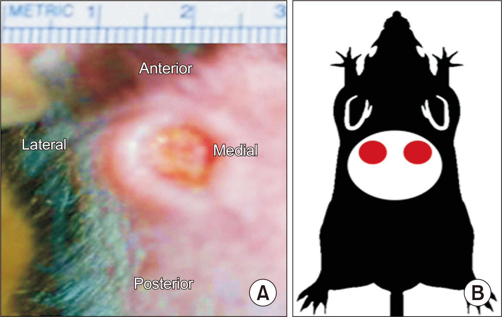
Fig. 1 Schematic diagram describing wound formation in diabetic rat. Two full thickness skin ulcers with same size (A) were made on the bilateral infrascapular regions (B).
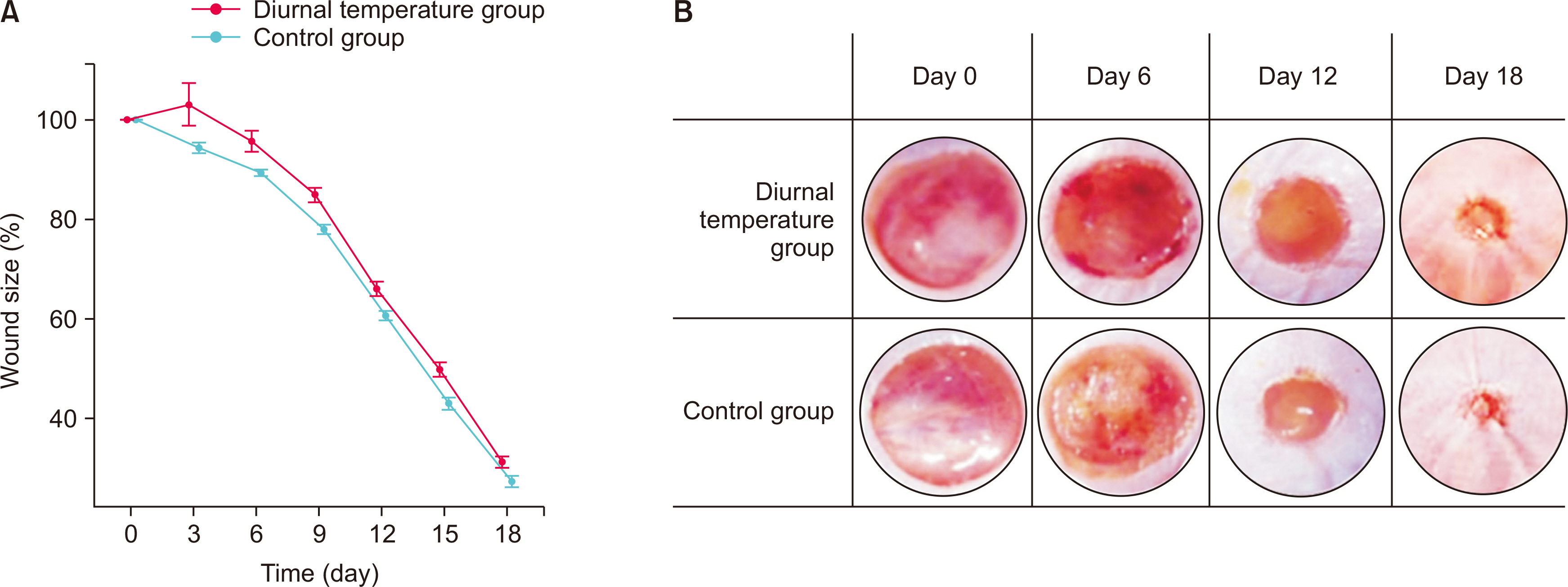
Fig. 2 Comparison of wound recovery in diurnal temperature group and control group. (A) Significantly delayed wound recovery was detected in the diurnal temperature group since three days after the wound formation. (B) Extent of wound recovery was evaluated by estimating the circumferential area of the wound.
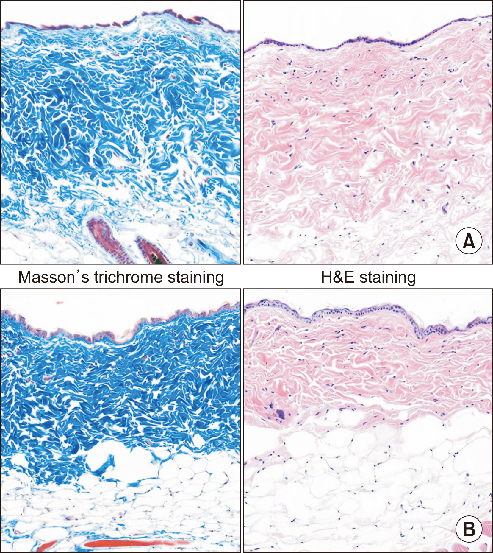
Fig. 3 Histologic analysis of the scar tissue for control group (A) and diurnal temperature group (B). Collagen fibers were stained with Masson’s trichrome staining (left; magnification, ×100) and wound cross section was stained with hematoxylin and eosin (H&E) staining (right; magnification, ×100). The control group showed higher degree of fibrosis and collagen fiber formation compared with the diurnal temperature group.
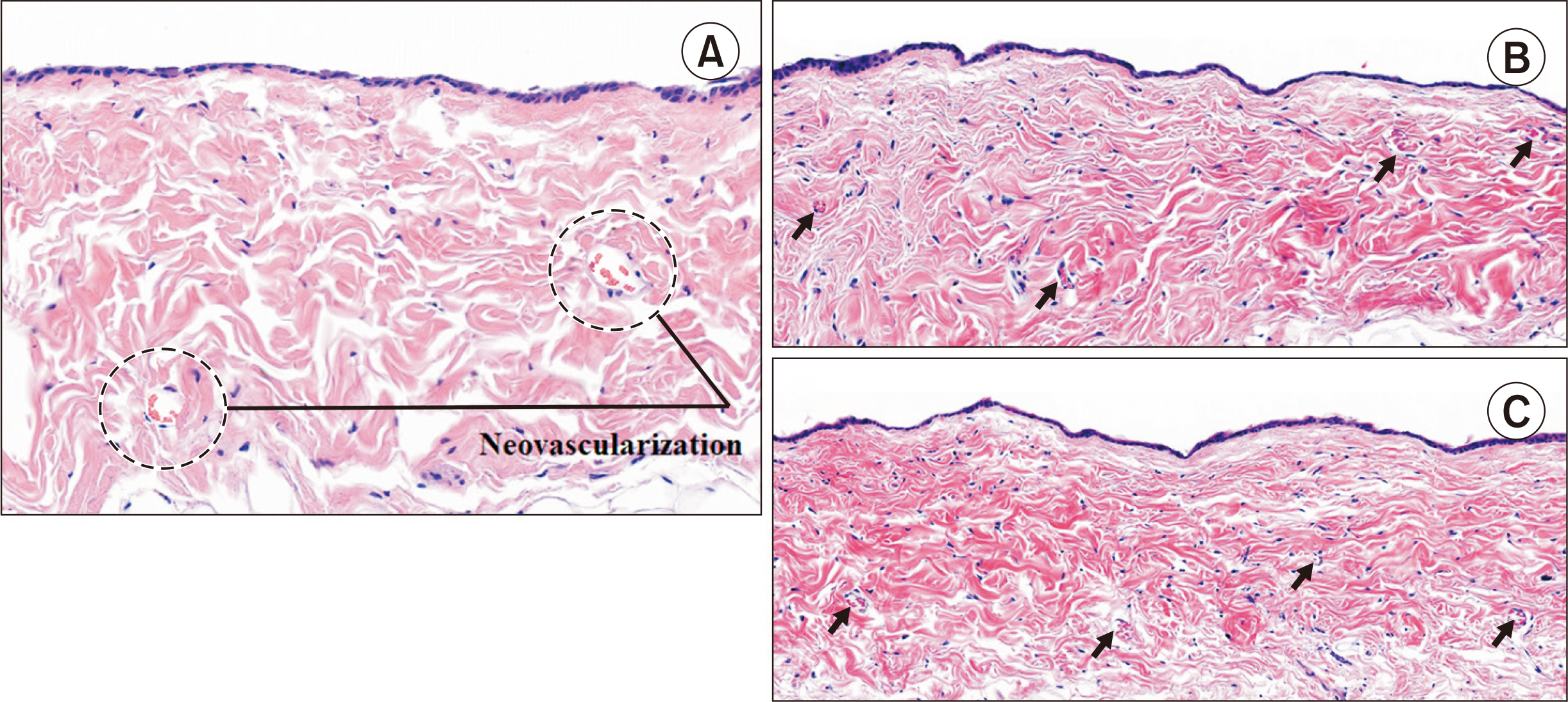
Fig. 4 (A) Neovascular formation of the wound tissue. Greater amount of neovascular formation (arrow) was found in control group (C) compared with diurnal temperature group (B) with statistically significance (H&E stain, ×100).
Reference
-
1. Keatinge WR, Coleshaw SR, Cotter F, Mattock M, Murphy M, Chelliah R. 1984; Increases in platelet and red cell counts, blood viscosity, and arterial pressure during mild surface cooling: factors in mortality from coronary and cerebral thrombosis in winter. Br Med J (Clin Res Ed). 289:1405–8. doi: 10.1136/bmj.289.6456.1405. DOI: 10.1136/bmj.289.6456.1405. PMID: 6437575. PMCID: PMC1443679.
Article2. Li J, Chen J, Kirsner R. 2007; Pathophysiology of acute wound healing. Clin Dermatol. 25:9–18. doi: 10.1016/j.clindermatol.2006.09.007. DOI: 10.1016/j.clindermatol.2006.09.007. PMID: 17276196.
Article3. Lavery LA, Higgins KR, Lanctot DR, Constantinides GP, Zamorano RG, Athanasiou KA, et al. 2007; Preventing diabetic foot ulcer recurrence in high-risk patients: use of temperature monitoring as a self-assessment tool. Diabetes Care. 30:14–20. doi: 10.2337/dc06-1600. DOI: 10.2337/dc06-1600. PMID: 17192326.
Article4. Lee SJ, Kim MJ, Jang SH, Lee KH, Han SH, Choi BY, et al. 2007; The difference of plantar temperature between non-DM and DM groups according to season. Korean J Clin Geri. 8:412–8.5. Kokate JY, Leland KJ, Held AM, Hansen GL, Kveen GL, Johnson BA, et al. 1995; Temperature-modulated pressure ulcers: a porcine model. Arch Phys Med Rehabil. 76:666–73. doi: 10.1016/s0003 9993(95)80637-7. DOI: 10.1016/S0003-9993(95)80637-7.
Article6. Menke NB, Ward KR, Witten TM, Bonchev DG, Diegelmann RF. 2007; Impaired wound healing. Clin Dermatol. 25:19–25. doi: 10.1016/. DOI: 10.1016/j.clindermatol.2006.12.005. PMID: 17276197.
Article7. Yager DR, Zhang LY, Liang HX, Diegelmann RF, Cohen IK. 1996; Wound fluids from human pressure ulcers contain elevated matrix metalloproteinase levels and activity compared to surgical wound fluids. J Invest Dermatol. 107:743–8. doi: 10.1111/1523-1747.ep12365637. DOI: 10.1111/1523-1747.ep12365637. PMID: 8875960.
Article8. Mast BA, Schultz GS. 1996; Interactions of cytokines, growth factors, and proteases in acute and chronic wounds. Wound Repair Regen. 4:411–20. doi: 10.1046/j.1524-475X.1996.40404.x. DOI: 10.1046/j.1524-475X.1996.40404.x. PMID: 17309691.
Article9. Kirsner RS, Eaglstein WH. 1993; The wound healing process. Dermatol Clin. 11:629–40. doi: 10.1016/S0733-8635(18)30216-X. DOI: 10.1016/S0733-8635(18)30216-X.
Article10. Enoch S, Leaper DJ. 2008; Basic science of wound healing. Surgery (Oxf). 26:31–7. doi: 10.1016/j.mpsur.2007.11.005. DOI: 10.1016/j.mpsur.2007.11.005.
Article11. Singer AJ, Clark RA. 1999; Cutaneous wound healing. N Engl J Med. 341:738–46. doi: 10.1056/NEJM199909023411006. DOI: 10.1056/NEJM199909023411006. PMID: 10471461.
Article12. Postlethwaite AE, Kang AH. 1976; Collagen-and collagen peptide-induced chemotaxis of human blood monocytes. J Exp Med. 143:1299307. doi: 10.1084/jem.143.6.1299. DOI: 10.1084/jem.143.6.1299. PMID: 1271012. PMCID: PMC2190221.
Article13. Lewis JS, Lee JA, Underwood JC, Harris AL, Lewis CE. 1999; Macrophage responses to hypoxia: relevance to disease mechanisms. J Leukoc Biol. 66:889–900. doi: 10.1002/jlb.66.6.889. DOI: 10.1002/jlb.66.6.889. PMID: 10614769.
Article14. Falanga V. 2004; The chronic wound: impaired healing and solutions in the context of wound bed preparation. Blood Cells Mol Dis. 32:8894. doi: 10.1016/j.bcmd.2003.09.020. DOI: 10.1016/j.bcmd.2003.09.020. PMID: 14757419.
Article15. Eming SA, Krieg T, Davidson JM. 2007; Inflammation in wound repair: molecular and cellular mechanisms. J Invest Dermatol. 127:514–25. doi: 10.1038/sj.jid.5700701. DOI: 10.1038/sj.jid.5700701. PMID: 17299434.
Article16. Liechty KW, Kim HB, Adzick NS, Crombleholme TM. 2000; Fetal wound repair results in scar formation in interleukin-10-deficient mice in a syngeneic murine model of scarless fetal wound repair. J Pediatr Surg. 35:866–72. doi: 10.1053/jpsu.2000.6868. discussion 872-3. DOI: 10.1053/jpsu.2000.6868. PMID: 10873028.
Article17. Stadelmann WK, Digenis AG, Tobin GR. 1998; Impediments to wound healing. Am J Surg. 176(2A Suppl):39S–47S. doi: 10.1016/s0002-9610(98)00184-6. DOI: 10.1016/S0002-9610(98)00184-6.
Article
- Full Text Links
-
- Actions
-
Cited
- CITED
-
- Close
- Share
-
- Similar articles
-
- Transcutaneous Oxygen Pressure to Predict Wound Healing in Mild Diabetic Feet
- Latest Approaches in the Treatment of Diabetic Foot Ulcers: An Integrative Analysis Based on the International Working Group on the Diabetic Foot (IWGDF) 2023 Guidelines
- Grafting Low-Temperature Micronized Adipose Tissue Niche to Treat Diabetic Foot Ulcers: A Pilot Study
- Application of Negative Pressure Wound Therapy in the Foot and Ankle Field
- Serum Collagen Level as a Predictor of Healing Wounds in Diabetic Foot Patients
