Comparison of Long Term Follow-up Chest CT Imaging in Adult and Pediatric Patients with Humidifier Disinfectant-related Lung Injury
- Affiliations
-
- 1Department of Radiology, Ulsan University Hospital, University of Ulsan College of Medicine, Ulsan, Korea
- 2Department of Occupational and Environmental Medicine, Inha University College of Medicine, Incheon, Korea
- 3Department of Preventive Medicine, Dong-A University Collage of Medicine, Busan, Korea
- 4Department of Occupational and Environmental Medicine, Kosin University College of Medicine, Busan, Korea
- 5Total Health Care Center, Kangbuk Samsung Hospital, School of Medicine, Sungkyungkwan University, Seoul, Korea
- 6Division of Pulmonary and Critical Care Medicine, Department of Internal Medicine, National Medical Center, Seoul, Korea
- 7Department of Occupational and Environmental Medicine, Ulsan University Hospital, University of Ulsan College of Medicine, Ulsan, Korea
- KMID: 2508803
- DOI: http://doi.org/10.3346/jkms.2020.35.e377
Abstract
- Background
To compare the chest computed tomography (CT) images of children and adults in families with clusters of humidifier disinfectant-related lung injury (HDLI) after cessation of exposure to humidifier disinfectant (HD).
Methods
We reviewed medical records of 19 families with 43 patients (21 adults, 22 children) among families, which had at least one adult and one child with HDLI. Each family was exposed to the same HD exposure environment.
Results
In adults, centrilobular nodules were predominant (95.2%) in chronic HDLI findings after cessation of exposure to HD, however, in children, normal pattern was most prevalent on chest CT (45.5%), followed by centrilobular nodule (36.4%), bizarre lung cysts (36.4%), and reticulation (13.6%).
Conclusion
Unlike the known chronic HDLI finding of adults, centrilobular nodules were only present in 36.4% of children. The frequency of bizarre lung cysts were significantly greater in children than that in adults after cessation of similar exposure to HD. Thus, bizarre lung cysts may be useful as another novel finding of chronic HDLI in children who have no history of pulmonary infection or other perinatal disorder such as hyaline membrane disease or other interstitial lung disease.
Figure
-
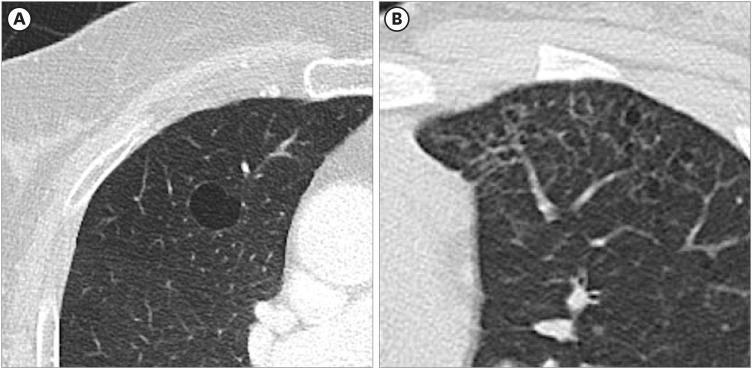
Fig. 1 Comparison of the shape of classic lung cyst and bizarre lung cyst. (A) The Fleischner Society described lung cyst as a round parenchymal lucency with a well-defined interface with normal lung. (B) A 11 years old girl with bizarre lung cysts (family No.19). Variable shaped lung cysts and relatively well-defined interface with normal lung.
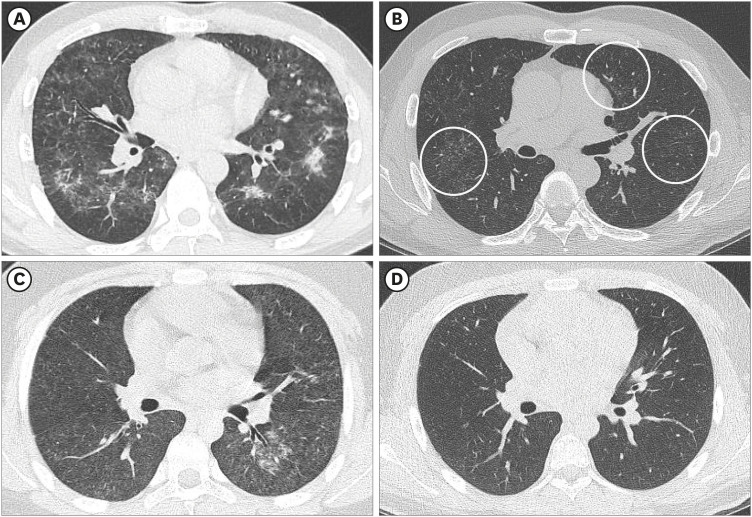
Fig. 2 Comparison of acute and chronic HDLI image between adult and child patients (family No. 3). (A, B) Chest CT of a 50-year-old man with 15 months' HD exposure. (A) Bilateral ill-defined centrilobular nodules, patchy consolidations with subpleural sparing indicates typical acute HDLI finding. (B) Two years after HD exposure cessation. Tiny, ill-defined centrilobular nodules were noted in bilateral lungs (circle). (C, D) Chest CT of a 13-year-old boy with HDLI exposure. (C) Diffuse, ill-defined centrilobular nodules, ground-glass opacity with subpleural sparing and some consolidations involving both lungs. (D) Two years after HD exposure cessation. Previous findings disappeared and showed normal lung parenchyma.HDLI = humidifier disinfectant-related lung injury, CT = computed tomography, HD = humidifier disinfectant.
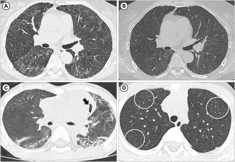
Fig. 3 Comparison of acute and chronic HDLI image between adult and child patients (family No. 7). (A, B) CT of a 63-year-old woman with 6 months HD exposure. (A) Bilateral ill-defined centrilobular nodules and ground-glass opacity with subpleural sparing indicates acute to subacute stage HDLI finding. (B) Two years after HD exposure cessation. Tiny, extensive centrilobular nodules were noted in bilateral lungs. (C, D) CT of a 4-year-old boy with acute HDLI exposure. (C) Bilateral, multifocal consolidations and ground-glass opacity involving both lung parenchyma. Pneumomediastinum also was noted (black arrow). (D) Two years after HD exposure cessation. Subtle, subpleural ill-defined centrilobular nodules remained in both upper lobes (circles).HDLI = humidifier disinfectant-related lung injury, CT = computed tomography, HD = humidifier disinfectant.
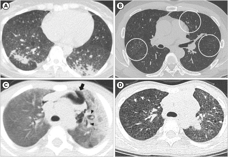
Fig. 4 Comparison of acute and chronic HDLI image between adult and child patients (family No. 6). (A, B) CT of a 35-year-old woman with 4 months HD exposure. (A) Bilateral ill-defined centrilobular nodules, consolidation and subpleural sparing indicates typical acute HDLI finding. (B) Two years after HD exposure cessation. Tiny, ill-defined centrilobular nodules were noted in the subpleural portion of the bilateral lower lobes (circles). (C, D) CT of a 2-year-old boy with HD exposure. (C) Bilateral extensive consolidation and ground-glass opacity involving both lungs. Pneumomediastinum (black arrow) and interstitial emphysema (black arrowheads). (D) Two years after HD exposure cessation. Numerous bizarre lung cysts (white arrowheads) are scattered in bilateral lungs.HDLI = humidifier disinfectant-related lung injury, CT = computed tomography, HD = humidifier disinfectant.
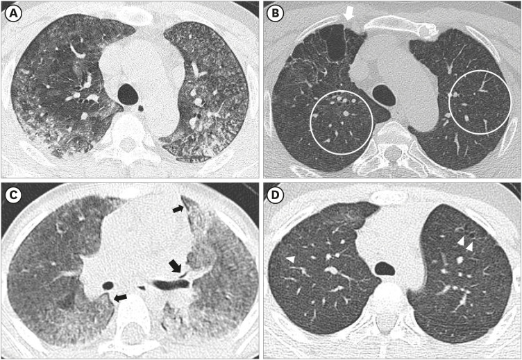
Fig. 5 Comparison of acute and chronic HDLI image between adult and child patients (family No. 10). (A, B) CT of a 47-year-old man with 20.3 months HD exposure. (A) Bilateral, extensive ground-glass opacity with subpleural sparing and irregular consolidations involving both lungs, indicates acute HDLI finding. (B) Four years after HD exposure cessation. Tiny, extensive centrilobular nodules were noted in bilateral lungs (circles). Due to previous lung damage, parenchymal distortion and bullae developed in the right upper lobe (white arrow). (C, D) CT of a 4-year-old boy with HD exposure. (C) Bilateral, extensive ground glass opacity and consolidation involve bilateral lungs. Interstitial emphysema was also noted (black arrow). (D) Four years after HD exposure cessation. Several bizarre lung cysts are noted in bilateral upper lobes (white arrowheads).HDLI = humidifier disinfectant-related lung injury, CT = computed tomography, HD = humidifier disinfectant.
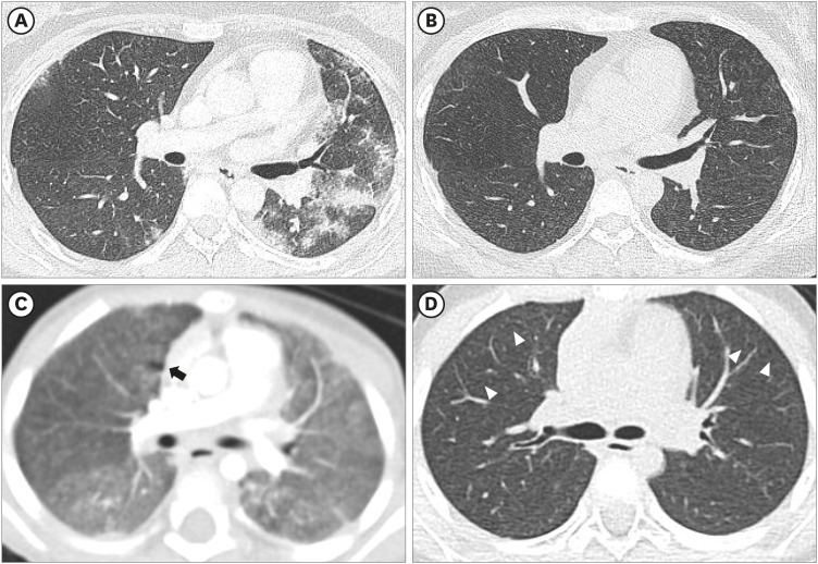
Fig. 6 Comparison of acute and chronic HDLI image between adult and child patients (family No. 11). (A, B) CT of a 34-year-old woman with 9.8 months HD exposure. (A) Ill-defined centrilobular nodules and some consolidations were noted in bilateral lungs. (B) Three years after HD exposure cessation. Tiny, extensive centrilobular nodules were noted in bilateral lungs. (C, D) CT of a 2-year-old girl with HD exposure. (C) Bilateral, extensive ground glass opacity and ill-defined centrilobular nodules involve bilateral lungs. Interstitial emphysema was also noted (black arrow). (D) Three years after HD exposure cessation. Subtle bizarre lung cysts were noted in bilateral upper lobes (white arrowheads).HDLI = humidifier disinfectant-related lung injury, CT = computed tomography, HD = humidifier disinfectant.
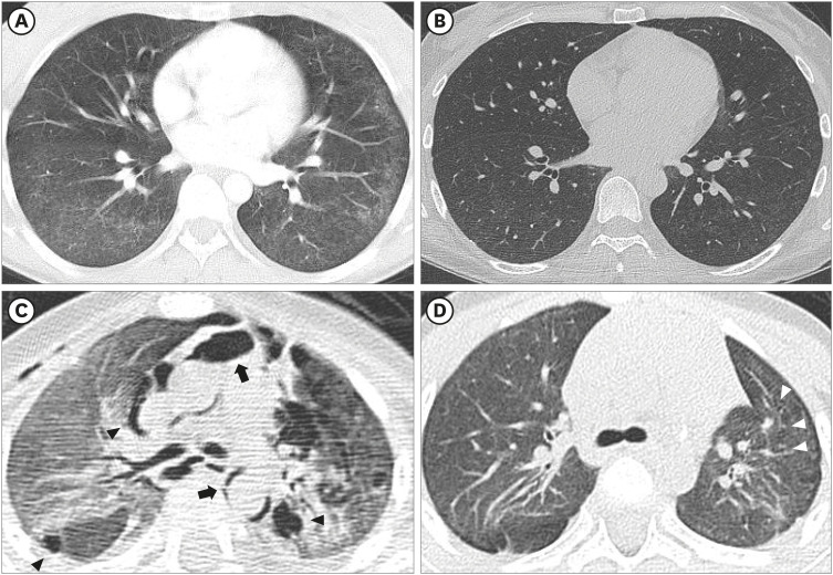
Fig. 7 Comparison of acute and chronic HDLI image between adult and child patients (family No. 16). (A, B) CT of a 34-year-old woman with 4 months' HD exposure. (A) Extensive, ill-defined tiny centrilobular nodules were noted in bilateral lungs, presumed to be subacute stage of HDLI. (B) Two years after HD exposure cessation. Very tiny centrilobular nodules remain in lung parenchyma. (C, D) CT of a 1-year-old boy with HDLI exposure. (C) Bilateral, extensive ground glass opacity and consolidations, indicates acute HDLI finding. Pneumomediastinum (black arrows) and interstitial emphysema (black arrowheads) were also found. (D) Two years after HD exposure cessation. Distortion of bilateral lungs suggest post-inflammatory parenchymal changes. Subtle bizarre lung cysts were noted in the left upper lobe (white arrowheads).HDLI = humidifier disinfectant-related lung injury, CT = computed tomography, HD = humidifier disinfectant.
Cited by 1 articles
-
Health Effects Associated With Humidifier Disinfectant Use: A Systematic Review for Exploration
Ji-Hun Song, Joonho Ahn, Min Young Park, Jaeyoung Park, Yu Min Lee, Jun-Pyo Myong, Jung-Wan Koo, Jongin Lee
J Korean Med Sci. 2022;37(33):e257. doi: 10.3346/jkms.2022.37.e257.
Reference
-
1. Park D, Leem J, Lee K, Lim H, Choi Y, Ahn JJ, et al. Exposure characteristics of familial cases of lung injury associated with the use of humidifier disinfectants. Environ Health. 2014; 13(1):70–76. PMID: 25178403.
Article2. Huh JW, Hong SB, Do KH, Koo HJ, Jang SJ, Lee MS, et al. Inhalation lung injury associated with humidifier disinfectants in adults. J Korean Med Sci. 2016; 31(12):1857–1862. PMID: 27822921.
Article3. Lee E, Seo JH, Kim HY, Yu J, Jhang WK, Park SJ, et al. Toxic inhalational injury-associated interstitial lung disease in children. J Korean Med Sci. 2013; 28(6):915–923. PMID: 23772158.
Article4. Lee MS, Kim HJ. Epidemiologic research on lung damage caused by humidifier disinfectants. Epidemiol Health. 2016; 38:e2016031. PMID: 27457061.
Article5. Hong SB, Kim HJ, Huh JW, Do KH, Jang SJ, Song JS, et al. A cluster of lung injury associated with home humidifier use: clinical, radiological and pathological description of a new syndrome. Thorax. 2014; 69(8):694–702. PMID: 24473332.6. Kim WY, Hong SB. Humidifier disinfectant-associated lung injury: six years after the tragic event. Tuberc Respir Dis (Seoul). 2017; 80(4):351–357. PMID: 28905528.
Article7. Koo HJ, Do KH, Chae EJ, Kim HJ, Song JS, Jang SJ, et al. Humidifier disinfectant-associated lung injury in adults: prognostic factors in predicting short-term outcome. Eur Radiol. 2017; 27(1):203–211. PMID: 27147415.
Article8. Yoon HM, Lee E, Lee JS, Do KH, Jung AY, Yoon CH, et al. Humidifier disinfectant-associated children's interstitial lung disease: computed tomographic features, histopathologic correlation and comparison between survivors and non-survivors. Eur Radiol. 2016; 26(1):235–243. PMID: 25991482.
Article9. Paek D, Koh Y, Park DU, Cheong HK, Do KH, Lim CM, et al. Nationwide study of humidifier disinfectant lung injury in South Korea, 1994–2011. Incidence and dose-response relationships. Ann Am Thorac Soc. 2015; 12(12):1813–1821. PMID: 26653190.
Article10. Choi JE, Hong SB, Do KH, Kim HJ, Chung S, Lee E, et al. Humidifier disinfectant lung injury, how do we approach the issues? Environ Health Toxicol. 2016; 31:e2016019. PMID: 27608716.
Article11. Ryu SH, Park DU, Lee E, Park S, Lee SY, Jung S, et al. Humidifier disinfectant and use characteristics associated with lung injury in Korea. Indoor Air. 2019; 29(5):735–747. PMID: 31278778.
Article12. Leem JH, Lee JH. Humidifier disinfectant-associated specific diseases should be called together as “humidifier disinfectant syndrome”. Environ Health Toxicol. 2017; 32:e2017017. PMID: 29026061.
Article13. Hansell DM, Bankier AA, MacMahon H, McLoud TC, Müller NL, Remy J. Fleischner Society: glossary of terms for thoracic imaging. Radiology. 2008; 246(3):697–722. PMID: 18195376.
Article14. Kim KW, Ahn K, Yang HJ, Lee S, Park JD, Kim WK, et al. Humidifier disinfectant-associated children's interstitial lung disease. Am J Respir Crit Care Med. 2014; 189(1):48–56. PMID: 24199596.15. Schittny JC. Development of the lung. Cell Tissue Res. 2017; 367(3):427–444. PMID: 28144783.
Article16. Odev K, Guler I, Altinok T, Pekcan S, Batur A, Ozbiner H. Cystic and cavitary lung lesions in children: radiologic findings with pathologic correlation. J Clin Imaging Sci. 2013; 3:60–70. PMID: 24605255.
Article17. Boddu P, Parimi V, Taddonio M, Kane JR, Yeldandi A. Pathologic and radiologic correlation of adult cystic lung disease: a comprehensive review. Patholog Res Int. 2017; 2017:3502438. PMID: 28270943.
Article18. Semple TR, Ashworth MT, Owens CM. Interstitial lung disease in children made easier…well, almost. Radiographics. 2017; 37(6):1679–1703. PMID: 29019755.
Article19. Selman M, Pardo A, King TE Jr. Hypersensitivity pneumonitis: insights in diagnosis and pathobiology. Am J Respir Crit Care Med. 2012; 186(4):314–324. PMID: 22679012.20. Spagnolo P, Rossi G, Cavazza A, Bonifazi M, Paladini I, Bonella F, et al. Hypersensitivity pneumonitis: a comprehensive review. J Investig Allergol Clin Immunol. 2015; 25(4):237–250.
- Full Text Links
-
- Actions
-
Cited
- CITED
-
- Close
- Share
-
- Similar articles
-
- Major concerns regarding lung injury and related health conditions caused by the use of humidifier disinfectant
- Problems with diagnostic criteria for humidifier disinfectant lung injury (HDLI): two cases of radiologically improved HDLI
- Frequency of Humidifier and Humidifier Disinfectant Usage in Gyeonggi Provine
- Mental health impact on a humidifier disinfectant disaster victim: a case report
- Toxic hepatitis after exposure to humidifier disinfectant: A case series report
