Late reconstruction of extensive orbital floor fracture with a patient-specific implant in a bombing victim
- Affiliations
-
- 1OMFS-IMPATH Research Group, Department of Imaging and Pathology, Faculty of Medicine, KU Leuven, Leuven, Belgium
- 2Department of Oral and Maxillofacial Surgery, University Hospitals Leuven, Leuven, Belgium
- KMID: 2508261
- DOI: http://doi.org/10.5125/jkaoms.2020.46.5.353
Abstract
- Fractures of the orbital floor and walls are among the most frequent maxillofacial fractures. Virtual three-dimensional (3D) planning and use of patientspecific implants (PSIs) could improve anatomic and functional outcomes in orbital reconstruction surgery. The presented case was a victim of a terrorist attack involving improvised explosive devices. This 58-year-old female suffered severe wounds caused by a single piece of metal from a bomb, shattering the left orbital floor and lateral orbital wall. Due to remaining hypotropia of the left eye compared to the right eye, late orbital floor reconstruction was carried out with a personalised 3D printed titanium implant. We concluded that this technique with PSI appears to be a viable method to correct complex orbital floor defects. Our research group noted good aesthetic and functional results one year after surgery. Due to the complexity of the surgery for a major bony defect of the orbital floor, it is important that the surgery be executed by experienced surgeons in the field of maxillofacial traumatology.
Keyword
Figure
-

Fig. 1 Overview of the overall treatment flow.
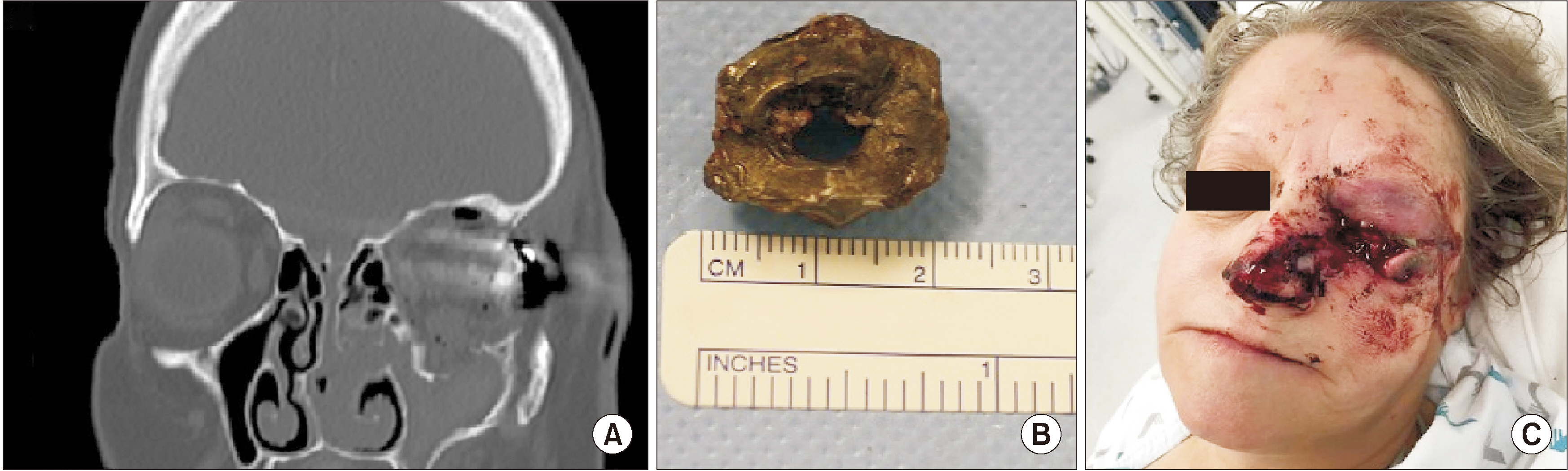
Fig. 2 Status immediately after the attacks. A. Initial computed tomography image with shattering of orbital bony structures. B. Shrapnel obtained after surgical removal. C. Facial lacerations.

Fig. 3 Status just before surgery and one year after the attacks. A. Interpupillary lines. B. Computed tomography image showing the residual defect of the orbital floor.
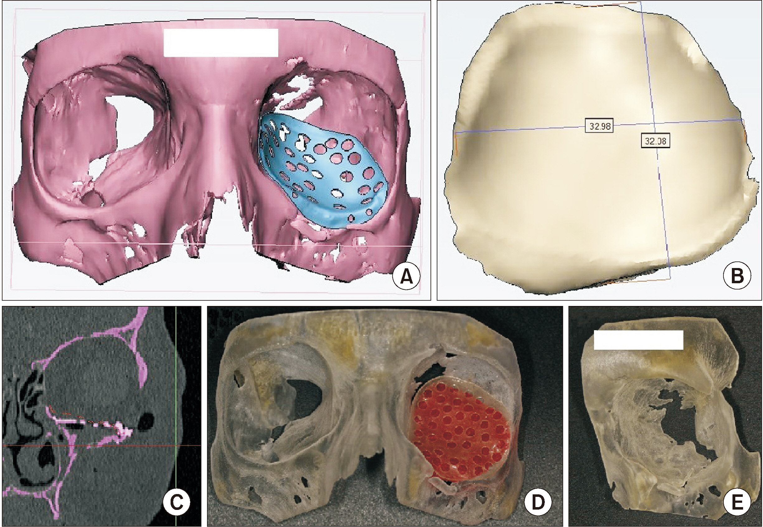
Fig. 4 Virtual three-dimensional planning. A. First version of the customized orbital floor reconstruction plate. B. Size of the defect in millimetres. C. Virtual position of the reconstruction plate on computed tomography scan. D. The stereolithographic model with a copy of the final titanium reconstruction plate. E. Magnification of the bony defect before surgery.
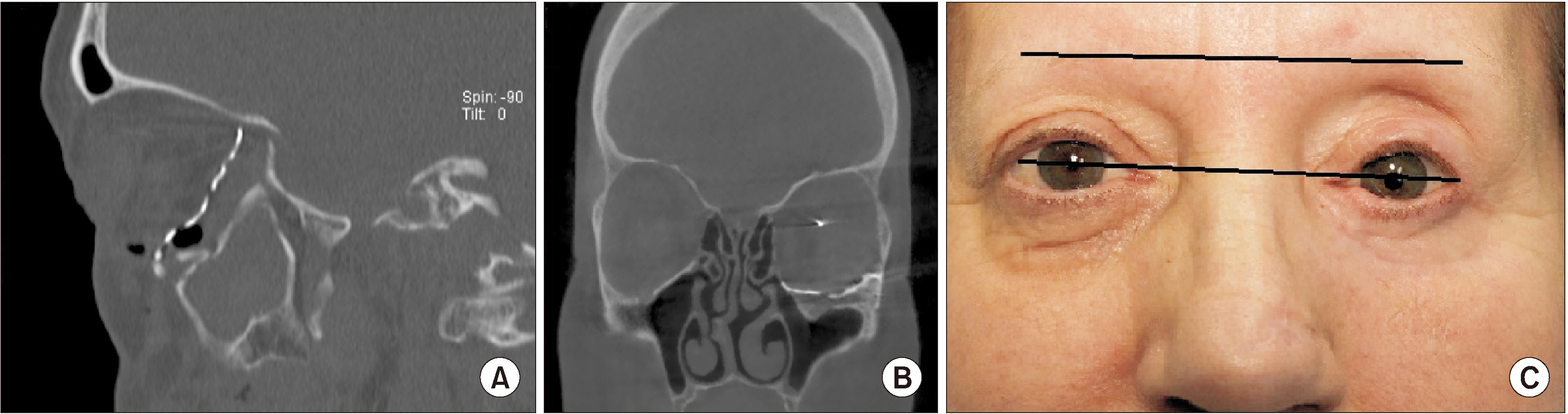
Fig. 5 Status one year after surgery and two years after the attacks. A. Computed tomography (CT) image showing current position of the patient-specific implant. B. CT image showing current status one year after surgery. C. Interpupillary lines one year after surgery.
Reference
-
References
1. Ellis E 3rd, Messo E. 2004; Use of nonresorbable alloplastic implants for internal orbital reconstruction. J Oral Maxillofac Surg. 62:873–81. https://doi.org/10.1016/j.joms.2003.12.025 . DOI: 10.1016/j.joms.2003.12.025. PMID: 15218569.
Article2. Bertol LS, Kindlein W Jr, da Silva FP, Aumund-Kopp C. 2010; Medical design: direct metal laser sintering of Ti-6Al-4V. Mater Des. 31:3982–8. https://doi.org/10.1016/j.matdes.2010.02.050 . DOI: 10.1016/j.matdes.2010.02.050.
Article3. de Oliveira FM, Salazar-Gamarra R, Öhman D, Nannmark U, Pecorari V, Dib LL. 2018; Quality of life assessment of patients utilizing orbital implant-supported prostheses. Clin Implant Dent Relat Res. 20:438–43. https://doi.org/10.1111/cid.12602 . DOI: 10.1111/cid.12602. PMID: 29508545.
Article4. Davies JC, Chan HHL, Bernstein JM, Goldstein DP, Irish JC, Gilbert RW. 2018; Orbital floor reconstruction: 3-Dimensional analysis shows comparable morphology of scapular and iliac crest bone grafts. J Oral Maxillofac Surg. 76:2011–8. https://doi.org/10.1016/j.joms.2018.03.034 . DOI: 10.1016/j.joms.2018.03.034. PMID: 29679587.
Article5. Ilankovan V, Jackson IT. 1992; Experience in the use of calvarial bone grafts in orbital reconstruction. Br J Oral Maxillofac Surg. 30:92–6. https://doi.org/10.1016/0266-4356(92)90077-v . DOI: 10.1016/0266-4356(92)90077-V. PMID: 1567810.
Article6. Zhang WB, Mao C, Liu XJ, Guo CB, Yu GY, Peng X. 2015; Outcomes of orbital floor reconstruction after extensive maxillectomy using the computer-assisted fabricated individual titanium mesh technique. J Oral Maxillofac Surg. 73:2065.e1–15. https://doi.org/10.1016/j.joms.2015.06.171 . DOI: 10.1016/j.joms.2015.06.171. PMID: 26188101.
Article7. Young SM, Sundar G, Lim TC, Lang SS, Thomas G, Amrith S. 2017; Use of bioresorbable implants for orbital fracture reconstruction. Br J Ophthalmol. 101:1080–5. https://doi.org/10.1136/bjophthalmol-2016-309330 . DOI: 10.1136/bjophthalmol-2016-309330. PMID: 27913446.
Article8. Al-Khdhairi OBH, Abdulrazaq SS. 2017; Is orbital floor reconstruction with titanium mesh safe? J Craniofac Surg. 28:e692–4. https://doi.org/10.1097/SCS.0000000000003864 . DOI: 10.1097/SCS.0000000000003864. PMID: 28885440.
Article9. Zimmerer RM, Ellis E 3rd, Aniceto GS, Schramm A, Wagner ME, Grant MP, et al. 2016; A prospective multicenter study to compare the precision of posttraumatic internal orbital reconstruction with standard preformed and individualized orbital implants. J Craniomaxillofac Surg. 44:1485–97. https://doi.org/10.1016/j.jcms.2016.07.014 . DOI: 10.1016/j.jcms.2016.07.014. PMID: 27519662.
Article10. Hwang WJ, Lee DH, Choi W, Hwang JH, Kim KS, Lee SY. 2017; Analysis of orbital volume measurements following reduction and internal fixation using absorbable mesh plates and screws for patients with orbital floor blowout fractures. J Craniofac Surg. 28:1664–9. https://doi.org/10.1097/SCS.0000000000003730 . DOI: 10.1097/SCS.0000000000003730. PMID: 28834830.
Article11. Oh TS, Jeong WS, Chang TJ, Koh KS, Choi JW. 2016; Customized orbital wall reconstruction using three-dimensionally printed rapid prototype model in patients with orbital wall fracture. J Craniofac Surg. 27:2020–4. https://doi.org/10.1097/SCS.0000000000003195 . DOI: 10.1097/SCS.0000000000003195. PMID: 28005746.
Article12. Purnell CA, Vaca EE, Ellis MF. 2018; Orbital fracture reconstruction using prebent, anatomic titanium plates: technical tips to avoid complications. J Craniofac Surg. 29:e515–7. https://doi.org/10.1097/SCS.0000000000004563 . DOI: 10.1097/SCS.0000000000004563. PMID: 29608480.
Article13. Fan B, Chen H, Sun YJ, Wang BF, Che L, Liu SY, et al. 2017; Clinical effects of 3-D printing-assisted personalized reconstructive surgery for blowout orbital fractures. Graefes Arch Clin Exp Ophthalmol. 255:2051–7. https://doi.org/10.1007/s00417-017-3766-y . DOI: 10.1007/s00417-017-3766-y. PMID: 28786025.
Article
- Full Text Links
-
- Actions
-
Cited
- CITED
-
- Close
- Share
-
- Similar articles
-
- A Case of Reconstruction of Orbital Floor Fracture by Grafting Parietal Bone
- Use of a Titanium Buttress to Prevent Implant Displacement in Extensive Orbital Blowout Fracture
- Results of Reconstruction of Orbital Wall Fracture With Bioresorbable Plate
- Simultaneous Transconjunctival and Transantral Approach for Repair of Blowout Fracture by using Medpor Orbital Implant
- Late Complication of a Silicone Implant Thirty Years after Orbital Fracture Reconstruction
