Removal of a Large, Intractable Common Bile Duct Stone by Direct Peroral Cholangioscopy Using Upper Gastrointestinal Endoscopy and Polypectomy Snare
- Affiliations
-
- 1Department of Internal Medicine, Kyungpook National University Hospital, Daegu, Korea
- 2Department of Internal Medicine, Kyungpook National University School of Medicine, Daegu, Korea
- KMID: 2507770
- DOI: http://doi.org/10.4166/kjg.2020.76.4.215
Abstract
- ERCP is the standard treatment for common bile duct stones. On the other hand, 10-15% of cases involve intractable common bile duct stones, which cannot be treated by conventional biliary sphincterotomy with a stone retrieval method. Large bile duct stones are typically managed by mechanical lithotripsy and endoscopic papillary large balloon dilatation. Peroral cholangioscopy techniques can be applied if this technique fails. In the present case, a 67-year-old woman had a large common bile duct stone that could not be retracted using the conventional ERCP stone extraction method. The common bile duct stone was eventually removed by direct peroral upper gastrointestinal endoscopy and a polypectomy snare.
Keyword
Figure
-
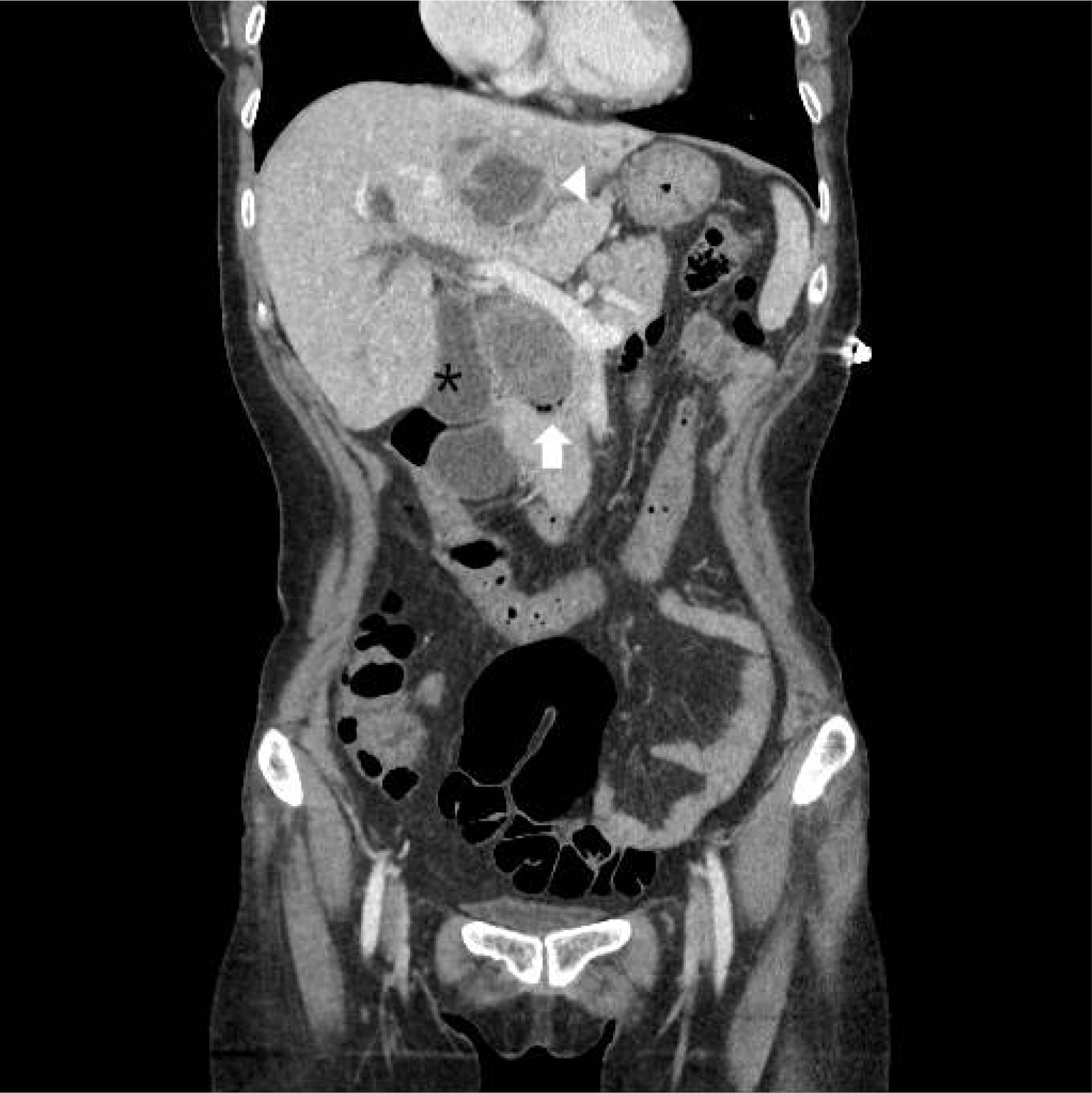
Fig. 1 Abdominal computed tomography on the admission day. The intrahepatic duct, common hepatic duct, and common bile duct (CBD) were dilated. The large CBD stone, size 54×32 mm, was observed in the CBD (arrow). A cystic lesion with peripheral rim enhancement, 50 mm in size, was observed in liver segment 3 (arrowhead). The gallbladder (asterisk) was dilated.

Fig. 2 Removal of common bile duct (CBD) stone by direct peroral cholangioscopy using upper gastrointestinal endoscopy and polypectomy snare. (A) A huge CBD stone showed brown pigment characteristics. The CBD was dilated significantly by the stone. (B) The CBD stone had been cracked into two pieces by grasp with snare polypectomy at the midline of the stone (arrow). (C) During cracking the stone by polypectomy snare, some fragmentations were extracted by the snare. (D) The upper gastrointestinal endoscope with snare was inserted inside the CBD.(E) After several attempts of the polypectomy snare technique, the stone was fragmented into several small pieces, which were extracted by basket and retrieval balloon.

Fig. 3 Follow-up cholangiogram by percutaneous transhepatic cholangioscopy before removal showed no remnant common bile duct stone.
Reference
-
1. Williams EJ, Green J, Beckingham I, et al. 2008; Guidelines on the management of common bile duct stones (CBDS). Gut. 57:1004–1021. DOI: 10.1136/gut.2007.121657. PMID: 18321943.
Article2. Copelan A, Kapoor BS. 2015; Choledocholithiasis: diagnosis and management. Tech Vasc Interv Radiol. 18:244–255. DOI: 10.1053/j.tvir.2015.07.008. PMID: 26615165.
Article3. Seitz U, Bapaye A, Bohnacker S, Navarrete C, Maydeo A, Soehendra N. 1998; Advances in therapeutic endoscopic treatment of common bile duct stones. World J Surg. 22:1133–1144. DOI: 10.1007/s002689900532. PMID: 9828721.
Article4. McHenry L, Lehman G. 2006; Difficult bile duct stones. Curr Treat Options Gastroenterol. 9:123–132. DOI: 10.1007/s11938-006-0031-6. PMID: 16539873.
Article5. Yasuda I, Itoi T. 2013; Recent advances in endoscopic management of difficult bile duct stones. Dig Endosc. 25:376–385. DOI: 10.1111/den.12118. PMID: 23650878.
Article6. Kim HJ, Choi HS, Park JH, et al. 2007; Factors influencing the technical difficulty of endoscopic clearance of bile duct stones. Gastrointest Endosc. 66:1154–1160. DOI: 10.1016/j.gie.2007.04.033. PMID: 17945223.
Article7. Doshi B, Yasuda I, Ryozawa S, Lee GH. 2018; Current endoscopic strategies for managing large bile duct stones. Dig Endosc. 30(Suppl 1):59–66. DOI: 10.1111/den.13019. PMID: 29658655.
Article8. Moon JH, Ko BM, Choi HJ, et al. 2009; Direct peroral cholangioscopy using an ultra-slim upper endoscope for the treatment of retained bile duct stones. Am J Gastroenterol. 104:2729–2733. DOI: 10.1038/ajg.2009.435. PMID: 19623165.
Article9. Kim HI, Moon JH, Choi HJ, et al. 2011; Holmium laser lithotripsy under direct peroral cholangioscopy by using an ultra-slim upper endoscope for patients with retained bile duct stones (with video). Gastrointest Endosc. 74:1127–1132. DOI: 10.1016/j.gie.2011.07.027. PMID: 21963070.
Article10. Katsinelos P, Pilpilidis I, Chatzimavroudis G, et al. 2009; Huge gastric bezoar caused by honeycomb, an unusual complication of health faddism: a case report. Cases J. 2:7077. DOI: 10.1186/1757-1626-2-7077. PMID: 19829904. PMCID: PMC2740191.
Article11. Tazuma S. 2006; Gallstone disease: epidemiology, pathogenesis, and classification of biliary stones (common bile duct and intrahepatic). Best Pract Res Clin Gastroenterol. 20:1075–1083. DOI: 10.1016/j.bpg.2006.05.009. PMID: 17127189.12. Sethi A, Chen YK, Austin GL, et al. 2011; ERCP with cholangiopancreatoscopy may be associated with higher rates of complications than ERCP alone: a single-center experience. Gastrointest Endosc. 73:251–256. DOI: 10.1016/j.gie.2010.08.058. PMID: 21106195.
Article13. Efthymiou M, Raftopoulos S, Antonio Chirinos J, May GR. 2012; Air embolism complicated by left hemiparesis after direct cholangioscopy with an intraductal balloon anchoring system. Gastrointest Endosc. 75:221–223. DOI: 10.1016/j.gie.2011.01.038. PMID: 21470606.
Article14. Hann A, Zizer E, Egger K, Allescher HD, Meining A. 2018; Fatal outcome due to CO2 emboli during direct cholangioscopy. Gut. 67:1378–1379. DOI: 10.1136/gutjnl-2017-313988. PMID: 28360098.
- Full Text Links
-
- Actions
-
Cited
- CITED
-
- Close
- Share
-
- Similar articles
-
- Early Bile Duct Cancer Detected by Direct Peroral Cholangioscopy with Narrow-Band Imaging after Bile Duct Stone Removal
- Bile-Duct Stone Removal under Direct Transnasal Cholangioscopy Using an Ultraslim Upper Endoscope
- Successful Removal of a Large Common Bile Duct Stone by Using Direct Peroral Cholangioscopy and Laser Lithotripsy in a Patient with Severe Kyphosis
- Recent update of therapeutic application of peroral cholangioscopy
- Application of Peroral Cholangioscopy in Biliary Diseases
