The estimation of age from elastic fibers in the tunica media of the aortic wall in a thai population: a preliminary study using aorta image analysis
- Affiliations
-
- 1PhD Degree Program in Anatomy, Department of Anatomy, Faculty of Medicine, Chiang Mai University, Chiang Mai, Thailand
- 2Department of Anatomy, Faculty of Medicine, Chiang Mai University, Chiang Mai, Thailand
- 3College of Arts Media and Technology, Chiang Mai University, Chiang Mai, Thailand
- 4Department of Statistics, Faculty of Science, Chiang Mai University, Chiang Mai, Thailand
- 5Forensic Osteology Research Center, Faculty of Medicine, Chiang Mai University, Chiang Mai, Thailand
- 6Excellence in Osteology Research and Training Center (ORTC), Chiang Mai University, Chiang Mai, Thailand
- KMID: 2507641
- DOI: http://doi.org/10.5115/acb.20.094
Abstract
- Image analysis has an increasing role in the identification of individuals in forensic application. Beside the bones, microstructural of arteries can be used in age estimation study. Aorta is the largest elastic artery which consists of many elastic fibers. Elastin in arterial wall highly resist to chemical and physical influence. The purposes of the study were to quantify elastic fibers in tunica media in each location of the aorta and examine the correlation between elastic fibers and age by using image analysis program. A total of 36 human aortas were dissected in 4 locations. The aortas were obtained from cadavers with an age range of 20 to 90 years. Specimens were stained with Elastic Van Gieson staining. Histological images were investigated about elastic fibers using light microscope with cellSens program and aorta image analysis was used for the evaluation of data. The results showed that the mean percentage density of elastic fibers in the ascending aorta and the aortic arch increased. However, the mean percentage density of elastic fibers decreased in the 31 to 40 years age group in the thoracic aorta and the abdominal aorta and decreased in each location of aorta continuously until 81 to 90 years. The abdominal aorta showed the highest correlation with age (r=0.732) followed by the thoracic aorta, the aortic arch and the ascending aorta, respectively. Changes in the percentage density of elastic fibers in the tunica media of the aortic wall can be used to add information to age estimation for identification purposes.
Keyword
Figure
-

Fig. 1 Image analysis steps. CIELAB, International Commission on Illumination.
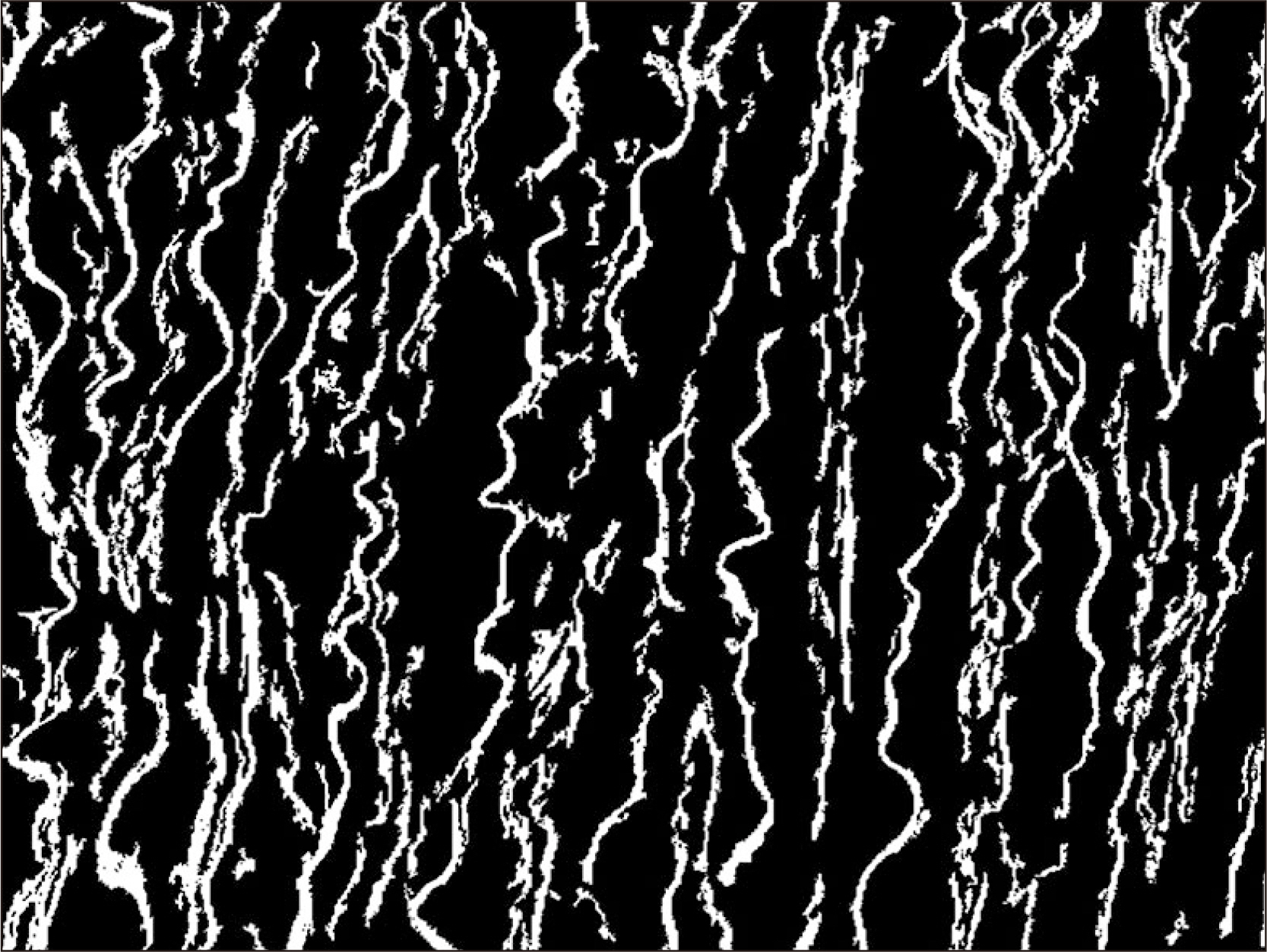
Fig. 2 Demonstration of the analyzed picture of the aortic segment using Aorta Image Analysis program. Percentage density of elastic fibers in tunica media of the aorta. White color, elastic fibers (region of interest); Black color, sections of no-interest.

Fig. 3 Example of elastic fibers in tunica media from the abdominal aorta at different ages (Elastic Van Gieson, ×40). (A) A 20 years of human aorta, (B) 43 years of human aorta, (C) 61 years of human aorta, (D) 81 years of human aorta. Scale Bars=20 μm (A–D).

Fig. 4 The bar chart showing mean percentage density of elastic fibers in various age groups.
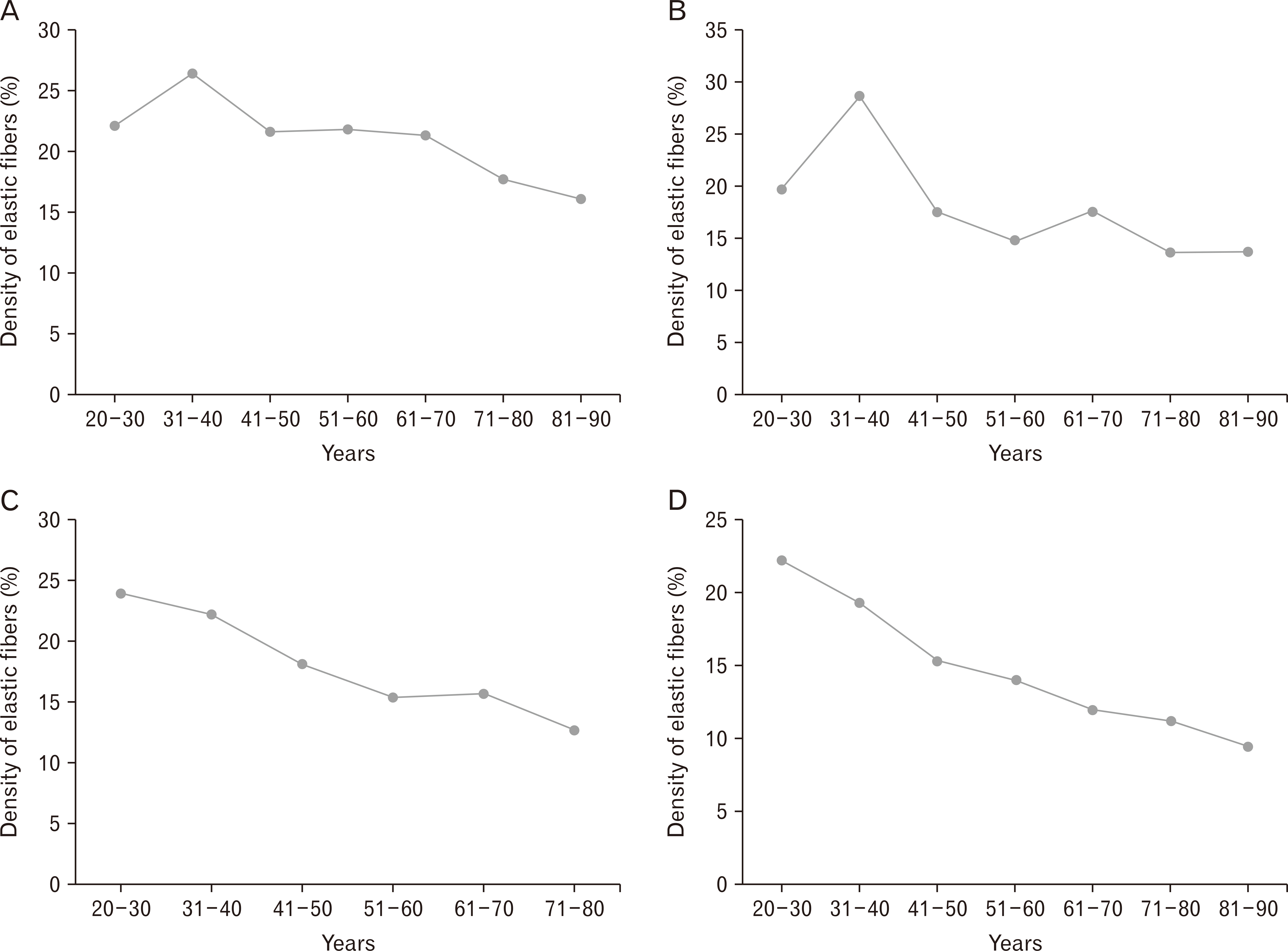
Fig. 5 The scatter plot showing mean percentage density of elastic fibers in various age groups with different locations of the aorta. (A) Ascending aorta, (B) aortic arch, (C) thoracic aorta, (D) abdominal aorta.
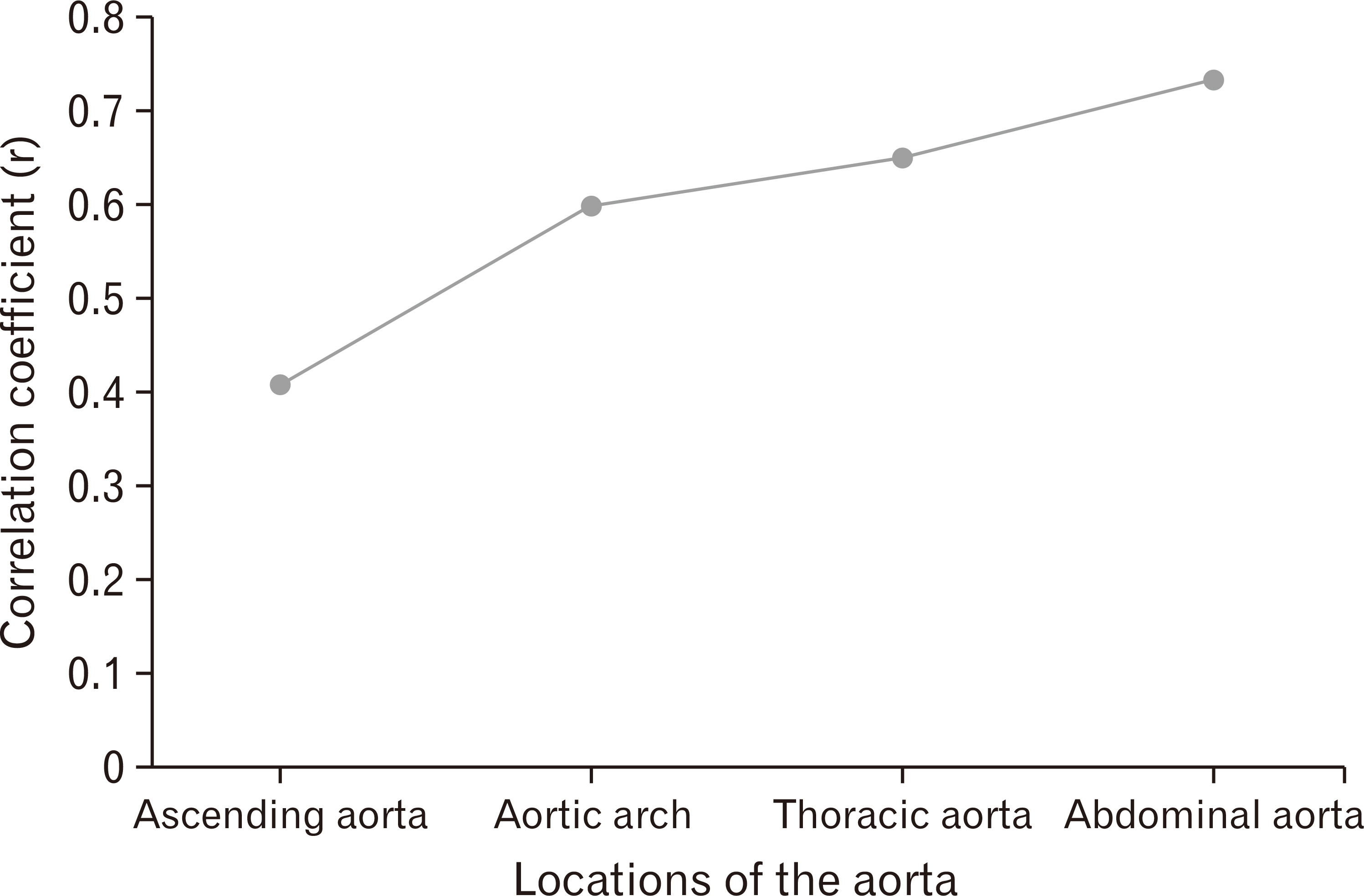
Fig. 6 The scatter plot showing the correlation coefficient with age in different locations of the aorta.
Cited by 1 articles
-
Does oral ciprofloxacin affect the structure of thoracic aorta in adult and senile male albino rats? A clue to fluoroquinolones-induced risk of aortic dissection
Ahmed Farid Al-Neklawy, Nagwa Ebrahim El-Nefiawy, Hagar Yousry Rady
Anat Cell Biol. 2022;55(1):79-91. doi: 10.5115/acb.21.170.
Reference
-
References
1. Prats-Montalbán JM, de Juan A, Ferrer A. 2011; Multivariate image analysis: a review with applications. Chemometr Intell Lab Syst. 107:1–23. DOI: 10.1016/j.chemolab.2011.03.002. PMID: 31823240.
Article2. Ritz-Timme S, Cattaneo C, Collins MJ, Waite ER, Schütz HW, Kaatsch HJ, Borrman HI. 2000; Age estimation: the state of the art in relation to the specific demands of forensic practise. Int J Legal Med. 113:129–36. DOI: 10.1007/s004140050283. PMID: 10876982.
Article3. Lynnerup N. 2013; Forensic anthropology and human identification. Scandinavian J Forensic Sci. 19:16–38. DOI: 10.2478/sjfs-2013-0005.
Article4. Webb RC, Inscho EW. Michael PL, editor. 2005. Age-related changes in the cardiovascular system. Hypertension in the elderly. Humana Press;Totowa: p. 11–21. DOI: 10.1007/978-1-59259-911-0_2. PMID: 15802027.
Article5. Gupta SD, Gupta SK, Pal DK, Sarawagi R, Gupta P. 2011; Microscopic study of aorta in relation of different age groups: an observational study. Int J Biol Med Res. 2:398–403.6. Lakatta EG, Levy D. 2003; Arterial and cardiac aging: major shareholders in cardiovascular disease enterprises: part II: the aging heart in health: links to heart disease. Circulation. 107:346–54. DOI: 10.1161/01.CIR.0000048893.62841.F7. PMID: 12538439.7. Collins JA, Munoz JV, Patel TR, Loukas M, Tubbs RS. 2014; The anatomy of the aging aorta. Clin Anat. 27:463–6. DOI: 10.1002/ca.22384. PMID: 24523152.
Article8. Shykoff BE. 2001; Arterial stiffness: clinical relevance, measurement, and treatment. Rev Cardiovasc Med. 2:29–40. DOI: 10.1042/CS20070080. PMID: 17623012.9. Xu X, Wang B, Ren C, Hu J, Greenberg DA, Chen T, Xie L, Jin K. 2017; Age-related impairment of vascular structure and functions. Aging Dis. 8:590–610. DOI: 10.14336/AD.2017.0430. PMID: 28966804. PMCID: PMC5614324.
Article10. Cheuk BL, Cheng SW. 2005; Expression of integrin alpha5beta1 and the relationship to collagen and elastin content in human suprarenal and infrarenal aortas. Vasc Endovascular Surg. 39:245–51. DOI: 10.1177/153857440503900305. PMID: 15920653.11. Samila ZJ, Carter SA. 1981; The effect of age on the unfolding of elastin lamellae and collagen fibers with stretch in human carotid arteries. Can J Physiol Pharmacol. 59:1050–7. DOI: 10.1139/y81-160. PMID: 7317828.
Article12. Komutrattananont P, Mahakkanukrauh P, Das S. 2019; Morphology of the human aorta and age-related changes: anatomical facts. Anat Cell Biol. 52:109–14. DOI: 10.5115/acb.2019.52.2.109. PMID: 31338225. PMCID: PMC6624342.
Article13. Gartner LP, Hiatt JL. 2014. Color atlas and text of histology. Wolters Kluwer Health/Lippincott Williams & Wilkins;Philadelphia:14. Mitchell JRA, Schwartz CJ, Pickering G. 1965. Arterial disease. Blackwell Scientific Publications;Oxford:15. Berillis P. 2013; The role of collagen in the aorta's structure. Open Circ Vasc J. 6:1–8. DOI: 10.2174/1877382601306010001.
Article16. Emmott A, Garcia J, Chung J, Lachapelle K, El-Hamamsy I, Mongrain R, Cartier R, Leask RL. 2016; Biomechanics of the ascending thoracic aorta: a clinical perspective on engineering data. Can J Cardiol. 32:35–47. DOI: 10.1016/j.cjca.2015.10.015. PMID: 26724509.
Article17. Tsamis A, Krawiec JT, Vorp DA. 2013; Elastin and collagen fibre microstructure of the human aorta in ageing and disease: a review. J R Soc Interface. 10:20121004. DOI: 10.1098/rsif.2012.1004. PMID: 23536538. PMCID: PMC3645409.
Article18. Ninomiya OH, Tavares Monteiro JA, Higuchi Mde L, Puech-Leão P, de Luccia N, Raghavan ML, da Silva ES. 2015; Biomechanical properties and microstructural analysis of the human nonaneurysmal aorta as a function of age, gender and location: an autopsy study. J Vasc Res. 52:257–64. DOI: 10.1159/000442979. PMID: 26799837.
Article19. Sherratt MJ. 2009; Tissue elasticity and the ageing elastic fibre. Age (Dordr). 31:305–25. DOI: 10.1007/s11357-009-9103-6. PMID: 19588272.
Article20. Schofield JD, Weightman B. 1978; New knowledge of connective tissue ageing. J Clin Pathol Suppl (R Coll Pathol). 12:174–90. DOI: 10.1136/jcp.31.Suppl_12.174.
Article21. Gosline JM. 1980; The elastic properties of rubber-like proteins and highly extensible tissues. Symp Soc Exp Biol. 34:332–57. PMID: 6894811.22. Quintas ML, Rodrigues CJ, Yoo JH, Rodrigues Junior AJ. 2000; Age related changes in the elastic fiber system of the interfoveolar ligament. Rev Hosp Clin Fac Med Sao Paulo. 55:83–6. DOI: 10.1590/S0041-87812000000300003. PMID: 10983010.
Article23. Frances C, Branchet MC, Boisnic S, Lesty CL, Robert L. 1990; Elastic fibers in normal human skin. Variations with age: a morphometric analysis. Arch Gerontol Geriatr. 10:57–67. DOI: 10.1016/0167-4943(90)90044-7. PMID: 15374522.
Article24. Robert C, Lesty C, Robert AM. 1988; Ageing of the skin: study of elastic fiber network modifications by computerized image analysis. Gerontology. 34:291–6. DOI: 10.1159/000212969. PMID: 2464530.
Article25. Toda T, Tsuda N, Nishimori I, Leszczynski DE, Kummerow FA. 1980; Morphometrical analysis of the aging process in human arteries and aorta. Acta Anat (Basel). 106:35–44. DOI: 10.1159/000145167. PMID: 7415788.
Article26. Hosoda Y, Minoshima I. 1965; Elastin content of the aorta and the pulmonary artery in the Japanese. Angiology. 16:325–32. DOI: 10.1177/000331976501600604. PMID: 14298305. PMCID: PMC6032080.
Article27. Schlatmann TJ, Becker AE. 1977; Histologic changes in the normal aging aorta: implications for dissecting aortic aneurysm. Am J Cardiol. 39:13–20. DOI: 10.1016/S0002-9149(77)80004-0. PMID: 831420.
Article28. Fritze O, Romero B, Schleicher M, Jacob MP, Oh DY, Starcher B, Schenke-Layland K, Bujan J, Stock UA. 2012; Age-related changes in the elastic tissue of the human aorta. J Vasc Res. 49:77–86. DOI: 10.1159/000331278. PMID: 22105095. PMCID: PMC7354228.
Article29. Greenwald SE. 2007; Ageing of the conduit arteries. J Pathol. 211:157–72. DOI: 10.1002/path.2101. PMID: 17200940.
Article30. Robert L, Robert AM, Fülöp T. 2008; Rapid increase in human life expectancy: will it soon be limited by the aging of elastin? Biogerontology. 9:119–33. DOI: 10.1007/s10522-007-9122-6. PMID: 18175202.
Article31. Yamada H, Sakata N, Wada H, Tashiro T, Tayama E. 2015; Age-related distensibility and histology of the ascending aorta in elderly patients with acute aortic dissection. J Biomech. 48:3267–73. DOI: 10.1016/j.jbiomech.2015.06.025. PMID: 26163750.
Article32. Myers VC, Lang WW. 1946; Some chemical changes in the human thoracic aorta accompanying the aging process. J Gerontol. 1(Pt 1 4):441–4. DOI: 10.1093/geronj/1.4_Part_1.441. PMID: 20273846.
Article33. Faber M, Møller-Hou G. 1952; The human aorta. V. Collagen and elastin in the normal and hypertensive aorta. Acta Pathol Microbiol Scand. 31:377–82. DOI: 10.1111/j.1699-0463.1952.tb00205.x. PMID: 12985344.34. Alex RB, Amma LKK. 2016; Microanatomical study of age changes in tunica media of ascending aorta. J Evol Med Dent Sci. 5:7409–12. DOI: 10.14260/Jemds/2016/1677.35. Foster LS. 1909; Changes occurring in the elastic fibers of the aorta with advancing Age. J Med Res. 21:297–311. DOI: 10.1007/s13239-017-0335-9. PMID: 19971920. PMCID: PMC2098995.36. Hosoda Y, Kawano K, Yamasawa F, Ishii T, Shibata T, Inayama S. 1984; Age-dependent changes of collagen and elastin content in human aorta and pulmonary artery. Angiology. 35:615–21. DOI: 10.1177/000331978403501001. PMID: 6497045.
Article37. Wolinsky H, Glagov S. 1969; Comparison of abdominal and thoracic aortic medial structure in mammals. Deviation of man from the usual pattern. Circ Res. 25:677–86. DOI: 10.1161/01.RES.25.6.677. PMID: 5364644.38. Osler W. 1892. The principles and practice of medicine: designed for the use of practitioners and students of medicine. D. Appleton and Company;New York:
Article39. Nichols WW, O'Rourke MF, Vlachopoulos C, Hoeck AP, Reneman RS. 2011. McDonald's blood flow in arteries: theoretic, experimental, and clinical principles. 6th ed. Hodder Arnold;London: p. 278.40. O'Rourke MF, Hashimoto J. 2007; Mechanical factors in arterial aging: a clinical perspective. J Am Coll Cardiol. 50:1–13. DOI: 10.1016/j.jacc.2006.12.050. PMID: 17601538. PMCID: PMC7188799.41. O'Rourke MF, Holloway C, O'Rourke J. 2014; The proximal thoracic aorta: keystone or Achilles' heel? J Am Coll Cardiol. 64:2630–2. DOI: 10.1016/j.jacc.2014.10.010. PMID: 25524342.42. Halloran BG, Davis VA, McManus BM, Lynch TG, Baxter BT. 1995; Localization of aortic disease is associated with intrinsic differences in aortic structure. J Surg Res. 59:17–22. DOI: 10.1006/jsre.1995.1126. PMID: 7630123.
Article43. Haskett D, Johnson G, Zhou A, Utzinger U, Vande Geest J. 2010; Microstructural and biomechanical alterations of the human aorta as a function of age and location. Biomech Model Mechanobiol. 9:725–36. DOI: 10.1007/s10237-010-0209-7. PMID: 20354753.
Article44. Avolio A, Jones D, Tafazzoli-Shadpour M. 1998; Quantification of alterations in structure and function of elastin in the arterial media. Hypertension. 32:170–5. DOI: 10.1161/01.HYP.32.1.170. PMID: 9674656.
Article
- Full Text Links
-
- Actions
-
Cited
- CITED
-
- Close
- Share
-
- Similar articles
-
- The effects of aging and atherosclerosis on elastin of human aortas; quantitative analysis of elastin-content and SEM analysis of elastolysis.
- Effects of adriamycin and candesartan on the collagen and elastin of the aorta in rats
- A Comparison of the Fraction of Collagen and Elastic Fibers by Image Analysis in Scleredema
- A Case Report of Expanding Abdominal Aneurysm and Annuloectasia in Marfan Syndrome
- Comparative Study of Elastic Fiber by Image Analysis System in Exposed and Nonexposed Human Skin
