Endoscopic Ultrasound-Guided Pancreatic Duct Drainage: Techniques and Literature Review of Transmural Stenting
- Affiliations
-
- 1Aoyama Hospital, Fujiidera, Osaka, Japan
- 2Second Department of Internal Medicine, Osaka Medical College, Takatsuki, Osaka, Japan
- KMID: 2507585
- DOI: http://doi.org/10.5946/ce.2020.173
Abstract
- Endoscopic ultrasound-guided pancreatic duct drainage (EUS-PD) has emerged as an option in patients with failure of retrograde access to the pancreatic duct (PD) because of difficulty in cannulation or surgically altered anatomy. This article provides a comprehensive review of the techniques and outcomes of EUS-PD, especially EUS-guided pancreatic transmural stenting. The clinical data derived from a total of 401 patients were reviewed in which the overall technical and clinical success rates were 339/401 (85%, range 63%–100%) and 328/372 (88%, range 76%–100%), respectively. Short-term adverse events occurred in 25% (102/401) of the cases, which included abdominal pain (n=45), acute pancreatitis (n=17), bleeding (n=10), and issues associated with pancreatic juice leakage such as perigastric or peripancreatic fluid collection (n=9). In conclusion, although EUS-PD remains a challenging procedure with a high risk of adverse events such as pancreatic juice leakage, perforation, and severe acute pancreatitis, the procedure seems to be a promising alternative for PD drainage in patients with altered anatomy or unsuccessful endoscopic retrograde pancreatography.
Keyword
Figure
-
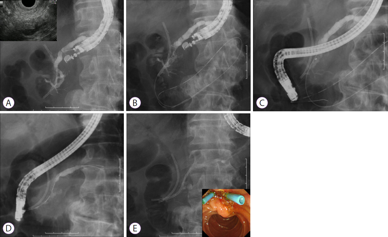
Fig. 1. Technique of endoscopic ultrasound-guided rendezvous. (A) The contrast agent is injected after puncturing the main pancreatic duct. (B) Attempts are made to advance the guidewire in an antegrade fashion across the stricture site into the intestine. (C) A duodenoscope is inserted into the level of the ampulla of Vater, and the guidewire is grasped. (D, E) After the guidewire is pulled into the duodenoscope, cannulation into the main pancreatic duct and stenting are performed in retrograde fashion.
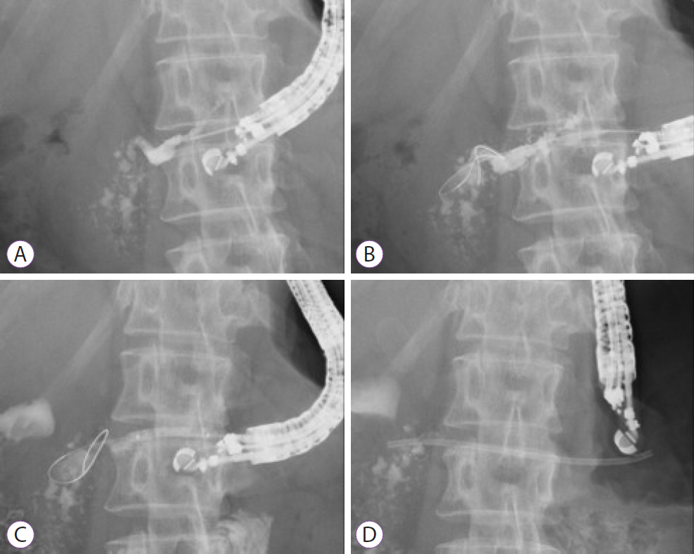
Fig. 2. Technical tips for endoscopic ultrasound-guided pancreatic transmural stenting. (A) The main pancreatic duct is punctured with a 19-G needle and confirmed by injection of contrast medium. (B) A guidewire is advanced into the main pancreatic duct. (C) The tract fistula is dilated using a balloon dilator. (D) Antegrade stent deployment is performed.

Fig. 3. REN (KANEKA Medics, Osaka, Japan). This balloon catheter is characterized by a 3 Fr ultra-tapered tip and coaxial guidewire followability.
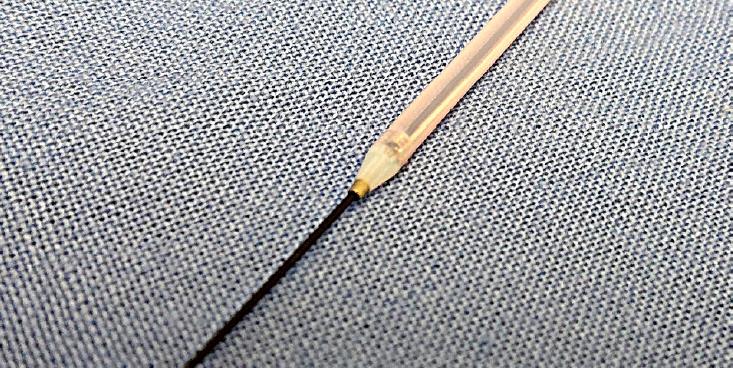
Fig. 4. Fine 025 (Medico’s HIRATA Inc., Osaka, Japan). The distal end of this diathermic dilator is only 3 Fr, and it contains a metal tip. This catheter is coaxial with a guidewire and can be useful for tract dilation in a severely fibrotic pancreas.
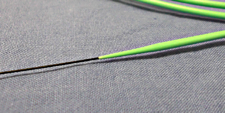
Fig. 5. ES dilator (Zeon Medical Co., Tokyo, Japan). This mechanical dilator can be pushed to a greater degree and exhibits only a small difference in the diameter of the inner lumen and the guidewire.
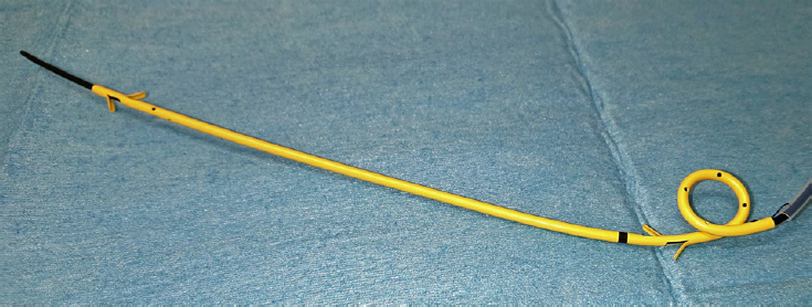
Fig. 6. TYPE IT (Gadelius Medical Co., Tokyo, Japan). This 7 Fr single pigtail type plastic stent has a total length of 20 cm and an effective length of 15 cm. The length and a pigtail anchor together with its four flanges are effective in preventing stent migration.
Cited by 1 articles
-
Endoscopic Ultrasound-guided Drainage in Pancreatobiliary Diseases
Tae Hyeon Kim, Hyung Ku Chon
Korean J Gastroenterol. 2022;79(5):203-209. doi: 10.4166/kjg.2022.064.
Reference
-
1. Bataille L, Deprez P. A new application for therapeutic EUS: main pancreatic duct drainage with a “pancreatic rendezvous technique”. Gastrointest Endosc. 2002; 55:740–743.
Article2. Oh D, Park DH, Song TJ, et al. Long-term outcome of endoscopic ultrasound-guided pancreatic duct drainage using a fully covered self-expandable metal stent for pancreaticojejunal anastomosis stricture. J Gastroenterol Hepatol. 2020; 35:994–1001.
Article3. Tessier G, Bories E, Arvanitakis M, et al. EUS-guided pancreatogastrostomy and pancreatobulbostomy for the treatment of pain in patients with pancreatic ductal dilatation inaccessible for transpapillary endoscopic therapy. Gastrointest Endosc. 2007; 65:233–241.
Article4. Hayat U, Freeman ML, Trikudanathan G, Azeem N, Amateau SK, Mallery J. Endoscopic ultrasound-guided pancreatic duct intervention and pancreaticogastrostomy using a novel cross-platform technique with small-caliber devices. Endosc Int Open. 2020; 8:E196–E202.
Article5. Uchida D, Kato H, Saragai Y, et al. Indications for endoscopic ultrasound-guided pancreatic drainage: for benign or malignant cases? Can J Gastroenterol Hepatol. 2018; 2018:8216109.
Article6. Matsunami Y, Itoi T, Sofuni A, et al. Evaluation of a new stent for EUS-guided pancreatic duct drainage: long-term follow-up outcome. Endosc Int Open. 2018; 6:E505–E512.
Article7. Will U, Reichel A, Fueldner F, Meyer F. Endoscopic ultrasonography-guided drainage for patients with symptomatic obstruction and enlargement of the pancreatic duct. World J Gastroenterol. 2015; 21:13140–13151.
Article8. Fujii LL, Topazian MD, Abu Dayyeh BK, et al. EUS-guided pancreatic duct intervention: outcomes of a single tertiary-care referral center experience. Gastrointest Endosc. 2013; 78:854–864.e1.
Article9. Itoi T, Kasuya K, Sofuni A, et al. Endoscopic ultrasonography-guided pancreatic duct access: techniques and literature review of pancreatography, transmural drainage and rendezvous techniques. Dig Endosc. 2013; 25:241–252.
Article10. Itoi T, Yasuda I, Kurihara T, Itokawa F, Kasuya K. Technique of endoscopic ultrasonography-guided pancreatic duct intervention (with videos). J Hepatobiliary Pancreat Sci. 2014; 21:E4–E9.
Article11. Chen YI, Levy MJ, Moreels TG, et al. An international multicenter study comparing EUS-guided pancreatic duct drainage with enteroscopy-assisted endoscopic retrograde pancreatography after Whipple surgery. Gastrointest Endosc. 2017; 85:170–177.
Article12. Dhir V, Isayama H, Itoi T, et al. Endoscopic ultrasonography-guided biliary and pancreatic duct interventions. Dig Endosc. 2017; 29:472–485.
Article13. Ogura T, Ohama H, Higuchi K. Endoscopic ultrasound-guided pancreatic transmural stenting and transmural intervention. Clin Endosc. 2020; 53:429–435.
Article14. Ogura T, Nishioka N, Yamada M, Ueshima K, Higuchi K. Endoscopic ultrasound-guided pancreatic duct drainage using a fine-gauge balloon catheter. Endoscopy. 2019; 51:E145–E146.
Article15. Dalal A, Patil G, Maydeo A. Six-year retrospective analysis of endoscopic ultrasonography-guided pancreatic ductal interventions at a tertiary referral center. Dig Endosc. 2020; 32:409–416.
Article16. Mandai K, Uno K, Yasuda K. Endoscopic ultrasound-guided pancreatic duct drainage using a novel fine-gauge electrocautery dilator. Endoscopy. 2019; 51:E388–E389.
Article17. Krafft MR, Nasr JY. Anterograde endoscopic ultrasound-guided pancreatic duct drainage: a technical review. Dig Dis Sci. 2019; 64:1770–1781.
Article18. Honjo M, Itoi T, Tsuchiya T, et al. Safety and efficacy of ultra-tapered mechanical dilator for EUS-guided hepaticogastrostomy and pancreatic duct drainage compared with electrocautery dilator (with video). Endosc Ultrasound. 2018; 7:376–382.
Article19. Kitamura K, Yamamiya A, Ishii Y, Nomoto T, Honma T, Yoshida H. Electrocautery vs non-electrocautery dilation catheters in endoscopic ultrasonography-guided pancreatic fluid collection drainage. World J Gastrointest Endosc. 2016; 8:458–465.20. Kahaleh M, Artifon EL, Perez-Miranda M, et al. Endoscopic ultrasonography guided biliary drainage: summary of consortium meeting, May 7th, 2011, Chicago. World J Gastroenterol. 2013; 19:1372–1379.
Article21. Fujii-Lau LL, Levy MJ. Endoscopic ultrasound-guided pancreatic duct drainage. J Hepatobiliary Pancreat Sci. 2015; 22:51–57.
Article22. Chapman CG, Waxman I, Siddiqui UD. Endoscopic ultrasound (EUS)-guided pancreatic duct drainage: the basics of when and how to perform EUS-guided pancreatic duct interventions. Clin Endosc. 2016; 49:161–167.
Article23. Park DH, Jang JW, Lee SS, Seo DW, Lee SK, Kim MH. EUS-guided biliary drainage with transluminal stenting after failed ERCP: predictors of adverse events and long-term results. Gastrointest Endosc. 2011; 74:1276–1284.
Article24. Khashab MA, Messallam AA, Penas I, et al. International multicenter comparative trial of transluminal EUS-guided biliary drainage via hepatogastrostomy vs. choledochoduodenostomy approaches. Endosc Int Open. 2016; 4:E175–E181.
Article25. Kanno Y, Ito K, Koshita S, et al. Efficacy of a newly developed dilator for endoscopic ultrasound-guided biliary drainage. World J Gastrointest Endosc. 2017; 9:304–309.
Article26. Amano M, Ogura T, Onda S, et al. Prospective clinical study of endoscopic ultrasound-guided biliary drainage using novel balloon catheter (with video). J Gastroenterol Hepatol. 2017; 32:716–720.
Article27. Tyberg A, Sharaiha RZ, Kedia P, et al. EUS-guided pancreatic drainage for pancreatic strictures after failed ERCP: a multicenter international collaborative study. Gastrointest Endosc. 2017; 85:164–169.
Article28. Park DH, Kim MH, Moon SH, Lee SS, Seo DW, Lee SK. Feasibility and safety of placement of a newly designed, fully covered self-expandable metal stent for refractory benign pancreatic ductal strictures: a pilot study (with video). Gastrointest Endosc. 2008; 68:1182–1189.
Article29. Oh D, Park DH, Cho MK, et al. Feasibility and safety of a fully covered self-expandable metal stent with antimigration properties for EUS-guided pancreatic duct drainage: early and midterm outcomes (with video). Gastrointest Endosc. 2016; 83:366–373.e2.
Article30. Bang JY, Varadarajulu S. Lumen-apposing metal stents for endoscopic ultrasonography-guided interventions. Dig Endosc. 2019; 31:619–626.
Article
- Full Text Links
-
- Actions
-
Cited
- CITED
-
- Close
- Share
-
- Similar articles
-
- Technical tips for endoscopic ultrasound-guided pancreatic duct access and drainage
- Endoscopic Ultrasound-Guided Pancreatic Transmural Stenting and Transmural Intervention
- Endoscopic Drainage of Pseudocysts
- Endoscopic Ultrasound-Guided Pancreatobiliary Endoscopy in Surgically Altered Anatomy
- Endoscopic Ultrasound-Guided Drainage of Pancreatic Fluid Collections (with Video)
