Deep Learning in Radiation Oncology
- Affiliations
-
- 1Proton Therapy Center, National Cancer Center, Goyang, Korea
- 2Department of Radiation Oncology, Yonsei Cancer Center, Yonsei University College of Medicine, Seoul, Korea
- KMID: 2507483
- DOI: http://doi.org/10.14316/pmp.2020.31.3.111
Abstract
- Deep learning (DL) is a subset of machine learning and artificial intelligence that has a deep neural network with a structure similar to the human neural system and has been trained using big data. DL narrows the gap between data acquisition and meaningful interpretation without explicit programming. It has so far outperformed most classification and regression methods and can automatically learn data representations for specific tasks. The application areas of DL in radiation oncology include classification, semantic segmentation, object detection, image translation and generation, and image captioning. This article tries to understand what is the potential role of DL and what can be more achieved by utilizing it in radiation oncology. With the advances in DL, various studies contributing to the development of radiation oncology were investigated com prehensively. In this article, the radiation treatment process was divided into six consecutive stages as follows: patient assessment, simulation, target and organs-at-risk segmentation, treatment planning, quality assurance, and beam delivery in terms of workflow. Studies using DL were classified and organized according to each radiation treatment process. State-of-the-art studies were identified, and the clinical utilities of those researches were examined. The DL model could provide faster and more accurate solutions to problems faced by oncologists. While the effect of a data-driven approach on improving the quality of care for cancer patients is evidently clear, implementing these methods will require cultural changes at both the professional and institutional levels. We believe this paper will serve as a guide for both clinicians and medical physicists on issues that need to be addressed in time.
Figure
-
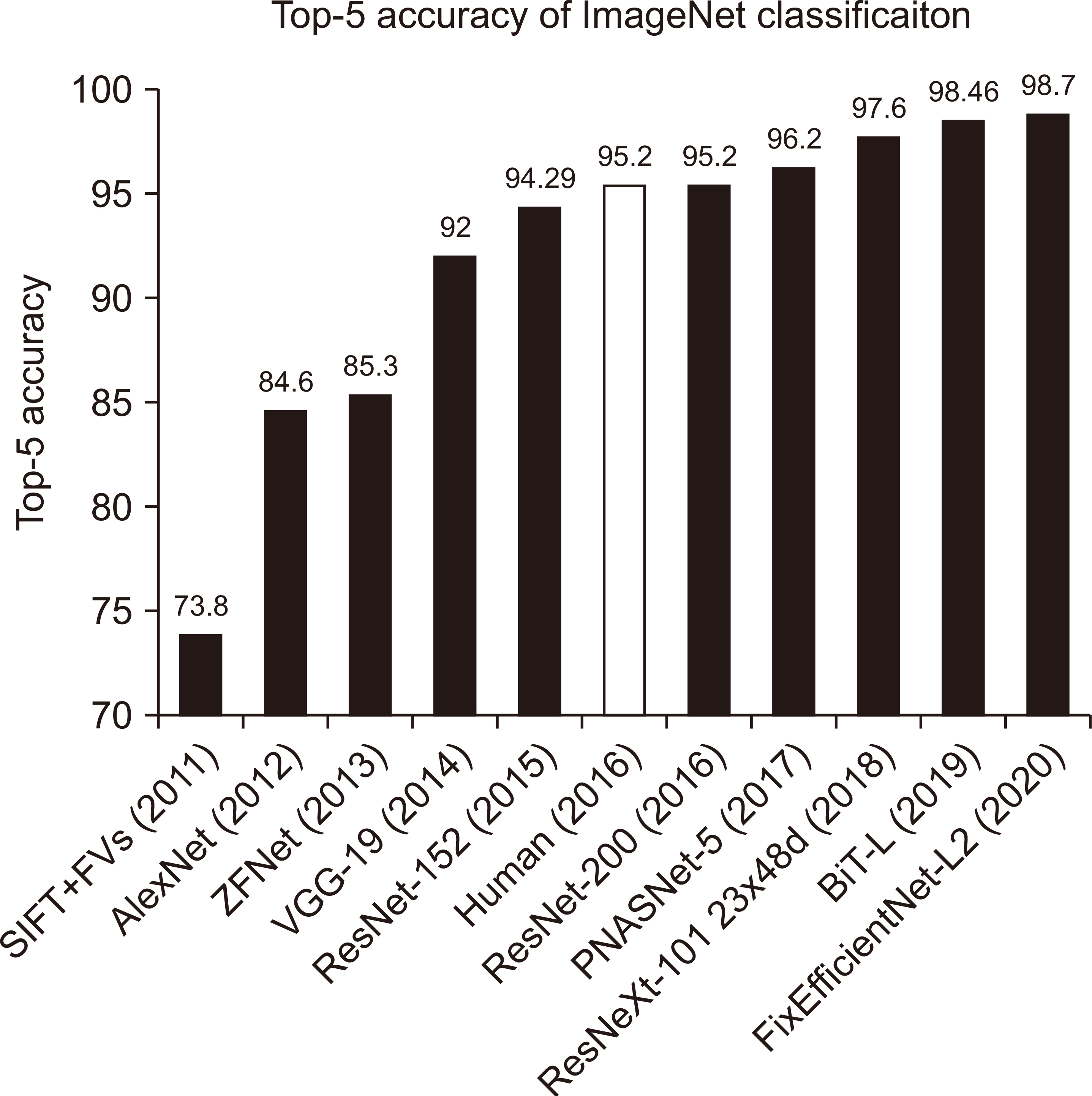
Fig. 1 Top accuracies for image classification models in ImageNet competitions over time.

Fig. 2 Example of prediction of a patient’s respiratory pattern using bilinear long short-term memory (LSTM; black), multilayer perceptron (MLP; blue), and ground truth (red).

Fig. 3 Example of a 3-dimensional lung volume of (a) manual segmentation and (b) DL-based autosegmentation using U-Net.

Fig. 4 Dose prediction for breast case: (a) optimized dose distribution by the treatment planning system, (b) predicted dose distribution by the deep learning model, and (c) dose difference between the optimized and predicted dose distributions.

Fig. 5 Results of FDNet: (a) total fluence map, (b) dose distribution calculated by the treatment planning system, (c) predicted dose distribution using FDNet, and (d) profiles at the middle of the total fluence map, predicted and calculated dose distribution. TPS, treatment-planning system; FDNet, fluence-to-dose network; CAX, central axis.

Fig. 6 Architecture and results of the proposed superresolution generative (pSRG) model for magnetic resonance imaging (MRI)-guided radiotherapy. LR, low resolution; HR, high resolution.
Reference
-
References
1. Goodfellow I, Bengio Y, Courville A. 2016. Deep learning. MIT Press;Massachusetts:2. Bini SA. 2018; Artificial intelligence, machine learning, deep learning, and cognitive computing: what do these terms mean and how will they impact health care? J Arthroplasty. 33:2358–2361. DOI: 10.1016/j.arth.2018.02.067. PMID: 29656964.
Article3. Garrido Á. 2017; Brain and artificial intelligence. Brain. 8:85–90.4. Turing AM. 1950; I.-Computing machinery and intelligence. Mind. 59:433–460. DOI: 10.1093/mind/LIX.236.433.
Article5. McCulloch WS, Pitts W. 1943; A logical calculus of the ideas immanent in nervous activity. Bull Math Biol. 5:15–133. DOI: 10.1007/BF02478259. PMID: 2185863.6. McCorduck P. 2004. Machines who think. A K Peters, Ltd.;Natick: DOI: 10.1201/9780429258985.7. Rosenblatt F. 1961. Principles of neurodynamics: perceptrons and the theory of brain mechanisms. Spartan Books;Washington D.C.: DOI: 10.21236/AD0256582.8. Howe J. 2007. Artificial intelligence at Edinburgh University: a perspective. the University of Edinburgh;Edinburgh: Available from: http://www.inf.ed.ac.uk/about/AIhistory.html. cited 2020 Jul 23.9. Russell SJ, Norvig P. 2003. Artificial intelligence: a modern approach. 2nd ed. Pearson Education, Inc.;Upper Saddle River:10. Nair V, Hinton GE. 2010. Rectified linear units improve restricted Boltzmann machines. Paper presented at: ICML'10: Proceedings of the 27th International Conference on International Conference on Machine Learning. Haifa, Israel; 2010 Jun 21-25. p. 807–814.11. Krizhevsky A, Sutskever I, Hinton GE. 2012; ImageNet classification with deep convolutional neural networks. 1097–1105. PMID: 2185863.12. He K, Zhang X, Ren S, Sun J. 2015. Deep residual learning for image recognition. arXiv.org;Ithaca: Available from: https://arxiv.org/abs/1512.03385. cited 2020 Jul 23.13. Goodfellow IJ, Pouget-Abadie J, Mirza M, Xu B, Warde-Farley D, Ozair S, et al. 2014. Generative adversarial networks. arXiv.org;Ithaca: Available from: https://arxiv.org/abs/1406.2661. cited 2020 Jul 23.14. Hochreiter S, Schmidhuber J. 1997; Long short-term memory. Neural Comput. 9:1735–1780. DOI: 10.1162/neco.1997.9.8.1735. PMID: 9377276.
Article15. Gers FA, Schmidhuber J. 2000. Recurrent nets that time and count. Paper presented at: the IEEE-INNS-ENNS International Joint Conference on Neural Networks. 2000. Neural Computing: New Challenges and Perspectives for the New Millennium. Como, Italy; 2000 Jul 27. New Challenges and Perspectives for the New Millennium;Neural Computing: DOI: 10.1109/IJCNN.2000.861302.16. Cho K, van Merrienboer B, Gulcehre C, Bahdanau D, Bougares F, Schwenk H, et al. 2014. Learning phrase representations using RNN encoder-decoder for statistical machine translation. arXiv.org;Ithaca: Available from: https://arxiv.org/abs/1406.1078. cited 2020 Jul 23. DOI: 10.3115/v1/D14-1179.17. Schuster M, Paliwal KK. 1997; Bidirectional recurrent neural networks. IEEE Trans Signal Process. 45:2673–2681. DOI: 10.1109/78.650093.
Article18. Krause B, Lu L, Murray I, Renals S. 2016. Multiplicative LSTM for sequence modelling. arXiv.org;Ithaca: Available from: https://arxiv.org/abs/1609.07959. cited 2020 Jul 23.19. Wu Y, Schuster M, Chen Z, Le QV, Norouzi M, Macherey W, et al. 2016. Google's neural machine translation system: bridging the gap between human and machine translation. arXiv.org;Ithaca: Available from: https://arxiv.org/abs/1609.08144. cited 2020 Jul 23.20. Cui S, Tseng HH, Pakela J, Ten Haken RK, El Naqa I. 2020; Introduction to machine and deep learning for medical physicists. Med Phys. 47:e127–e147. DOI: 10.1002/mp.14140. PMID: 32418339. PMCID: PMC7331753.
Article21. Hanley J, Debois MM, Mah D, Mageras GS, Raben A, Rosenzweig K, et al. 1999; Deep inspiration breath-hold technique for lung tumors: the potential value of target immobilization and reduced lung density in dose escalation. Int J Radiat Oncol Biol Phys. 45:603–611. DOI: 10.1016/S0360-3016(99)00154-6. PMID: 10524412.
Article22. Kubo HD, Hill BC. 1996; Respiration gated radiotherapy treatment: a technical study. Phys Med Biol. 41:83–91. DOI: 10.1088/0031-9155/41/1/007. PMID: 8685260.
Article23. Ramsey CR, Scaperoth D, Arwood D, Oliver AL. 1999; Clinical efficacy of respiratory gated conformal radiation therapy. Med Dosim. 24:115–119. DOI: 10.1016/S0958-3947(99)00006-0. PMID: 10379508.
Article24. Shirato H, Shimizu S, Kunieda T, Kitamura K, van Herk M, Kagei K, et al. 2000; Physical aspects of a real-time tumor-tracking system for gated radiotherapy. Int J Radiat Oncol Biol Phys. 48:1187–1195. DOI: 10.1016/S0360-3016(00)00748-3. PMID: 11072178.
Article25. de Kruijff RM. 2020; FLASH radiotherapy: ultra-high dose rates to spare healthy tissue. Int J Radiat Biol. 96:419–423. DOI: 10.1080/09553002.2020.1704912. PMID: 31829765.
Article26. Sun WZ, Jiang MY, Ren L, Dang J, You T, Yin FF. 2017; Respiratory signal prediction based on adaptive boosting and multi-layer perceptron neural network. Phys Med Biol. 62:6822–6835. DOI: 10.1088/1361-6560/aa7cd4. PMID: 28665297. PMCID: PMC5555420.
Article27. Wang R, Liang X, Zhu X, Xie Y. 2018; A feasibility of respiration prediction based on deep Bi-LSTM for real-time tumor tracking. IEEE Access. 6:51262–51268. DOI: 10.1109/ACCESS.2018.2869780.
Article28. Chaikh A, Thariat J, Thureau S, Tessonnier T, Kammerer E, Fontbonne C, et al. 2020; [Construction of radiobiological models as TCP (tumor control probability) and NTCP (normal tissue complication probability): from dose to clinical effects prediction]. Cancer Radiother. 24:247–257. French. DOI: 10.1016/j.canrad.2019.12.004. PMID: 32220563.29. Luo Y, Chen S, Valdes G. 2020; Machine learning for radiation outcome modeling and prediction. Med Phys. 47:e178–e184. DOI: 10.1002/mp.13570. PMID: 32418338.
Article30. Arefan D, Mohamed AA, Berg WA, Zuley ML, Sumkin JH, Wu S. 2020; Deep learning modeling using normal mammograms for predicting breast cancer risk. Med Phys. 47:110–118. DOI: 10.1002/mp.13886. PMID: 31667873. PMCID: PMC6980268.
Article31. Szegedy C, Liu W, Jia Y, Sermanet P, Reed S, Anguelov D, et al. 2014. Going deeper with convolutions. arXiv.org;Ithaca: Available from: https://arxiv.org/abs/1409.4842. cited 2020 Jul 23. DOI: 10.1109/CVPR.2015.7298594.32. Li H, Boimel P, Janopaul-Naylor J, Zhong H, Xiao Y, Ben-Josef E, et al. 2019; Deep convolutional neural networks for imaging data based survival analysis of rectal cancer. Proc IEEE Int Symp Biomed Imaging. 2019:846–849. DOI: 10.1109/ISBI.2019.8759301. PMID: 31929858. PMCID: PMC6955095.
Article33. Tait LM, Hoffman D, Benedict S, Valicenti R, Mayadev JS. 2016; The use of MRI deformable image registration for CT-based brachytherapy in locally advanced cervical cancer. Brachytherapy. 15:333–340. DOI: 10.1016/j.brachy.2016.01.002. PMID: 26882845.
Article34. Qi M, Li Y, Wu A, Jia Q, Li B, Sun W, et al. 2020; Multi-sequence MR image-based synthetic CT generation using a generative adversarial network for head and neck MRI-only radiotherapy. Med Phys. 47:1880–1894. DOI: 10.1002/mp.14075. PMID: 32027027.
Article35. Yang C, Liu F, Ahunbay E, Chang YW, Lawton C, Schultz C, et al. 2014; Combined online and offline adaptive radiation therapy: a dosimetric feasibility study. Pract Radiat Oncol. 4:e75–e83. DOI: 10.1016/j.prro.2013.02.012. PMID: 24621436.
Article36. Chen L, Liang X, Shen C, Jiang S, Wang J. 2020; Synthetic CT generation from CBCT images via deep learning. Med Phys. 47:1115–1125. DOI: 10.1002/mp.13978. PMID: 31853974. PMCID: PMC7067667.
Article37. Long J, Shelhamer E, Darrell T. 2014. Fully convolutional networks for semantic segmentation. arXiv.org;Ithaca: Available from: https://arxiv.org/abs/1411.4038. cited 2020 Jul 23. DOI: 10.1109/CVPR.2015.7298965.38. Zhou Z, He Z, Jia Y. 2020; AFPNet: A 3D fully convolutional neural network with atrous-convolution feature pyramid for brain tumor segmentation via MRI images. Neurocomputing. 402:235–244. DOI: 10.1016/j.neucom.2020.03.097.
Article39. Ronneberger O, Fischer P, Brox T. 2015. U-net: convolutional networks for biomedical image segmentation. arXiv.org;Ithaca: Available from: https://arxiv.org/abs/1505.04597. cited 2020 Jul 23. DOI: 10.1007/978-3-319-24574-4_28.40. Rachmadi MF, Valdés-Hernández MDC, Agan MLF, Di Perri C, Komura T. Alzheimer's Disease Neuroimaging Initiative. 2018; Segmentation of white matter hyperintensities using convolutional neural networks with global spatial information in routine clinical brain MRI with none or mild vascular pathology. Comput Med Imaging Graph. 66:28–43. DOI: 10.1016/j.compmedimag.2018.02.002. PMID: 29523002.
Article41. Zhu W, Huang Y, Zeng L, Chen X, Liu Y, Qian Z, et al. 2019; AnatomyNet: deep learning for fast and fully automated whole-volume segmentation of head and neck anatomy. Med Phys. 46:576–589. DOI: 10.1002/mp.13300. PMID: 30480818.
Article42. Ahn SH, Yeo AU, Kim KH, Kim C, Goh Y, Cho S, et al. 2019; Comparative clinical evaluation of atlas and deep-learning-based auto-segmentation of organ structures in liver cancer. Radiat Oncol. 14:213. DOI: 10.1186/s13014-019-1392-z. PMID: 31775825. PMCID: PMC6880380.
Article43. Quan TM, Hildebrand DGC, Jeong WK. 2016. FusionNet: a deep fully residual convolutional neural network for image segmentation in connectomics. arXiv.org;Ithaca: Available from: https://arxiv.org/abs/1612.05360. cited 2020 Jul 23.44. Vinod SK, Min M, Jameson MG, Holloway LC. 2016; A review of interventions to reduce inter-observer variability in volume delineation in radiation oncology. J Med Imaging Radiat Oncol. 60:393–406. DOI: 10.1111/1754-9485.12462. PMID: 27170216.
Article45. Rios Piedra EA, Taira RK, El-Saden S, Ellingson BM, Bui AAT, Hsu W. 2016; Assessing variability in brain tumor segmentation to improve volumetric accuracy and characterization of change. IEEE EMBS Int Conf Biomed Health Inform. 2016:380–383. DOI: 10.1109/BHI.2016.7455914. PMID: 28670648. PMCID: PMC5489257.46. Apolle R, Appold S, Bijl HP, Blanchard P, Bussink J, Faivre-Finn C, et al. 2019; Inter-observer variability in target delineation increases during adaptive treatment of head-and-neck and lung cancer. Acta Oncol. 58:1378–1385. DOI: 10.1080/0284186X.2019.1629017. PMID: 31271079.
Article47. Chabane KTW. 2018. Interobserver variation of prostate delineation on CT and MR by radiation oncologists, radiologists and urologists at the Universitas annex oncology department [dissertation]. University of the Free State;Bloemfontein:48. Franco P, Arcadipane F, Trino E, Gallio E, Martini S, Iorio GC, et al. 2018; Variability of clinical target volume delineation for rectal cancer patients planned for neoadjuvant radiotherapy with the aid of the platform Anatom-e. Clin Transl Radiat Oncol. 11:33–39. DOI: 10.1016/j.ctro.2018.06.002. PMID: 29928706. PMCID: PMC6008279.
Article49. Cabrera GG, Ehrgott M, Mason AJ, Raith A. 2018; A matheuristic approach to solve the multiobjective beam angle optimization problem in intensity-modulated radiation therapy. 25:243–268. DOI: 10.1111/itor.12241. PMID: 29928706. PMCID: PMC6008279.50. Breedveld S, Storchi PR, Voet PW, Heijmen BJ. 2012; iCycle: integrated, multicriterial beam angle, and profile optimization for generation of coplanar and noncoplanar IMRT plans. Med Phys. 39:951–963. DOI: 10.1118/1.3676689. PMID: 22320804.
Article51. Taasti VT, Hong L, Shim JSA, Deasy JO, Zarepisheh M. 2020; Automating proton treatment planning with beam angle selection using Bayesian optimization. Med Phys. 47:3286–3296. DOI: 10.1002/mp.14215. PMID: 32356335. PMCID: PMC7429260.
Article52. Sadeghnejad Barkousaraie A, Ogunmolu O, Jiang S, Nguyen D. 2020; A fast deep learning approach for beam orientation optimization for prostate cancer treated with intensity-modulated radiation therapy. Med Phys. 47:880–897. DOI: 10.1002/mp.13986. PMID: 31868927.53. Quan EM, Li X, Li Y, Wang X, Kudchadker RJ, Johnson JL, et al. 2012; A comprehensive comparison of IMRT and VMAT plan quality for prostate cancer treatment. Int J Radiat Oncol Biol Phys. 83:1169–1178. DOI: 10.1016/j.ijrobp.2011.09.015. PMID: 22704703. PMCID: PMC3805837.
Article54. Hoffmann M, Pacey J, Goodworth J, Laszcyzk A, Ford R, Chick B, et al. 2019; Analysis of a volumetric-modulated arc therapy (VMAT) single phase prostate template as a class solution. Rep Pract Oncol Radiother. 24:92–96. DOI: 10.1016/j.rpor.2018.10.009. PMID: 30505239. PMCID: PMC6251366.
Article55. Chen X, Men K, Li Y, Yi J, Dai J. 2019; A feasibility study on an automated method to generate patient-specific dose distributions for radiotherapy using deep learning. Med Phys. 46:56–64. DOI: 10.1002/mp.13262. PMID: 30367492.
Article56. Barragán-Montero AM, Nguyen D, Lu W, Lin MH, Norouzi-Kandalan R, Geets X, et al. 2019; Three-dimensional dose prediction for lung IMRT patients with deep neural networks: robust learning from heterogeneous beam configurations. Med Phys. 46:3679–3691. DOI: 10.1002/mp.13597. PMID: 31102554.57. Galib SM, Lee HK, Guy CL, Riblett MJ, Hugo GD. 2020; A fast and scalable method for quality assurance of deformable image registration on lung CT scans using convolutional neural networks. Med Phys. 47:99–109. DOI: 10.1002/mp.13890. PMID: 31663137.
Article58. Nyflot MJ, Thammasorn P, Wootton LS, Ford EC, Chaovalitwongse WA. 2019; Deep learning for patient-specific quality assurance: identifying errors in radiotherapy delivery by radiomic analysis of gamma images with convolutional neural networks. Med Phys. 46:456–464. DOI: 10.1002/mp.13338. PMID: 30548601.
Article59. Interian Y, Rideout V, Kearney VP, Gennatas E, Morin O, Cheung J, et al. 2018; Deep nets vs expert designed features in medical physics: an IMRT QA case study. Med Phys. 45:2672–2680. DOI: 10.1002/mp.12890. PMID: 29603278.
Article60. Cheon W, Kim SJ, Hwang UJ, Min BJ, Han Y. 2019; Feasibility study of the fluence-to-dose network (FDNet) for patient-specific IMRT quality assurance. J Korean Phys Soc. 75:724–734. DOI: 10.3938/jkps.75.724.
Article61. Kim KH, Do WJ, Park SH. 2018; Improving resolution of MR images with an adversarial network incorporating images with different contrast. Med Phys. 45:3120–3131. DOI: 10.1002/mp.12945. PMID: 29729006.62. Chun J, Zhang H, Gach HM, Olberg S, Mazur T, Green O, et al. 2019; MRI super-resolution reconstruction for MRI-guided adaptive radiotherapy using cascaded deep learning: in the presence of limited training data and unknown translation model. Med Phys. 46:4148–4164. DOI: 10.1002/mp.13717. PMID: 31309585.
Article63. Cheon W, Lee J, Min BJ, Han Y. 2019; Super-resolution model for high-precision in vivo proton range verification using a stereo gamma camera: a feasibility study. J Korean Phys Soc. 75:617–627. DOI: 10.3938/jkps.75.617.
Article64. Lu Y, Kowarschik M, Huang X, Xia Y, Choi JH, Chen S, et al. 2019; A learning-based material decomposition pipeline for multi-energy x-ray imaging. Med Phys. 46:689–703. DOI: 10.1002/mp.13317. PMID: 30508253.
Article65. Cheon W, Kim SJ, Kim K, Lee M, Lee J, Jo K, et al. 2019; Feasibility of two-dimensional dose distribution deconvolution using convolution neural networks. Med Phys. 46:5833–5847. DOI: 10.1002/mp.13869. PMID: 31621917.
Article66. Grau C, Hoyer M. 2005; High-precision radiotherapy. Eur Oncol Rev. 40–44. DOI: 10.17925/EOH.2005.0.0.40.
Article
- Full Text Links
-
- Actions
-
Cited
- CITED
-
- Close
- Share
-
- Similar articles
-
- Optimizing e-learning in oncology during the COVID-19 pandemic and beyond
- Current status of deep learning applications in abdominal ultrasonography
- Deep Learning in Dental Radiographic Imaging
- Radiomics and Deep Learning from Research to Clinical Workflow: Neuro-Oncologic Imaging
- Deep Learning in MR Motion Correction:a Brief Review and a New Motion Simulation Tool (view2Dmotion)
