Three-dimensional surgical accuracy between virtually planned and actual surgical movements of the maxilla in two-jaw orthognathic surgery
- Affiliations
-
- 1Department of Orthodontics, School of Dentistry, Kyungpook National University, Daegu, Korea
- 2Private Practice, Seoul, Korea
- 3Department of Orthodontics, School of Dentistry, Seoul National University, Seoul, Korea
- KMID: 2506464
- DOI: http://doi.org/10.4041/kjod.2020.50.5.293
Abstract
Objective
To investigate the three-dimensional (3D) surgical accuracy between virtually planned and actual surgical movements (SM) of the maxilla in twojaw orthognathic surgery.
Methods
The sample consisted of 15 skeletal Class III patients who underwent two-jaw orthognathic surgery performed by a single surgeon using a virtual surgical simulation (VSS) software. The 3D cone-beam computed tomography (CBCT) images were obtained before (T0) and after surgery (T1). After merging the dental cast image onto the T0 CBCT image, VSS was performed. SM were classified into midline correction (anterior and posterior), advancement, setback, anterior elongation, and impaction (total and posterior). The landmarks were the midpoint between the central incisors, the mesiobuccal cusp tip (MBCT) of both first molars, and the midpoint of the two MBCTs. The amount and direction of SM by VSS and actual surgery were measured using 3D coordinates of the landmarks. Discrepancies less than 1 mm between VSS and T1 landmarks indicated a precise outcome. The surgical achievement percentage (SAP, [amount of movement in actual surgery/ amount of movement in VSS] × 100) (%) and precision percentage (PP, [number of patients with precise outcome/number of total patients] × 100) (%) were compared among SM types using Fisher’s exact and Kruskal–Wallis tests.
Results
Overall mean discrepancy between VSS and actual surgery, SAP, and PP were 0.13 mm, 89.9%, and 68.3%, respectively. There was no significant difference in the SAP and PP values among the seven SM types (all p > 0.05).
Conclusions
VSS could be considered as an effective tool for increasing surgical accuracy.
Keyword
Figure
-
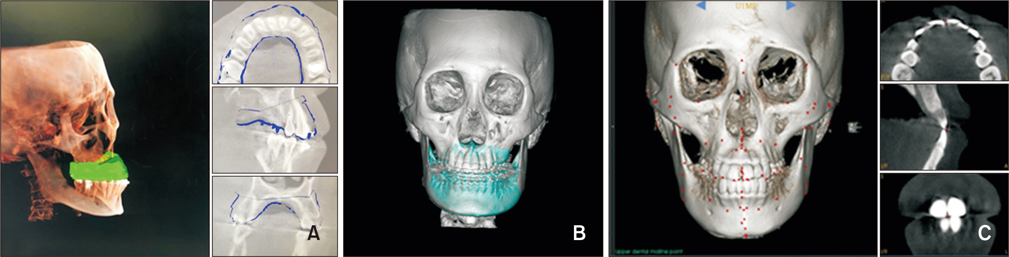
Figure 1 The process of merging between the scanned dentition and the three-dimensional cone-beam computed tomography (3D-CBCT) image and landmark digitization. A, Merging of the scanned dentition with 3D-CBCT image using three sectional views of the sagittal, frontal, and axial planes. B, Result of fine-tuned merging between dentition and 3D-CBCT images. C, Landmark digitization to identify the midpoint between the maxillary central incisors at the occlusal level (U1MP) and the mesiobuccal cusp tips (MBCTs) of the maxillary right and left first molar crowns using the three sectional views of the sagittal, frontal, and axial planes. The mean coordinate value from the two digitized MBCTs was automatically calculated by the ON3D software (3D ONS Inc., Seoul, Korea).
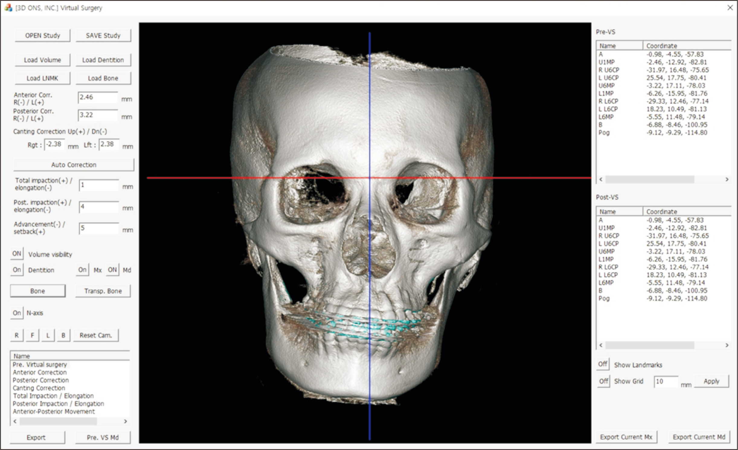
Figure 2 Virtual surgery modules of the ON3D software (3D ONS Inc., Seoul, Korea).
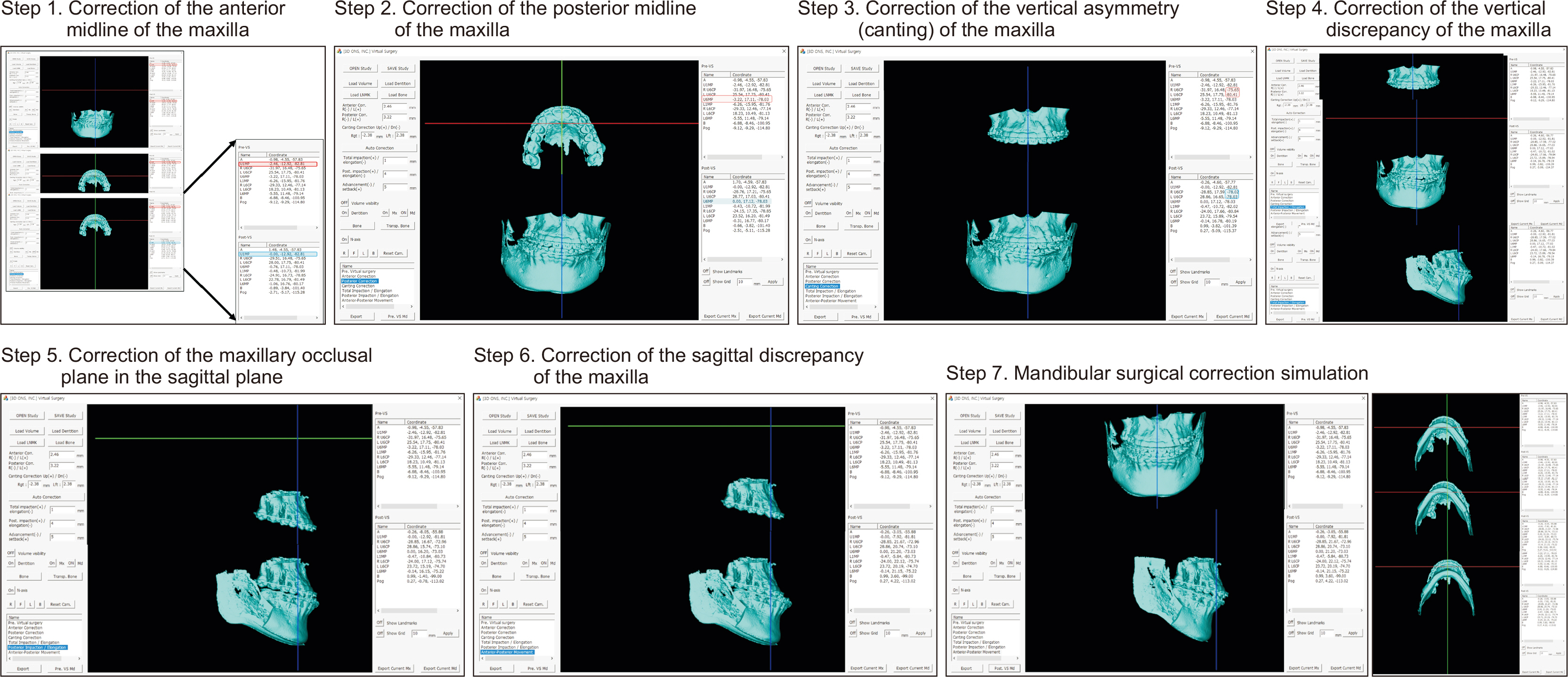
Figure 3 The step-by-step procedure of virtual surgical simulation. Step 1, Correction of the anterior midline of the maxilla. Step 2, Correction of the posterior midline of the maxilla. Step 3, Correction of the vertical asymmetry (canting) of the maxilla. Step 4, Correction of the vertical discrepancy of the maxilla. Step 5, Correction of the maxillary occlusal plane in the sagittal plane. Step 6, Correction of the sagittal discrepancy of the maxilla. Step 7, Correction of the mandible.
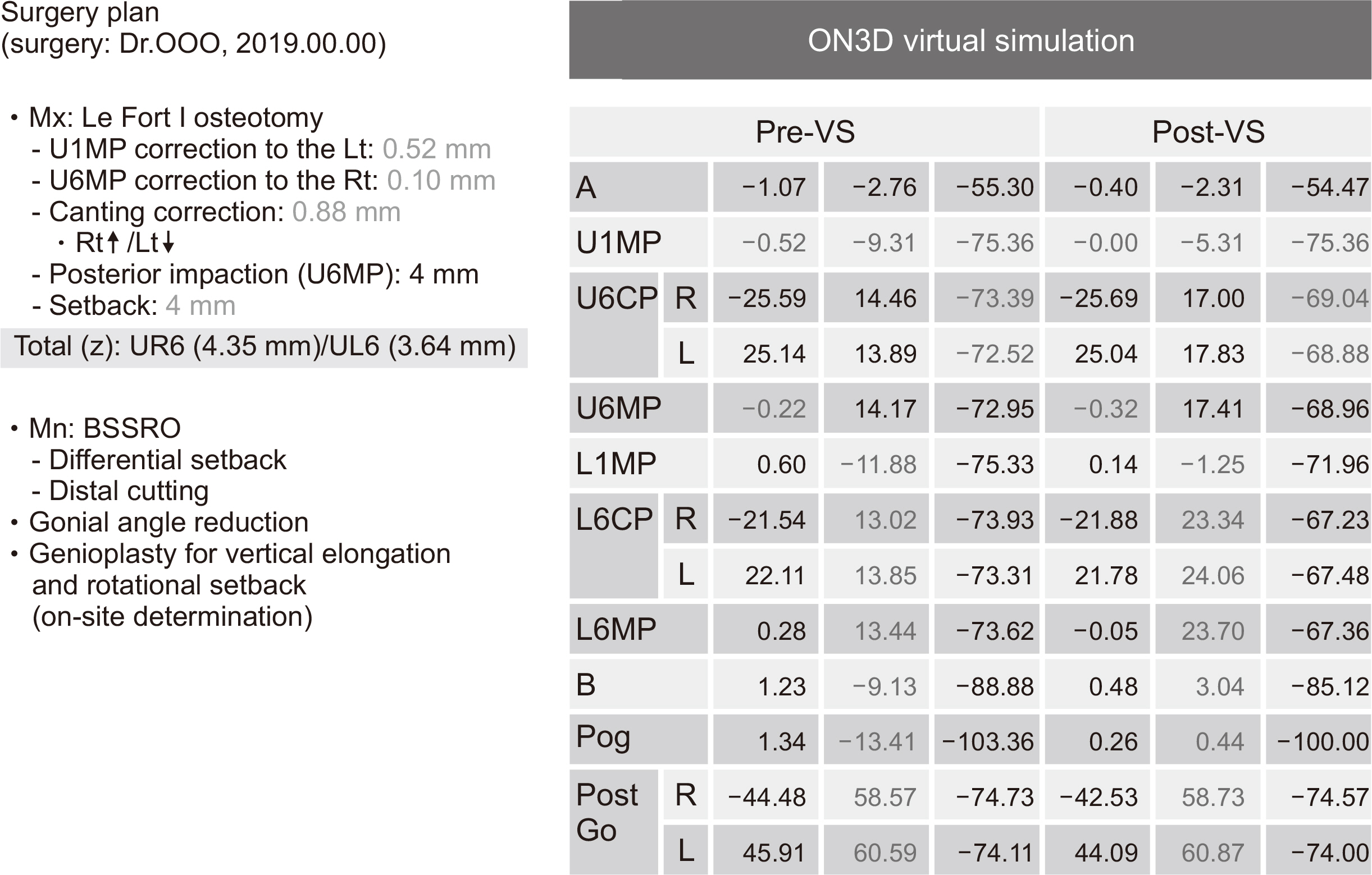
Figure 4 An example of surgical plan and coordinate values of the selected landmarks at the pre- and post-virtual surgery (VS). Mx, maxilla; Mn, mandible; Lt, left; Rt, right; BSSRO, bilateral sagittal split ramus osteotomy; U1MP, the midpoint between the maxillary central incisors at the occlusal level; U6MP, the midpoint between the two mesiobuccal cusp tips of the maxillary right and left first molar crowns; UR6, the mesiobuccal cusp tip of the maxillary right first molar crown; UL6, the mesiobuccal cusp tip of the maxillary left first molar crown; U6CP, the mesiobuccal cusp tip of the maxillary first molar crown; L1MP, the midpoint between the mandibular central incisors at the occlusal level; L6CP, the mesiobuccal cusp tip of the mandibular first molar crown; L6MP, the midpoint between the two mesiobuccal cusp tips of the mandibular right and left first molar crowns; A, A point; B, B point; Pog, pogonion; Go, gonion.
Cited by 2 articles
-
Characterization of facial asymmetry phenotypes in adult patients with skeletal Class III malocclusion using three-dimensional computed tomography and cluster analysis
Sang-Woon Ha, Su-Jung Kim, Jin-Young Choi, Seung-Hak Baek
Korean J Orthod. 2022;52(2):85-101. doi: 10.4041/kjod.2022.52.2.85.Evaluation of mandibular condyle position in Class III patients after bimaxillary orthognathic surgery: A cone-beam computed tomography study
Osman Küçükçakır, Nilüfer Ersan, Yunus Ziya Arslan, Erol Cansız
Korean J Orthod. 2024;54(4):247-256. doi: 10.4041/kjod23.188.
Reference
-
1. Jacobson R, Sarver DM. 2002; The predictability of maxillary repositioning in LeFort I orthognathic surgery. Am J Orthod Dentofacial Orthop. 122:142–54. DOI: 10.1067/mod.2002.125576. PMID: 12165768.
Article2. Semaan S, Goonewardene MS. 2005; Accuracy of a LeFort I maxillary osteotomy. Angle Orthod. 75:964–73.3. Gil JN, Claus JD, Manfro R. 2007; Predictability of maxillary repositioning during bimaxillary surgery: accuracy of a new technique. Int J Oral Maxillofac Surg. 36:296–300. DOI: 10.1016/j.ijom.2006.10.015. PMID: 17240117.
Article4. Choi JY, Choi JP, Baek SH. 2009; Surgical accuracy of maxillary repositioning according to type of surgical movement in two-jaw surgery. Angle Orthod. 79:306–11. DOI: 10.2319/030608-136.1. PMID: 19216601.
Article5. Stokbro K, Aagaard E, Torkov P, Bell RB, Thygesen T. 2016; Surgical accuracy of three-dimensional virtual planning: a pilot study of bimaxillary orthognathic procedures including maxillary segmentation. Int J Oral Maxillofac Surg. 45:8–18. DOI: 10.1016/j.ijom.2015.07.010. PMID: 26250603.
Article6. Gaber RM, Shaheen E, Falter B, Araya S, Politis C, Swennen GRJ, et al. 2017; A systematic review to uncover a universal protocol for accuracy assessment of 3-dimensional virtually planned orthognathic surgery. J Oral Maxillofac Surg. 75:2430–40. DOI: 10.1016/j.joms.2017.05.025. PMID: 28646644.
Article7. Song KG, Baek SH. 2009; Comparison of the accuracy of the three-dimensional virtual method and the conventional manual method for model surgery and intermediate wafer fabrication. Oral Surg Oral Med Oral Pathol Oral Radiol Endod. 107:13–21. DOI: 10.1016/j.tripleo.2008.06.002. PMID: 18755612.
Article8. De Riu G, Virdis PI, Meloni SM, Lumbau A, Vaira LA. 2018; Accuracy of computer-assisted orthognathic surgery. J Craniomaxillofac Surg. 46:293–8. DOI: 10.1016/j.jcms.2017.11.023. PMID: 29275075.
Article9. Cho HJ. 2009; A three-dimensional cephalometric analysis. J Clin Orthod. 43:235–52. discussion 235quiz 273DOI: 10.5040/9781474293754.0223. PMID: 19458456.10. Metzger MC, Hohlweg-Majert B, Schwarz U, Teschner M, Hammer B, Schmelzeisen R. 2008; Manufacturing splints for orthognathic surgery using a three-dimensional printer. Oral Surg Oral Med Oral Pathol Oral Radiol Endod. 105:e1–7. DOI: 10.1016/j.tripleo.2007.07.040. PMID: 18230371.
Article11. Aboul-Hosn Centenero S, Hernández-Alfaro F. 2012; 3D planning in orthognathic surgery: CAD/CAM surgical splints and prediction of the soft and hard tissues results - our experience in 16 cases. J Craniomaxillofac Surg. 40:162–8. DOI: 10.1016/j.jcms.2011.03.014. PMID: 21458285.
Article12. Hernández-Alfaro F, Guijarro-Martínez R. 2013; New protocol for three-dimensional surgical planning and CAD/CAM splint generation in orthognathic surgery: an in vitro and in vivo study. Int J Oral Maxillofac Surg. 42:1547–56. DOI: 10.1016/j.ijom.2013.03.025. PMID: 23768749.
Article13. Stokbro K, Aagaard E, Torkov P, Bell RB, Thygesen T. 2014; Virtual planning in orthognathic surgery. Int J Oral Maxillofac Surg. 43:957–65. DOI: 10.1016/j.ijom.2014.03.011. PMID: 24746388.
Article14. Baan F, Liebregts J, Xi T, Schreurs R, de Koning M, Bergé S, et al. 2016; A new 3D tool for assessing the accuracy of bimaxillary surgery: the OrthoGnathicAnalyser. PLoS One. 11:e0149625. DOI: 10.1371/journal.pone.0149625. PMID: 26901524. PMCID: PMC4762705.
Article15. Shaheen E, Sun Y, Jacobs R, Politis C. 2017; Three-dimensional printed final occlusal splint for orthognathic surgery: design and validation. Int J Oral Maxillofac Surg. 46:67–71. DOI: 10.1016/j.ijom.2016.10.002. PMID: 27815012.
Article16. Van den Bempt M, Liebregts J, Maal T, Bergé S, Xi T. 2018; Toward a higher accuracy in orthognathic surgery by using intraoperative computer navigation, 3D surgical guides, and/or customized osteosynthesis plates: a systematic review. J Craniomaxillofac Surg. 46:2108–19. DOI: 10.1016/j.jcms.2018.10.012. PMID: 30420150.
Article17. Tankersley AC, Nimmich MC, Battan A, Griggs JA, Caloss R. 2019; Comparison of the planned versus actual jaw movement using splint-based virtual surgical planning: how close are we at achieving the planned outcomes? J Oral Maxillofac Surg. 77:1675–80. DOI: 10.1016/j.joms.2019.03.004. PMID: 30959009.
Article18. Lin HH, Lo LJ. 2015; Three-dimensional computer-assisted surgical simulation and intraoperative navigation in orthognathic surgery: a literature review. J Formos Med Assoc. 114:300–7. DOI: 10.1016/j.jfma.2015.01.017. PMID: 25744942.19. Mangano FG, Admakin O, Bonacina M, Biaggini F, Farronato D, Lerner H. 2020; Accuracy of 6 desktop 3D printers in dentistry: a comparative in vitro study. Eur J Prosthodont Restor Dent. 28:75–85. DOI: 10.1922/EJPRD_2050Mangano11. PMID: 32347671.20. Choi JH, Mah J. 2010; A new method for superimposition of CBCT volumes. J Clin Orthod. 44:303–12. DOI: 10.1007/s00784-020-03539-3. PMID: 20831099.21. Gkantidis N, Schauseil M, Pazera P, Zorkun B, Katsaros C, Ludwig B. 2015; Evaluation of 3-dimensional superimposition techniques on various skeletal structures of the head using surface models. PLoS One. 10:e0118810. DOI: 10.1371/journal.pone.0118810. PMID: 25706151. PMCID: PMC4338241.
Article22. Tucker S, Cevidanes LH, Styner M, Kim H, Reyes M, Proffit W, et al. 2010; Comparison of actual surgical outcomes and 3-dimensional surgical simulations. J Oral Maxillofac Surg. 68:2412–21. DOI: 10.1016/j.joms.2009.09.058. PMID: 20591553. PMCID: PMC2945446.
Article23. Hsu SS, Gateno J, Bell RB, Hirsch DL, Markiewicz MR, Teichgraeber JF, et al. 2013; Accuracy of a computer-aided surgical simulation protocol for orthognathic surgery: a prospective multicenter study. J Oral Maxillofac Surg. 71:128–42. DOI: 10.1016/j.joms.2012.03.027. PMID: 22695016. PMCID: PMC3443525.
Article24. Shehab MF, Barakat AA, AbdElghany K, Mostafa Y, Baur DA. 2013; A novel design of a computer-generated splint for vertical repositioning of the maxilla after Le Fort I osteotomy. Oral Surg Oral Med Oral Pathol Oral Radiol. 115:e16–25. DOI: 10.1016/j.oooo.2011.09.035. PMID: 23312923.
Article
- Full Text Links
-
- Actions
-
Cited
- CITED
-
- Close
- Share
-
- Similar articles
-
- READER’S FORUM
- Two-jaw surgery by use of Surgical Jaw Relator
- Fabrication of surgical splint by using of surgical jaw relator
- The effects of orthognathic surgery on auditory function
- A comparative study on the degree of relapse following one jaw surgery and two jaw surgery in skeletal Class III patients
