Camouflage treatment of posterior bite collapse in a patient with skeletal asymmetry by using posterior maxillary segmental osteotomy
- Affiliations
-
- 1Department of Orthodontics, Kyung Hee University Dental Hospital, Seoul, Korea
- 2Department of Dentistry, Graduate School, Kyung Hee University, Seoul, Korea
- 3Private Practice, Seoul, Korea
- 4Department of Oral & Maxillofacial Surgery, Kyung Hee University School of Dentistry, Seoul, Korea
- 5Department of Orthodontics, Kyung Hee University School of Dentistry, Seoul, Korea
- KMID: 2504145
- DOI: http://doi.org/10.4041/kjod.2020.50.4.278
Abstract
- Orthodontic treatment of posterior bite collapse due to early loss of molars and the consequent drift of adjacent teeth is complicated. When the posterior bite collapse occurs in patients with facial asymmetry, both transverse and vertical compensation are necessary for camouflage orthodontic treatment. In such cases, posterior maxillary segmental osteotomy (PMSO) can be an effective alternative procedure that simplifies the orthodontic treatment and shows long-term stability through dental compensation within the alveolar bone housing. This case report aimed to describe the orthodontic treatment of maxillary occlusal plane canting caused by severely extruded maxillary teeth in a patient with skeletal facial asymmetry that was corrected with PMSO along with protraction of the lower second molar to replace the space of the extracted first molar. The treatment duration was 18 months, and stable results were obtained after 2 years of retention.
Figure
-
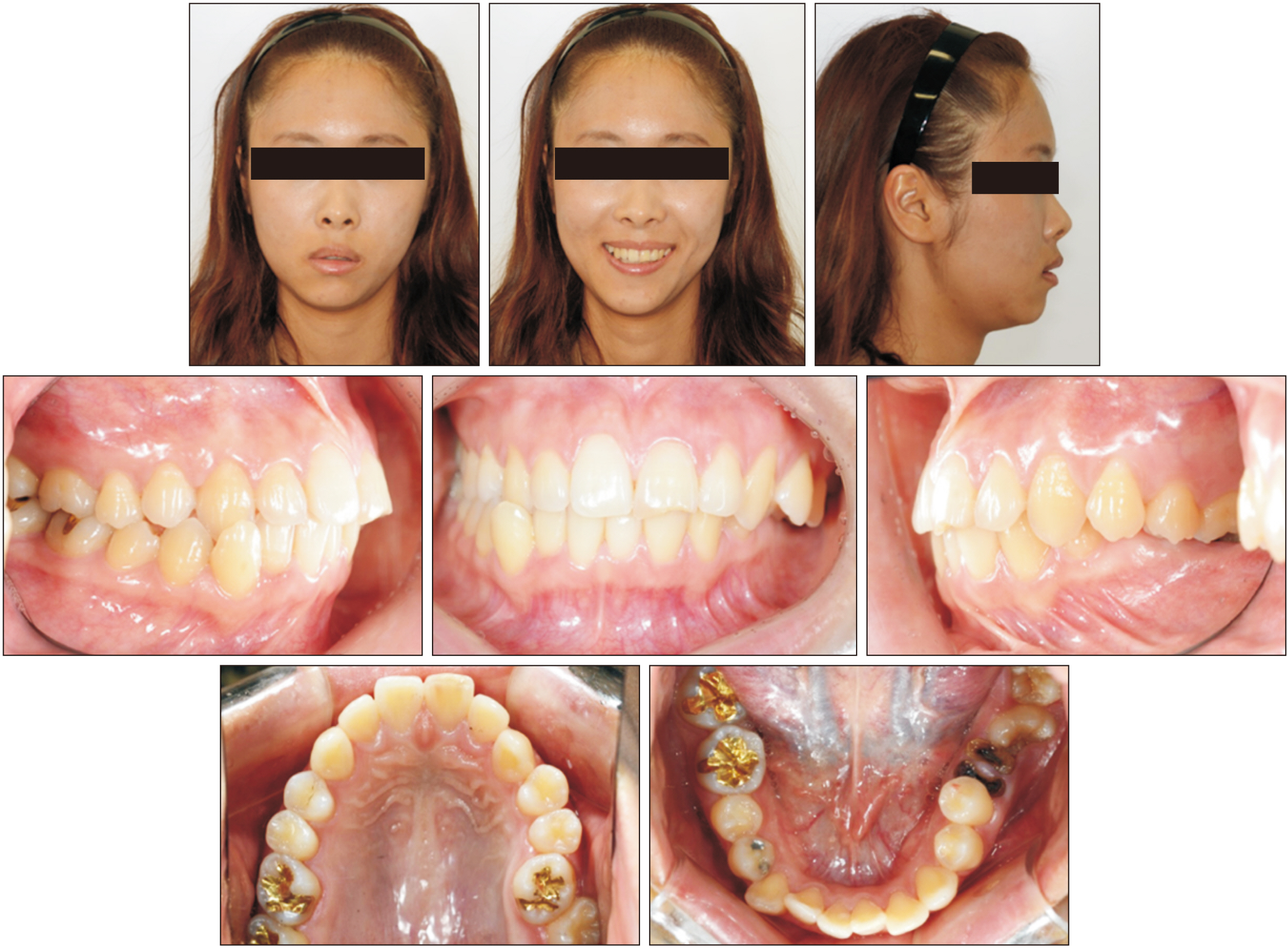
Figure 1 Pretreatment facial and intraoral photographs.
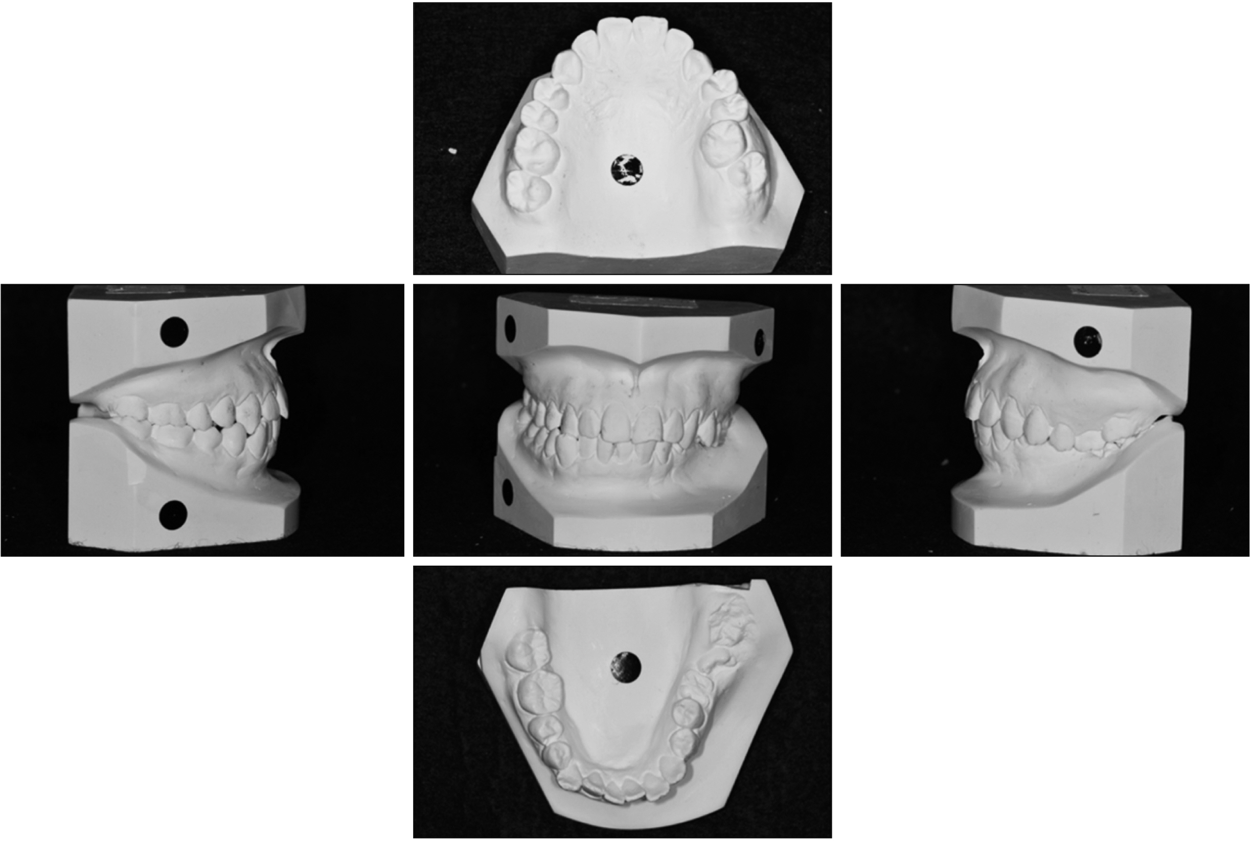
Figure 2 Pretreatment cast models.

Figure 3 Pretreatment radiographs. A, Lateral cephalogram; B, panoramic radiograph; and C, cone-beam computed tomography images.
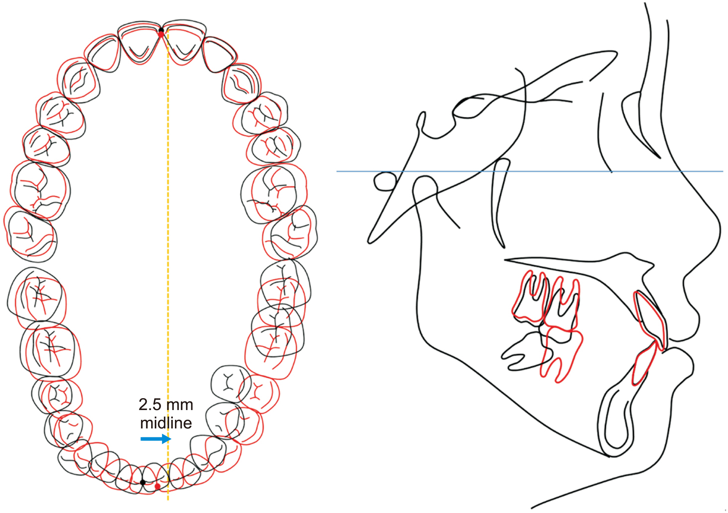
Figure 4 Cephalometric and dental visual treatment objective (VTO): the posterior teeth on the cephalometric VTO represent the collapsed side only (left). Posterior maxillary segmental osteotomy is planned on the left maxillary arch. In the mandibular arch, uprighting and protraction of the mandibular left molars and transverse compensation is planned. Black, Pretreatment; red, VTO.
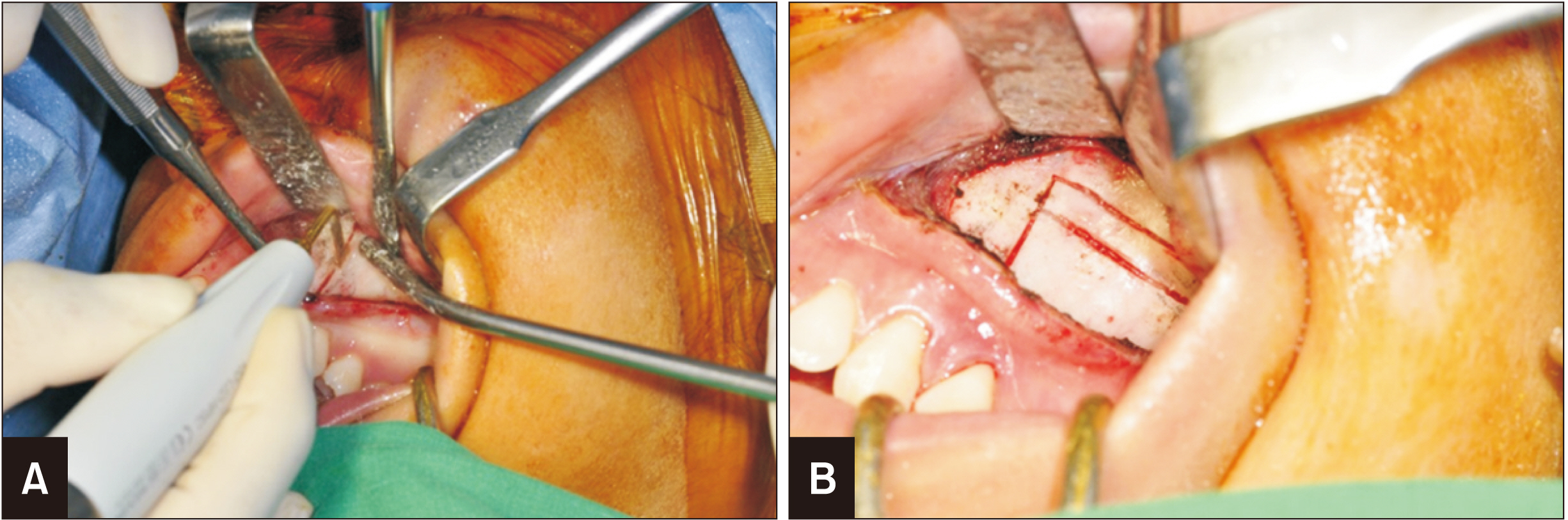
Figure 5 Posterior maxillary segmental osteotomy procedure with a vertical bone cut (A) and a horizontal bone cut (B).

Figure 6 Treatment progression 2 weeks (A), 6 weeks (B), 10 weeks (C), 5 months (D), 11 months (E), and 16 months (F) after posterior maxillary segmental osteotomy.
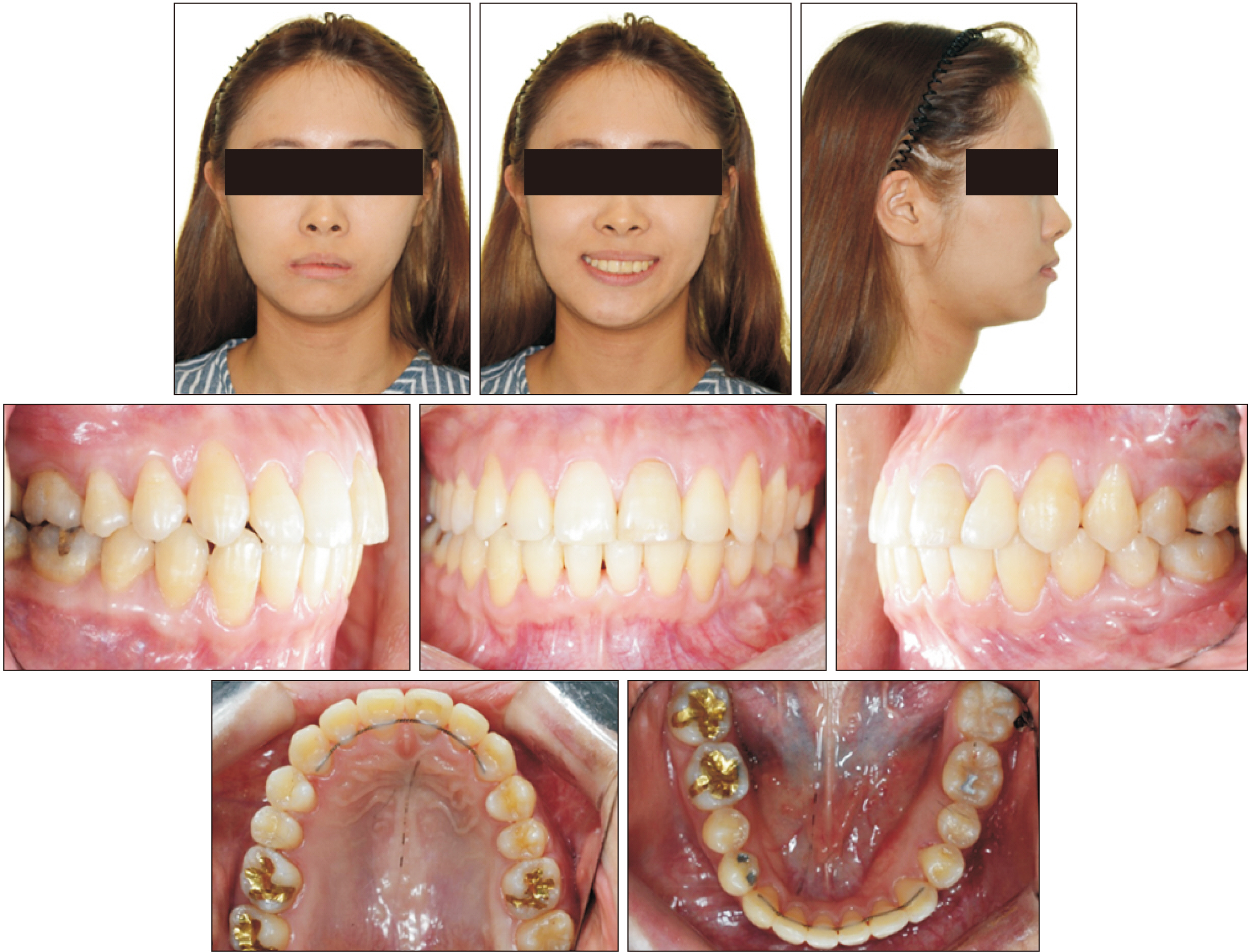
Figure 7 Post-treatment facial and intraoral photographs.
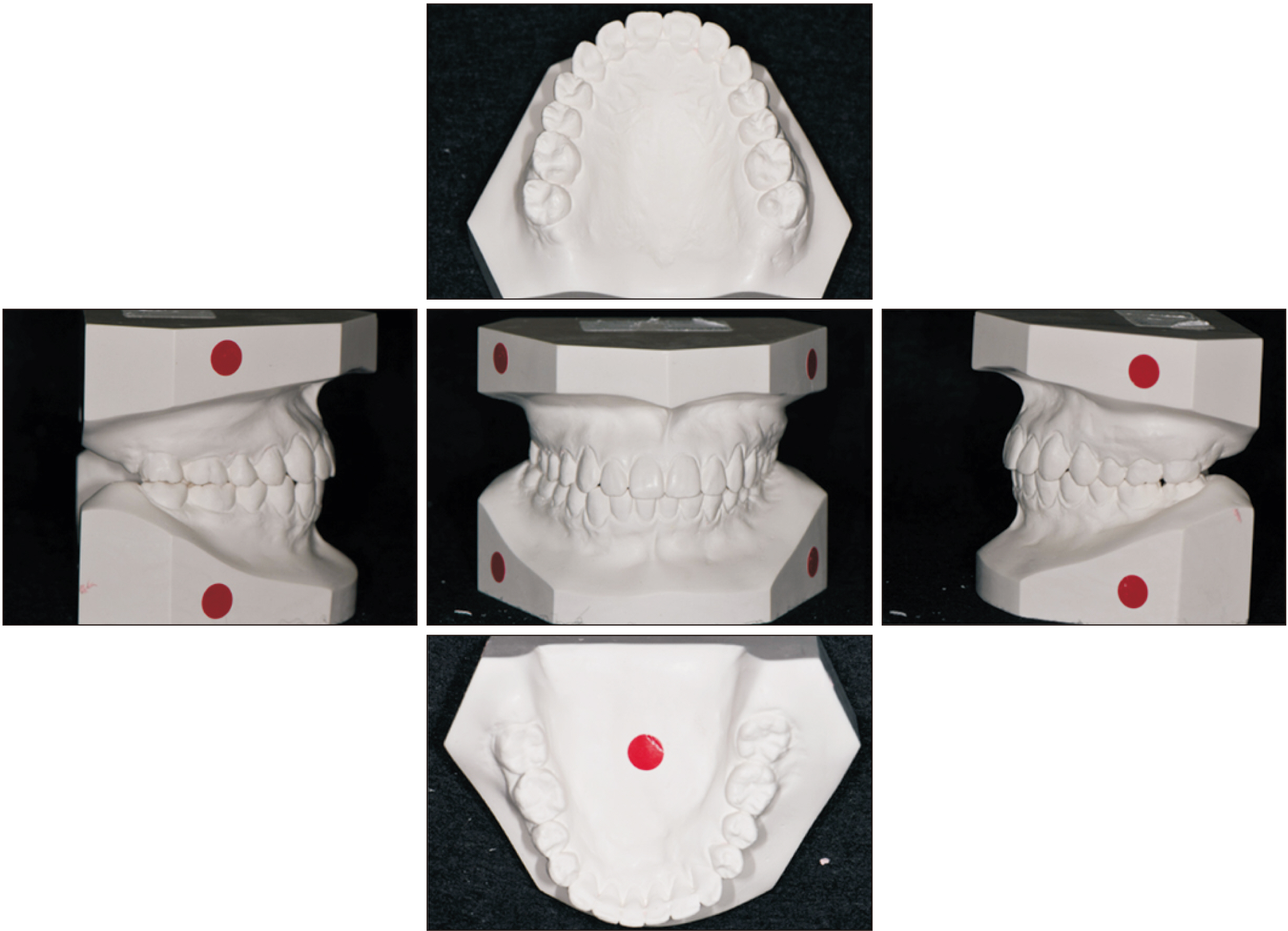
Figure 8 Post-treatment cast models.
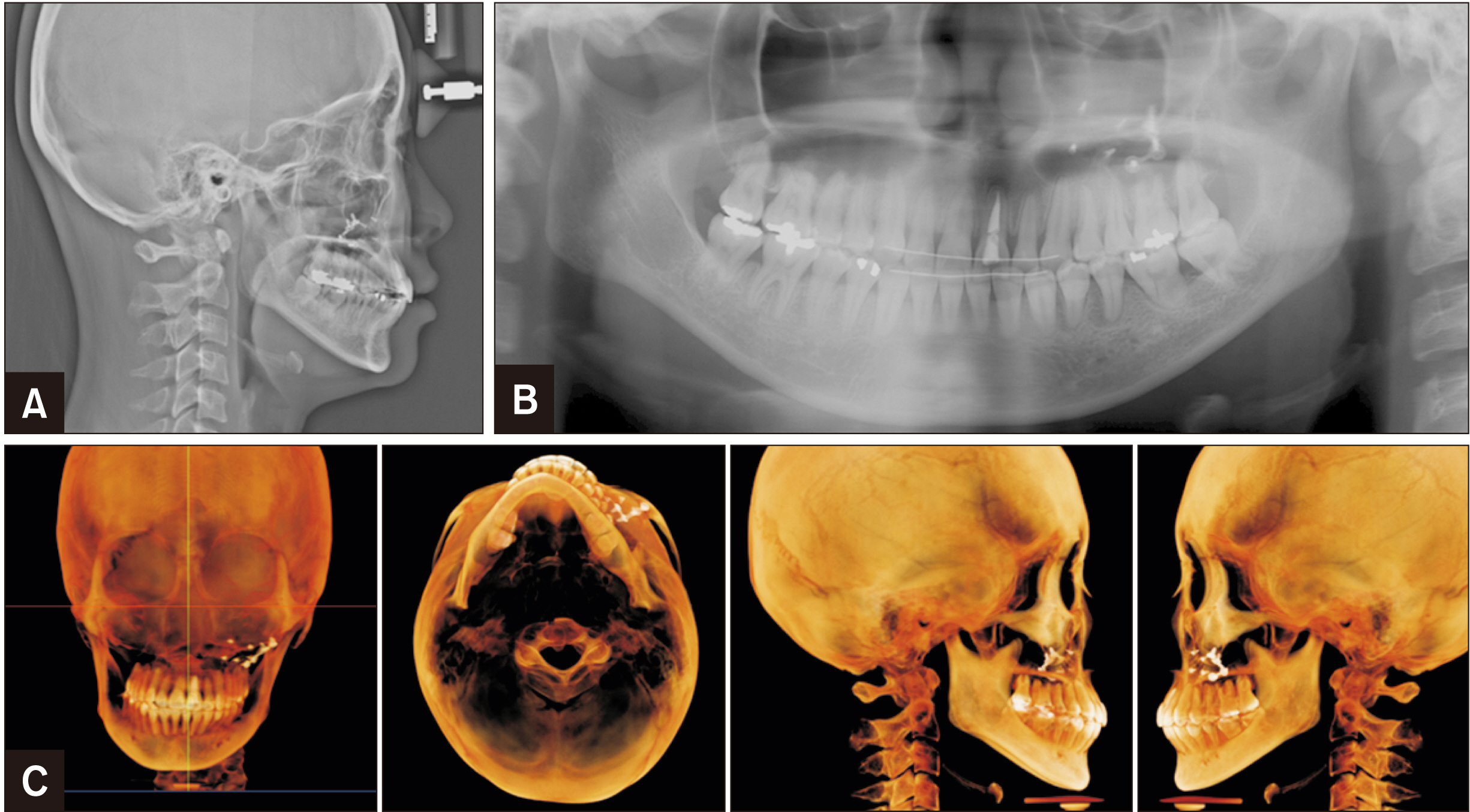
Figure 9 Post-treatment radiographs. A, Lateral cephalogram; B, panoramic radiograph; and C, cone-beam computed tomography images.

Figure 10 Cephalometric superimpositions between the pretreatment (black lines) and post-treatment stages (red lines): overall, maxilla, and mandible. For the mandibular molars, the dotted line represents the left second molar, and the solid line represents the right first molar.
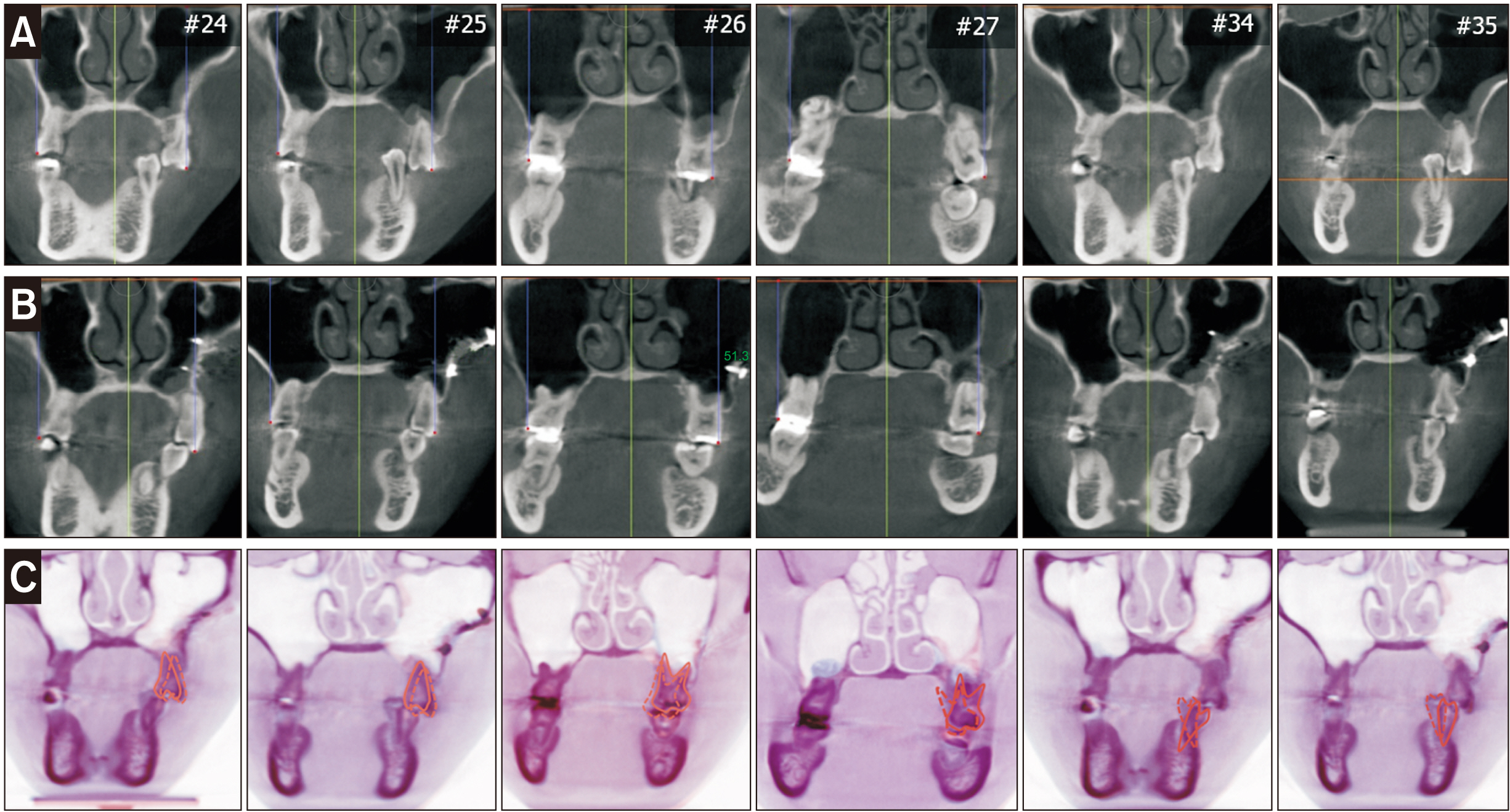
Figure 11 Changes in vertical and transverse positions of maxillary posterior teeth and mandibular left premolars. A, Pretreatment; B, post-treatment; and C, superimposition. Solid line, Pretreatment; dotted line, post-treatment.

Figure 12 Temporomandibular joint position and morphology assessed using cone-beam computed tomography. A, Pretreatment; B, post-treatment.
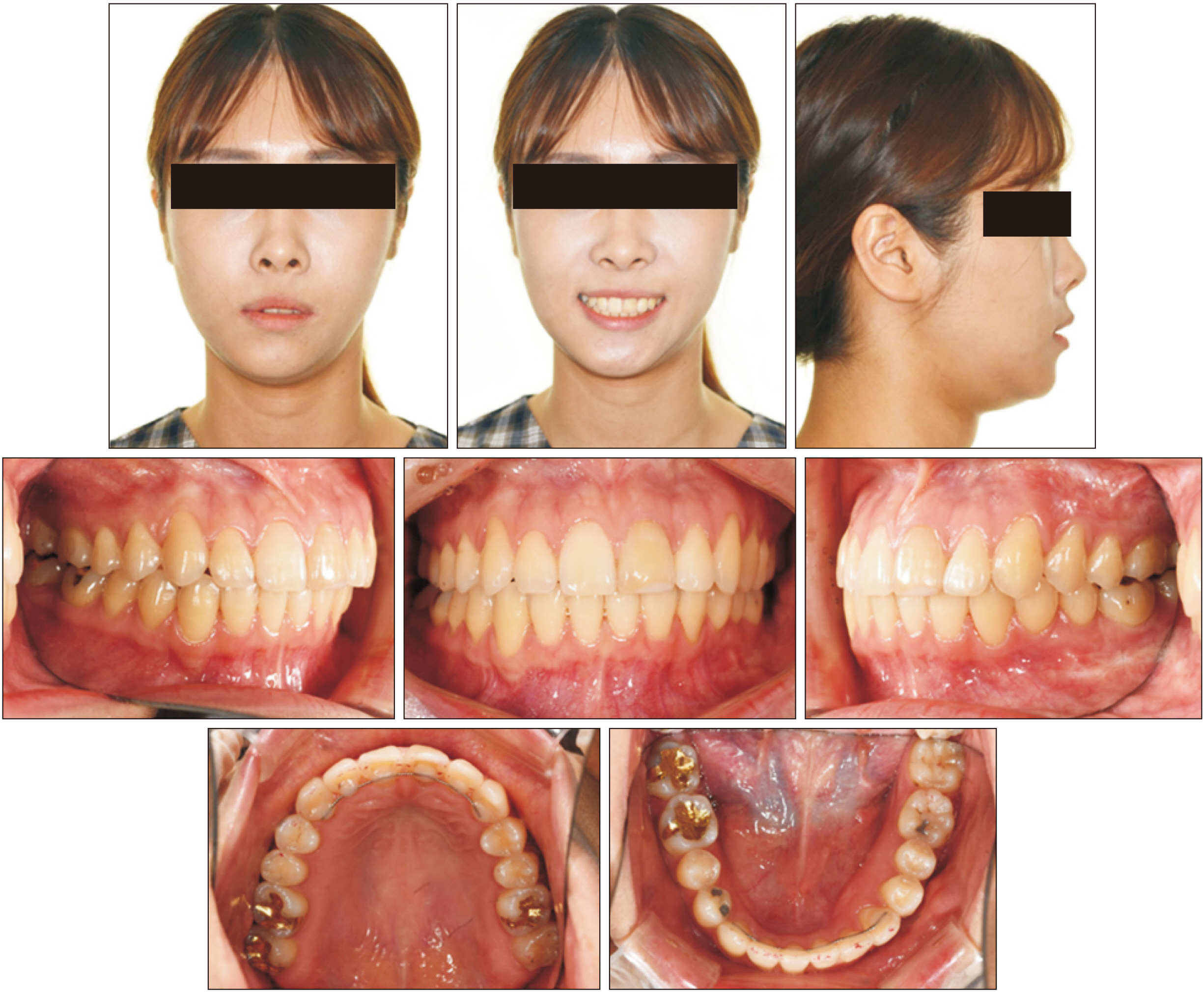
Figure 13 Post-retention (2 years) facial and intraoral photographs.

Figure 14 Lateral cephalogram at 2-year retention (A), and cephalometric superimpositions between post-treatment (red lines) and post-retention stages (green lines): overall, maxilla, and mandible (B).
Reference
-
1. Shifman A, Laufer BZ, Chweidan H. 1998; Posterior bite collapse--revisited. J Oral Rehabil. 25:376–85. DOI: 10.1046/j.1365-2842.1998.00243.x. PMID: 9639163.2. Dersot JM, Giovannoli JL. 1989; [Posterior bite collapse. 1. Etiology and diagnosis]. J Parodontol. 8:187–94. French. PMID: 2639188.3. Sekiya T, Nakamura Y, Oikawa T, Ishii H, Hirashita A, Seto K. 2010; Elimination of transverse dental compensation is critical for treatment of patients with severe facial asymmetry. Am J Orthod Dentofacial Orthop. 137:552–62. DOI: 10.1016/j.ajodo.2007.10.064. PMID: 20362918.
Article4. Kim KA, Lee JW, Park JH, Kim BH, Ahn HW, Kim SJ. 2017; Targeted presurgical decompensation in patients with yaw-dependent facial asymmetry. Korean J Orthod. 47:195–206. DOI: 10.4041/kjod.2017.47.3.195. PMID: 28523246. PMCID: PMC5432441.
Article5. Jeon YJ, Kim YH, Son WS, Hans MG. 2006; Correction of a canted occlusal plane with miniscrews in a patient with facial asymmetry. Am J Orthod Dentofacial Orthop. 130:244–52. DOI: 10.1016/j.ajodo.2006.04.016. PMID: 16905071.
Article6. Kang YG, Nam JH, Park YG. 2010; Use of rhythmic wire system with miniscrews to correct occlusal-plane canting. Am J Orthod Dentofacial Orthop. 137:540–7. DOI: 10.1016/j.ajodo.2008.04.032. PMID: 20362916.
Article7. Daimaruya T, Takahashi I, Nagasaka H, Umemori M, Sugawara J, Mitani H. 2003; Effects of maxillary molar intrusion on the nasal floor and tooth root using the skeletal anchorage system in dogs. Angle Orthod. 73:158–66.8. Yao CC, Lee JJ, Chen HY, Chang ZC, Chang HF, Chen YJ. 2005; Maxillary molar intrusion with fixed appliances and mini-implant anchorage studied in three dimensions. Angle Orthod. 75:754–60. DOI: 10.1043/0003-3219(2005)75[754:MMIWFA]2.0.CO;2. PMID: 16279822.9. Park YC, Lee SY, Kim DH, Jee SH. 2003; Intrusion of posterior teeth using mini-screw implants. Am J Orthod Dentofacial Orthop. 123:690–4. DOI: 10.1016/S0889-5406(03)00047-7. PMID: 12806352.
Article10. Meningaud JP, Pitak-Arnnop P, Corcos L, Bertrand JC. 2006; Posterior maxillary segmental osteotomy for mandibular implants placement: case report. Oral Surg Oral Med Oral Pathol Oral Radiol Endod. 102:e1–3. DOI: 10.1016/j.tripleo.2006.03.013. PMID: 17052616.
Article11. West RA, Epker BN. 1972; Posterior maxillary surgery its place in the treatment of dentofacial deformities. J Oral Surg. 30:562–3.12. Bell WH, Levy BM. 1971; Revascularization and bone healing after posterior maxillary osteotomy. J Oral Surg. 29:313–20. PMID: 4995705.13. Rosen PS, Forman D. 1999; The role of orthognathic surgery in the treatment of severe dentoalveolar extrusion. J Am Dent Assoc. 130:1619–22. DOI: 10.14219/jada.archive.1999.0101. PMID: 10573942.
Article14. O'Ryan F, Schendel S. 1989; Nasal anatomy and maxillary surgery. II. Unfavorable nasolabial esthetics following the Le Fort I osteotomy. Int J Adult Orthodon Orthognath Surg. 4:75–84.15. Kim C, Kim M, Kim D. 1999. Upward repositioning of the extruded posterior maxillary alveolar segment by one-stage segmental osteotomies. Paper presented at: 14th International Conference on Oral and Maxillofacial Surgery. 1999 Apr 24-29; Washington DC, USA. Chicago. ICOMS;p. 13. DOI: 10.1016/S0901-5027(99)80712-7.
Article16. Sherwood KH, Burch J, Thompson W. 2003; Intrusion of supererupted molars with titanium miniplate anchorage. Angle Orthod. 73:597–601. DOI: 10.1043/0003-3219(2003)073<0597:IOSMWT>2.0.CO;2. PMID: 14580029.17. Erverdi N, Keles A, Nanda R. 2004; The use of skeletal anchorage in open bite treatment: a cephalometric evaluation. Angle Orthod. 74:381–90. DOI: 10.1043/0003-3219(2004)074<0381:TUOSAI>2.0.CO;2. PMID: 15264651.18. Kusayama M, Motohashi N, Kuroda T. 2003; Relationship between transverse dental anomalies and skeletal asymmetry. Am J Orthod Dentofacial Orthop. 123:329–37. DOI: 10.1067/mod.2003.41. PMID: 12637905.
Article19. Solow B. 1980; The dentoalveolar compensatory mechanism: background and clinical implications. Br J Orthod. 7:145–61. DOI: 10.1179/bjo.7.3.145. PMID: 6934010.
Article20. Anhoury PS. 2009; Nonsurgical treatment of an adult with mandibular asymmetry and unilateral posterior crossbite. Am J Orthod Dentofacial Orthop. 135:118–26. DOI: 10.1016/j.ajodo.2006.12.024. PMID: 19121511.
Article21. Meskin LH, Brown LJ. 1988; Prevalence and patterns of tooth loss in U.S. employed adult and senior populations, 1985-86. J Dent Educ. 52:686–91. DOI: 10.1002/j.0022-0337.1988.52.12.tb02263.x. PMID: 3263994.
Article22. Roberts WE, Arbuckle GR, Analoui M. 1996; Rate of mesial translation of mandibular molars using implant-anchored mechanics. Angle Orthod. 66:331–8. DOI: 10.1043/0003-3219(1996)066<0331:ROMTOM>2.3.CO;2. PMID: 8893103.23. Raveli TB, Raveli DB, de Mathias Almeida KC, Pinto ADS. 2017; Molar uprighting: a considerable and safe decision to avoid prosthetic treatment. Open Dent J. 11:466–75. DOI: 10.2174/1874210601711010466. PMID: 29114332. PMCID: PMC5646130.
Article24. Proffit WR, Turvey TA, Phillips C. 2007; The hierarchy of stability and predictability in orthognathic surgery with rigid fixation: an update and extension. Head Face Med. 3:21. DOI: 10.1186/1746-160X-3-21. PMID: 17470277. PMCID: PMC1876453.
Article25. Jung SK, Kim TW. 2015; Treatment of unilateral posterior crossbite with facial asymmetry in a female patient with transverse discrepancy. Am J Orthod Dentofacial Orthop. 148:154–64. DOI: 10.1016/j.ajodo.2014.09.023. PMID: 26124038.
Article26. Kai R, Umeki D, Sekiya T, Nakamura Y. 2016; Defining the location of the dental midline is critical for oral esthetics in camouflage orthodontic treatment of facial asymmetry. Am J Orthod Dentofacial Orthop. 150:1028–38. DOI: 10.1016/j.ajodo.2015.10.035. PMID: 27894524.
Article
- Full Text Links
-
- Actions
-
Cited
- CITED
-
- Close
- Share
-
- Similar articles
-
- Treatment of anterior open bite by posterior maxillary segmental osteotomy and miniplates: a case report
- Le Fort I Osteotomy and Posterior Maxillary Segmental Osteotomy for Correction of Malunioned Maxilla
- Treatment of anterior open bite with bimaxillary anterior segmental osteotomy and genioplasty
- Functional evaluation of orthopedic and orthodontic treatment in a patient with unilateral posterior crossbite and facial asymmetry
- The surgical correction of post-traumatic malocclusion
