Management of peri-implantitis associated with tear-like implant fracture: case reports
- Affiliations
-
- 1Department of Periodontology, Daejeon Dental Hospital, Wonkwang University College of Dentistry, Daejeon, Republic of Korea
- 2Institute of Wonkwang Dental Research, Wonkwang University College of Dentistry, iksan, Republic of Korea
- KMID: 2503810
- DOI: http://doi.org/10.14368/jdras.2020.36.2.138
Abstract
- Implant fracture is rare, but one of the most serious problem in implantation. Treatment of implant fracture can be different according to the extent of the fracture and on the state of the surrounding prosthetic restoration. Maintaining or submerging implant after treatment of peri-implantitis can be useful options for cases of tear-like fracture on the coronal area of an implant.
Keyword
Figure
-
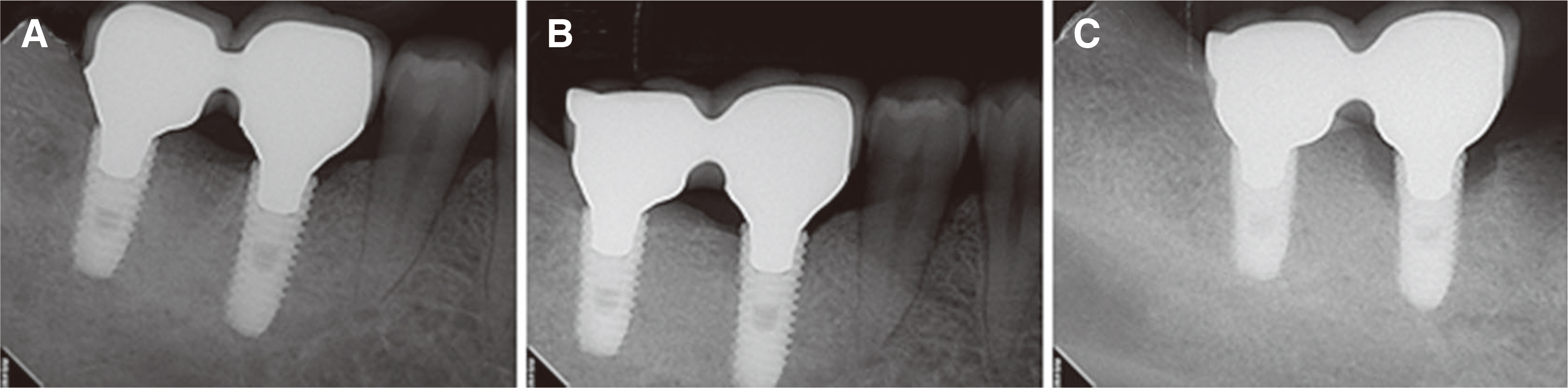
Fig. 1 Periapical radiography of case 1. (A) Prosthesis delivery on December 2013. (B) There are no major clinical symptoms detected at 4-years follow up. (C) Rapid and aggressive peri-implant bone loss on the #46 implant occurred with prosthesis agitation 4 month after last follow up.
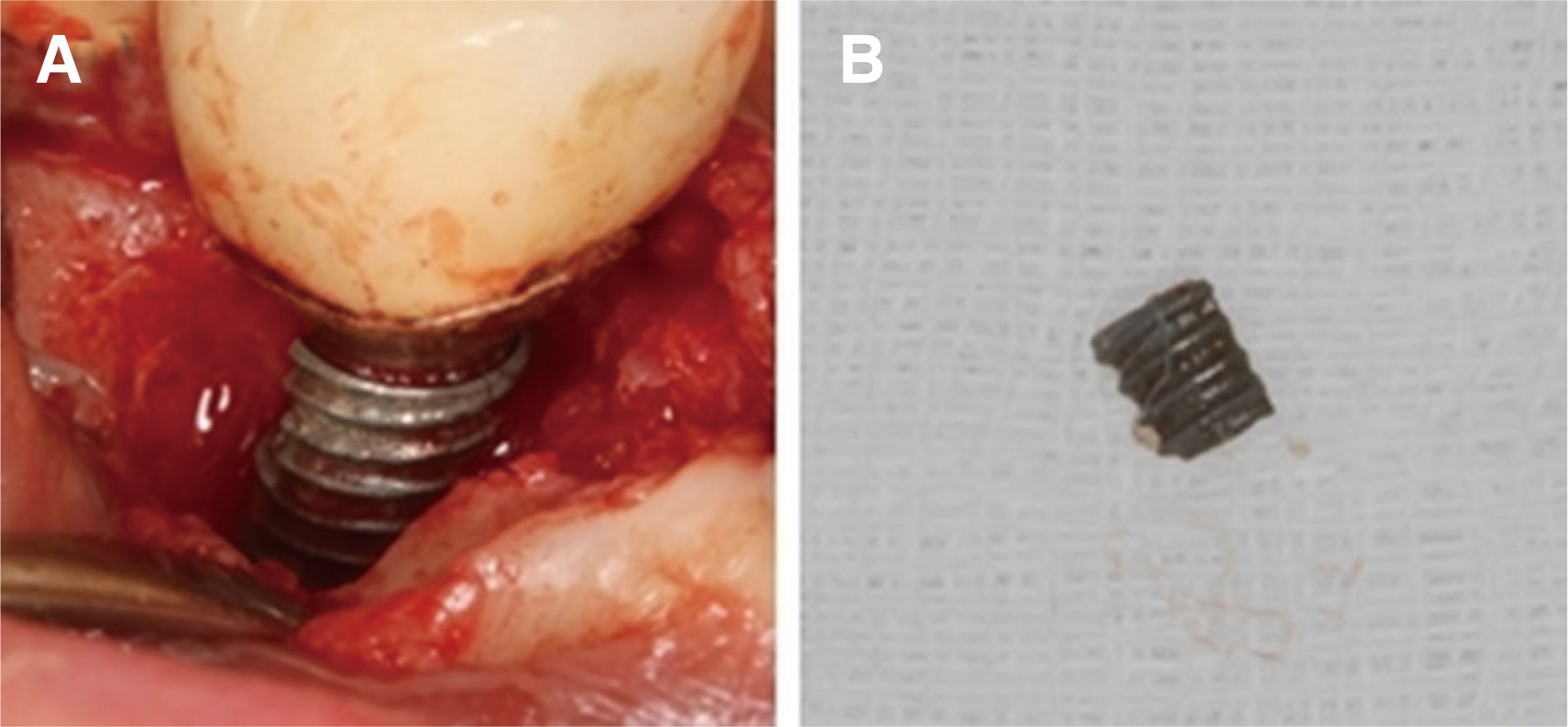
Fig. 2 (A) Intraoral photograph of peri-implnatitis surgery of case 1: Peri-implant bone loss with buccal and mesial implant fracture lines were observed. After removing the fragment and surface decontamination, xenobone grafting combination with enamel matrix derivates were performed around the defect area. (B) Removed fractured fragement. Fragment size: 2 x 3 mm.
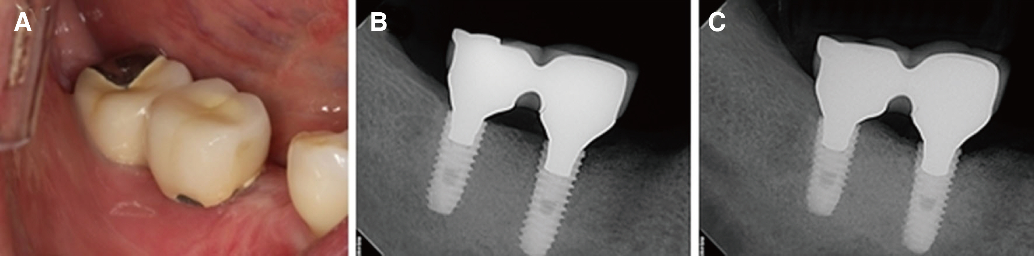
Fig. 3 (A) Intraoral photograph of case 1: no specific sign at 3 month follow up. (B) Periapical radiography: 3 month follow up after peri-implantitis surgery with indicating defect fill. (C) On 15 month follow up periapical radiography, surgical site is maintained stable without any abnormal sign.
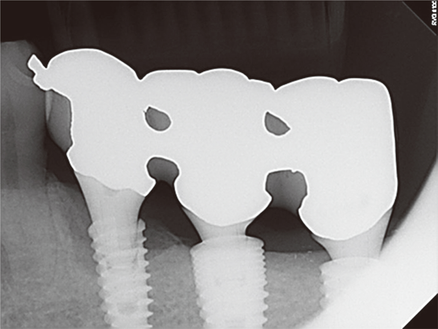
Fig. 4 Periapical radiography of case 2: Periimplant bone loss on the #35 implant was detected approximately 5 years after implantation.
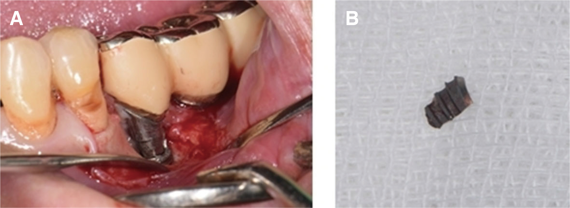
Fig. 5 (A) Intraoral photograph of peri-implnatitis surgery of case 2: Buccal bone loss and buccal implant fractures were observed. After removing the fragment, mechanical debridement and chemical decontamination were performed. (B) Removed fractured fragement. Fragement size: 1.5 x 3 mm.
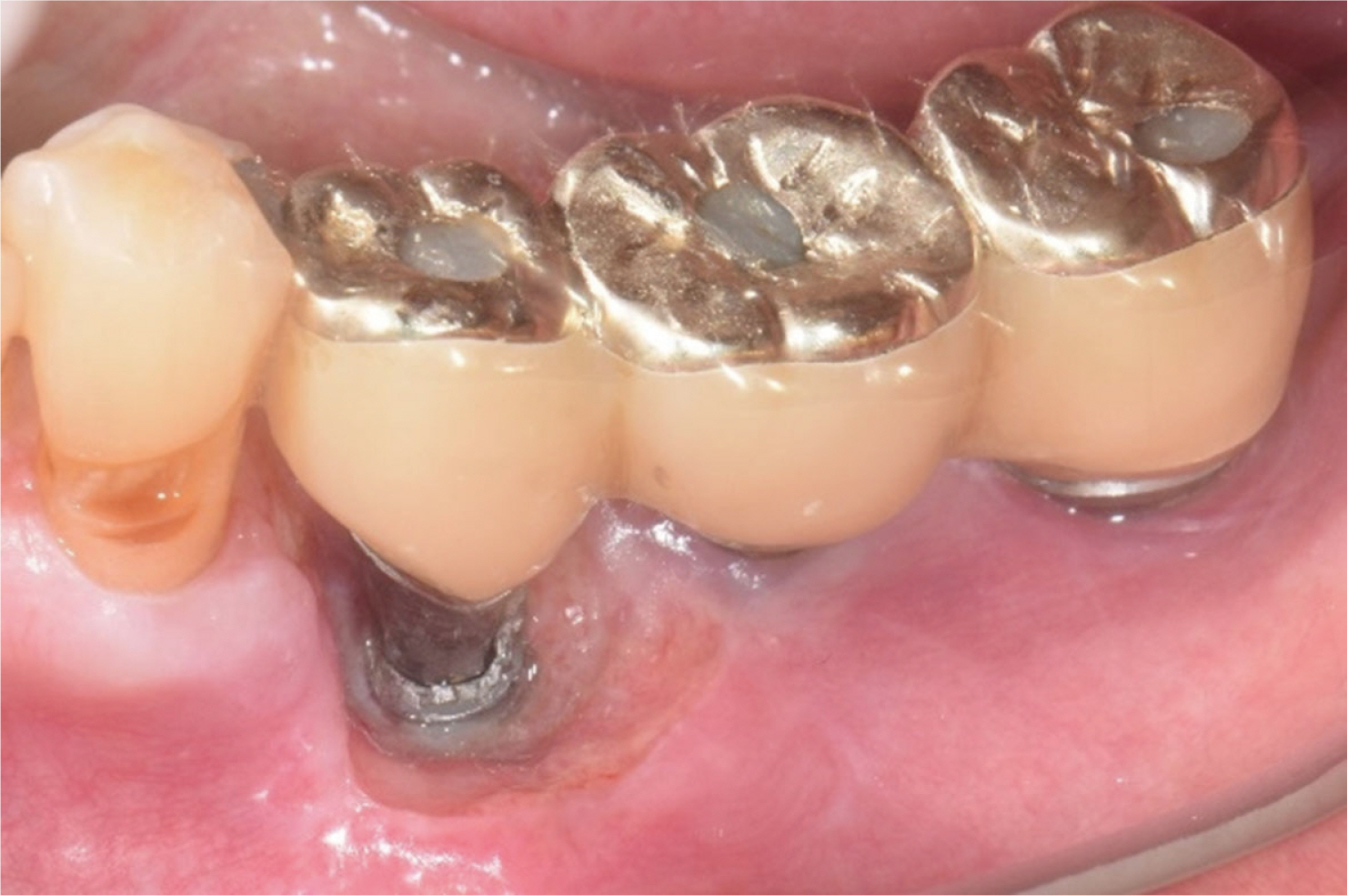
Fig. 6 Intraoral photograph of case 2: 2 weeks after periimplantitis surgery, healing with buccal fractured line is exposed. After surgery, the implant functioned stably for 5 months.
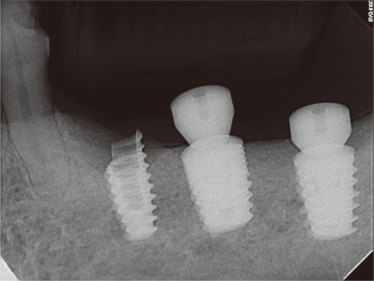
Fig. 7 Periapical radiography of case 2: A radiograph with a healing abutment on #36, 37 implants to fix the prosthesis. 5 months after peri-implantitis surgery, due to fractured part, mobility and retention loss of restoration occurred. It was planned to remain submerged a #35 implant and removed it later if necessary.
Reference
-
References
1. Balshi TJ. 1996; An analysis and management of fractured implants: a clinical report. Int J Oral Maxillofac Implants. 11:660–6. PMID: 8908866.2. Eckert SE, Meraw SJ, Cal E, Ow RK. 2000; Analysis of incidence and associated factors with fractured implants: a retrospective study. Int J Oral Maxillofac Implants. 15:662–7. PMID: 11055133.3. Goodacre C, Goodacre B. 2017; Fixed vs removable complete arch implant prostheses: a literature review of prosthodontic outcomes. Eur J Oral Implantol. 10(Suppl 1):13–34. PMID: 28944366.4. Jung RE, Pjetursson BE, Glauser R, Zembic A, Zwahlen M, Lang NP. 2008; A systematic review of the 5-year survival and complication rates of implant-supported single crowns. Clin Oral Implants Res. 19:119–30. DOI: 10.1111/j.1600-0501.2007.01453.x. PMID: 18067597.5. Adell R, Lekholm U, Rockler B, Brånemark PI. 1981; A 15-year study of osseointegrated implants in the treatment of the edentulous jaw. Int J Oral Surg. 10:387–416. DOI: 10.1016/S0300-9785(81)80077-4.6. Mendonca G, Mendonca DBS, Fernandes-Neto AJ, Neves FD. 2009; Management of fractured dental implants: a case report. Implant Dent. 18:10–6. DOI: 10.1097/ID.0b013e318192cafe. PMID: 19212233.7. Stacchi C, Berton F, Perinetti G, Frassetto A, Lombardi T, Khoury A, Andolsek F, Di Lenarda R. 2016; Risk factors for peri-implantitis: Effect of history of periodontal disease and smoking habits. A systematic review and meta-analysis. J Oral Maxillofac Res. 7:e3. DOI: 10.5037/jomr.2016.7303. PMCID: PMC5100643. PMID: 27833728.8. Roccuzzo M, Bonino L, Dalmasso P, Aglietta M. 2014; Long-term results of a three arms prospective cohort study on implants in periodontally compromised patients: 10-year data around sandblasted and acid-etched (SLA) surface. Clin Oral Implants Res. 25:1105–12. DOI: 10.1111/clr.12227. PMID: 23865554.9. Roos-Jansåker AM, Lindahl C, Renvert H, Renvert S. 2006; Nine- to fourteen-year follow-up of implant treatment. Part I: implant loss and associations to various factors. J Clin Periodontol. 33:283–9. 68DOI: 10.1111/j.1600-051X.2006.00907.x. PMID: 16553637.10. Tagger-Green N, Horwitz J, Machtei EE, Peled M. 2002; Implant fracture: a complication of treatment with dental implants-review of the literature. Refuat Hapeh Vehashinayim. 19:19–24.11. de Moraes SLD, Verri FR, Santiago JF Jr, de Faria Almeida DA, de Mello CC, Pellizzer EP. 2013; A 3-D finite element study of the influence of crown-implant ratio on stress distribution. Braz Dent J. 24:635–41. DOI: 10.1590/0103-6440201302287. PMID: 24474362.12. Shinogaya T, Bakke M, Thomsen CE, Vilmann A, Sodeyama A, Matsumoto M. 2001; Effects of ethnicity, gender and age on clenching force and load distribution. Clin Oral Investig. 5:63–8. DOI: 10.1007/s007840000099. PMID: 11355102.13. 1994. Abstracts. J Prosthet Dent. 72:In : Proceedings of the 4th International Symposium on Implant Dentistry: Focus on Esthetics; January 27-29, 1994; San Diego, California. p. 623–34. DOI: 10.1016/0022-3913(94)90295-X.14. Misch CE, Suzuki JB, Misch-Dietsh FM, Bidez MW. 2005; A positive correlation between occlusal trauma and peri-implant bone loss: literature support. Implant Dent. 14:108–16. DOI: 10.1097/01.id.0000165033.34294.db. PMID: 15968181.15. Sánchez-Pérez A, Moya-Villaescusa MJ, Jornet-Garcia A, Gomez S. 2010; Etiology, risk factors and management of implant fractures. Med Oral Patol Oral Cir Bucal. 15:e504–8. DOI: 10.4317/medoral.15.e504. PMID: 20038899.
- Full Text Links
-
- Actions
-
Cited
- CITED
-
- Close
- Share
-
- Similar articles
-
- Risk factors of peri-implantitis: a narrative review
- Unusual bone regeneration following resective surgery and decontamination of peri-implantitis: a 6-year follow-up
- Full mouth rehabilitation in a patient with peri-implantitis: A case report
- Retrospective analysis of keratinized tissue augmentation using a xenogeneic collagen matrix for resolving peri-implant mucositis and peri-implantitis
- Prevalence and risk factors of peri-implant mucositis and peri-implantitis after at least 7 years of loading
