Observation of an extracted premolar 2.5 years after mineral trioxide aggregate apexification using micro-computed tomography
- Affiliations
-
- 1Department of Conservative Dentistry, Gangnam Severance Hospital, Yonsei University College of Dentistry, Seoul, Korea
- 2Department of Orthodontics, Gangnam Severance Hospital, Yonsei University College of Dentistry, Seoul, Korea
- 3Department of Conservative Dentistry, Yonsei University College of Dentistry, Seoul, Korea
- KMID: 2503507
- DOI: http://doi.org/10.5395/rde.2020.45.e4
Abstract
- Although numerous studies have been conducted on apexification using mineral trioxide aggregate (MTA), direct observation of extracted human teeth after the procedure has been rarely reported. This case report describes a mandibular premolar treated 2.5 years ago and extracted recently for orthodontic treatment. The tubercle of the right mandibular premolar of a 12-year-old boy with dens evaginatus was fractured and the pulp was exposed. The tooth was diagnosed with pulp necrosis and asymptomatic periapical abscess. During the first visit, copious irrigation was performed with 2.5% sodium hypochlorite. Calcium hydroxide paste was placed as an intracanal medicament. The sinus tract had disappeared at the second visit after 3 weeks. MTA was applied on to the bleeding point as a 4-mm-thick layer, followed by a 3-mm-thick gutta-percha filling and resin core build-up. After 2.5 years, the tooth and three other premolars were extracted for orthodontic treatment. The right and left mandibular premolars were scanned with micro-computed tomography to determine the root shape and canal anatomy. Irregular root growth was observed and the root outline of the right mandibular premolar differed from that of the contralateral tooth. Apexification with MTA leads to the formation of roots with irregular morphology, without any pulpal space.
Figure
-

Figure 1 Series of periapical radiographs taken during the treatment of tooth #45. (A) A preoperative periapical radiograph showing an immature root with periapical radiolucency with tooth #45. A gutta-percha cone was used to trace the periapical lesion to tooth #45. (B) A postoperative periapical radiograph showing apexification with tooth #45. (C) A periapical radiograph taken 6 months after the treatment demonstrating the resolution of the previous periapical radiolucency. (D) A periapical radiograph taken 1.5 years after the treatment showing the formation of the root apex. (E) A preoperative panoramic view. (F) A panoramic view taken 2.5 years after the root canal treatment with tooth #45.op, operative.

Figure 2 Reconstructed 3D images of teeth #45 and #35 obtained by micro-computed tomography. (A) Reconstructed 3D image of tooth #35. (B) Reconstructed 3D image of tooth #45.3D, three-dimensional; MTA, mineral trioxide aggregate; GP, gutta-percha.
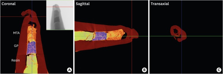
Figure 3 Representative colored images of tooth #45 obtained by micro-computed tomography (micro-CT). (A) A coronal image of micro-CT of tooth #45 showing composite resin, gutta-percha (GP), and mineral trioxide aggregate (MTA) filling in the canal. An irregularly shaped root tip was observed. (B) A sagittal image of tooth #45. (C) A transaxial view of tooth #45.
Cited by 1 articles
-
Incorporation of amoxicillin-loaded microspheres in mineral trioxide aggregate cement: an
in vitro study
Fábio Rocha Bohns, Vicente Castelo Branco Leitune, Isadora Martini Garcia, Bruna Genari, Nélio Bairros Dornelles, Silvia Stanisçuaski Guterres, Fabrício Aulo Ogliari, Mary Anne Sampaio de Melo, Fabrício Mezzomo Collares
Restor Dent Endod. 2020;45(4):e50. doi: 10.5395/rde.2020.45.e50.
Reference
-
1. Simon S, Rilliard F, Berdal A, Machtou P. The use of mineral trioxide aggregate in one-visit apexification treatment: a prospective study. Int Endod J. 2007; 40:186–197. PMID: 17305695.
Article2. Witherspoon DE, Small JC, Regan JD, Nunn M. Retrospective analysis of open apex teeth obturated with mineral trioxide aggregate. J Endod. 2008; 34:1171–1176. PMID: 18793914.
Article3. Farhad AR, Shokraneh A, Shekarchizade N. Regeneration or replacement? A case report and review of literature. Dent Traumatol. 2016; 32:71–79. PMID: 26134932.
Article4. Huang GT. A paradigm shift in endodontic management of immature teeth: conservation of stem cells for regeneration. J Dent. 2008; 36:379–386. PMID: 18420332.
Article5. Hargreaves KM, Diogenes A, Teixeira FB. Treatment options: biological basis of regenerative endodontic procedures. Pediatr Dent. 2013; 35:129–140. PMID: 23635981.
Article6. Shabahang S. Treatment options: apexogenesis and apexification. J Endod. 2013; 39(Supplement):S26–S29. PMID: 23439042.
Article7. Alobaid AS, Cortes LM, Lo J, Nguyen TT, Albert J, Abu-Melha AS, Lin LM, Gibbs JL. Radiographic and clinical outcomes of the treatment of immature permanent teeth by revascularization or apexification: a pilot retrospective cohort study. J Endod. 2014; 40:1063–1070. PMID: 25069909.
Article8. Fouad AF, Verma P. Healing after regenerative procedures with and without pulpal infection. J Endod. 2014; 40(Supplement):S58–S64. PMID: 24698695.
Article9. Linsuwanont P, Sinpitaksakul P, Lertsakchai T. Evaluation of root maturation after revitalization in immature permanent teeth with nonvital pulps by cone beam computed tomography and conventional radiographs. Int Endod J. 2017; 50:836–846. PMID: 27689773.
Article10. Jung IY, Lee SJ, Hargreaves KM. Biologically based treatment of immature permanent teeth with pulpal necrosis: a case series. J Endod. 2008; 34:876–887. PMID: 18571000.
Article11. Nosrat A, Kolahdouzan A, Hosseini F, Mehrizi EA, Verma P, Torabinejad M. Histologic outcomes of uninfected human immature teeth treated with regenerative endodontics: 2 case reports. J Endod. 2015; 41:1725–1729. PMID: 26259646.
Article12. Peng C, Zhao Y, Wang W, Yang Y, Qin M, Ge L. Histologic findings of a human immature revascularized/regenerated tooth with symptomatic irreversible pulpitis. J Endod. 2017; 43:905–909. PMID: 28416306.
Article13. Shimizu E, Jong G, Partridge N, Rosenberg PA, Lin LM. Histologic observation of a human immature permanent tooth with irreversible pulpitis after revascularization/regeneration procedure. J Endod. 2012; 38:1293–1297. PMID: 22892754.
Article14. Bose R, Nummikoski P, Hargreaves K. A retrospective evaluation of radiographic outcomes in immature teeth with necrotic root canal systems treated with regenerative endodontic procedures. J Endod. 2009; 35:1343–1349. PMID: 19801227.
Article15. Holden DT, Schwartz SA, Kirkpatrick TC, Schindler WG. Clinical outcomes of artificial root-end barriers with mineral trioxide aggregate in teeth with immature apices. J Endod. 2008; 34:812–817. PMID: 18570985.
Article16. Yassen GH, Chu TM, Eckert G, Platt JA. Effect of medicaments used in endodontic regeneration technique on the chemical structure of human immature radicular dentin: an in vitro study. J Endod. 2013; 39:269–273. PMID: 23321244.17. Hoshino E, Kurihara-Ando N, Sato I, Uematsu H, Sato M, Kota K, Iwaku M. In-vitro antibacterial susceptibility of bacteria taken from infected root dentine to a mixture of ciprofloxacin, metronidazole and minocycline. Int Endod J. 1996; 29:125–130. PMID: 9206436.
Article18. Windley W 3rd, Teixeira F, Levin L, Sigurdsson A, Trope M. Disinfection of immature teeth with a triple antibiotic paste. J Endod. 2005; 31:439–443. PMID: 15917683.
- Full Text Links
-
- Actions
-
Cited
- CITED
-
- Close
- Share
-
- Similar articles
-
- Regenerative Endodontic Procedure in Korean Children and Adolescents: A Case Report
- Chemical characteristics of mineral trioxide aggregate and its hydration reaction
- One-visit Apexification Using MTA and Reattachment of a Crown-root Fractured Tooth with Severe Coronal Damage: A Case Report
- Endodontic management of central incisor associated with large periapical lesion and fused supernumerary root: a conservative approach
- Treatment of non-vital immature teeth with amoxicillin-containing triple antibiotic paste resulting in apexification
