The lumbar multifidus is characterised by larger type I muscle fibres compared to the erector spinae
- Affiliations
-
- 1Department of Rehabilitation Sciences and Physiotherapy, Hasselt University, Rehabilitation Research Centre, Faculty of Rehabilitation Sciences, Diepenbeek, Belgium
- 2Department of Cardio and Internal Systems, Hasselt University, Biomedical Research Institute, Faculty of Medicine and Life Sciences, Diepenbeek, Belgium
- KMID: 2503444
- DOI: http://doi.org/10.5115/acb.20.009
Abstract
- The metabolic capacity of a muscle is one of the determinants of muscle function. Muscle fiber type characteristics give an indication about this metabolic capacity. Therefore it might be expected that the lumbar multifidus (MF) as a local stabilizer contains higher proportions of slow type I fibers, compared to the erector spinae (ES) as a global mobilizer. The aim of this study is to determine the muscle fiber characteristics of the ES and MF to provide insight into their structural and metabolic characteristics, and thereby the functional capacity of both muscles. Muscle fiber type characteristics in the ES and MF were investigated with an immunofluorescence staining of the myosin heavy chain isoforms. In both the ES and MF, type I muscle fibers are predominantly present. The cross-sectional area (CSA) of type I muscle fibers is significantly larger in the lumbar MF compared to the ES. However, the mean muscle fiber type percentage for type I was not significantly different, which resulted in an insignificant difference in relative cross-sectional area (RCSA) for type I. No significant differences were found for all other muscle fiber types. This may indicate that the MF displays muscle fiber type characteristics that tend to be more appropriate to maintain stability of the spine. However, because we could not demonstrate significant differences in RCSA between ES and MF, we cannot firmly state that there are functional differences between the ES an MF based only on structural characteristics.
Keyword
Figure
-

Fig. 1 Representative immunofluore scence image of the lumbar ES and MF muscle. Muscle cross-sections were in cubated with primary antibody against laminin (red), MHC I (blue), MHC IIa (green) and MHC IIx (red). ES, erector spinae; MF, multifidus; MHC, myosin heavy chain.
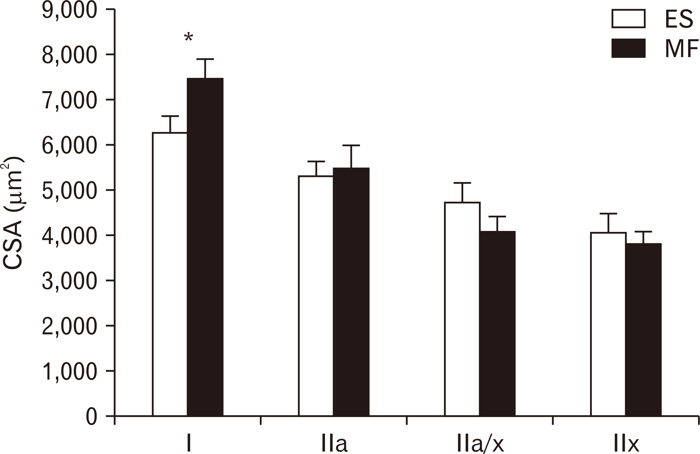
Fig. 2 CSA of the different muscle fibre types. CSA of type I, type IIa, type IIa/x and type IIx in the lumbar ES (white) and MF (black) muscle. Values are presented as mean±SE. CSA, cross-sectional area; ES, erector spinae; MF, multifidus; SE, standard error. *P=0.0053 ES vs. MF.
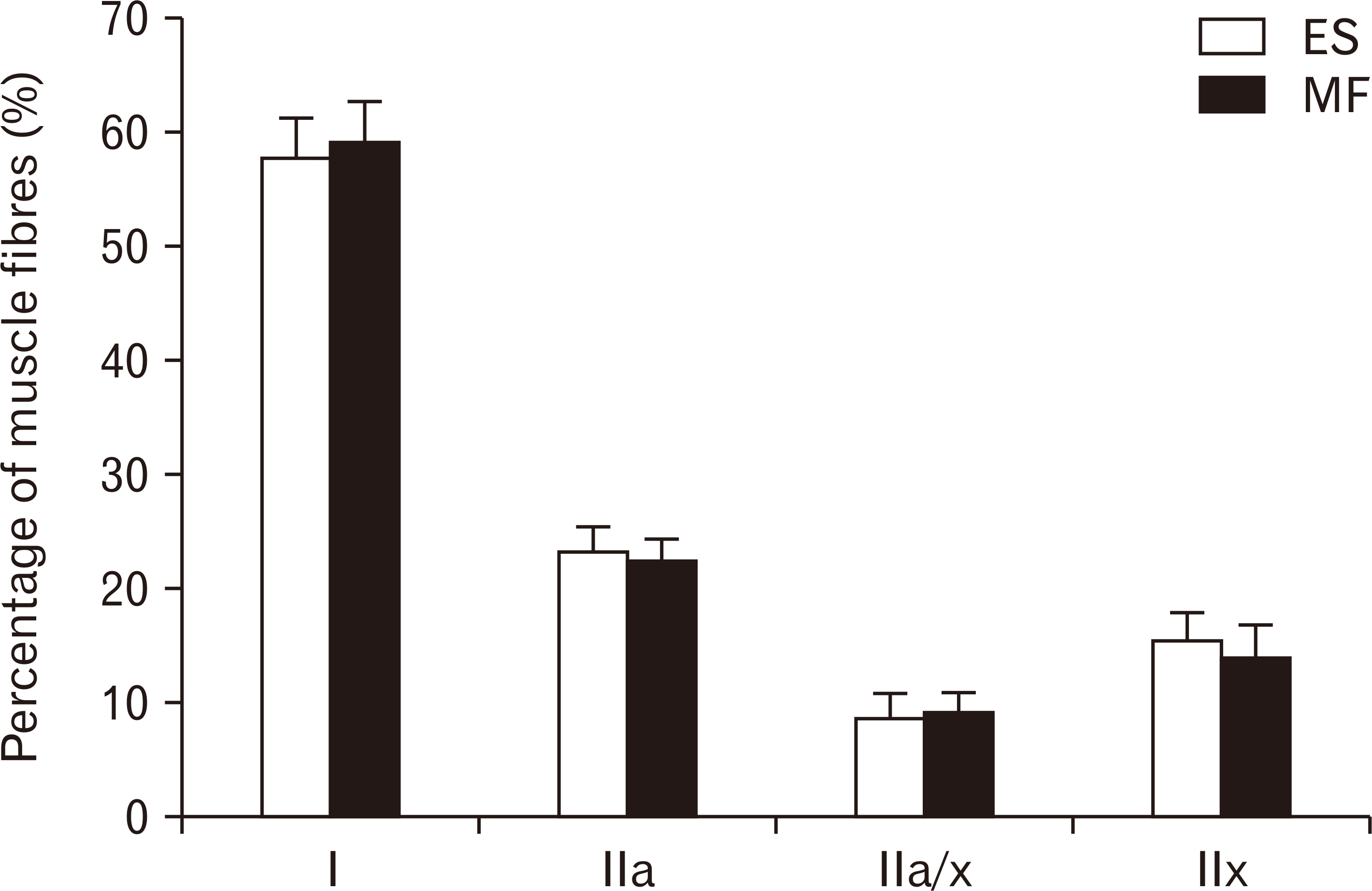
Fig. 3 Number of the different muscle fibres. Number of type I, type IIa, type IIa/x and type IIx in the lumbar ES (white) and MF (black) muscle. Values are presented as mean±SE. ES, erector spinae; MF, multifidus; SE, standard error.Number of the different muscle fibres. Number of type I, type IIa, type IIa/x and type IIx in the lumbar ES (white) and MF (black) muscle. Values are presented as mean±SE. ES, erector spinae; MF, multifidus; SE, standard error.
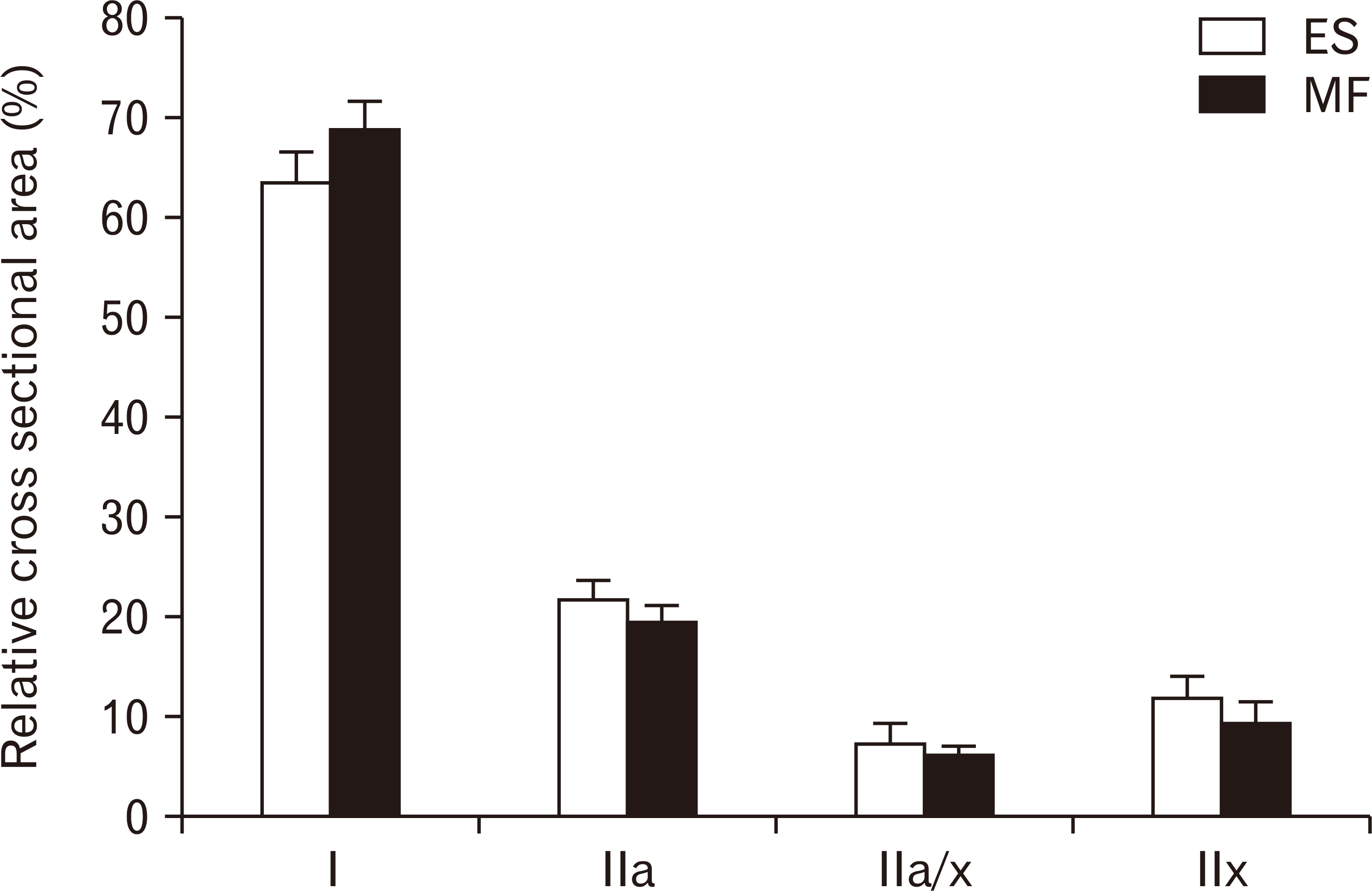
Fig. 4 Relative RCSA of the different muscle fibres. RCSA of type I, type IIa, type IIa/x and type IIx in the lumbar ES (white) and MF (black) muscle. Values are presented as mean±SE. ES, erector spinae; MF, multifidus; RCSA, relative cross-sectional area; SE, standard error.
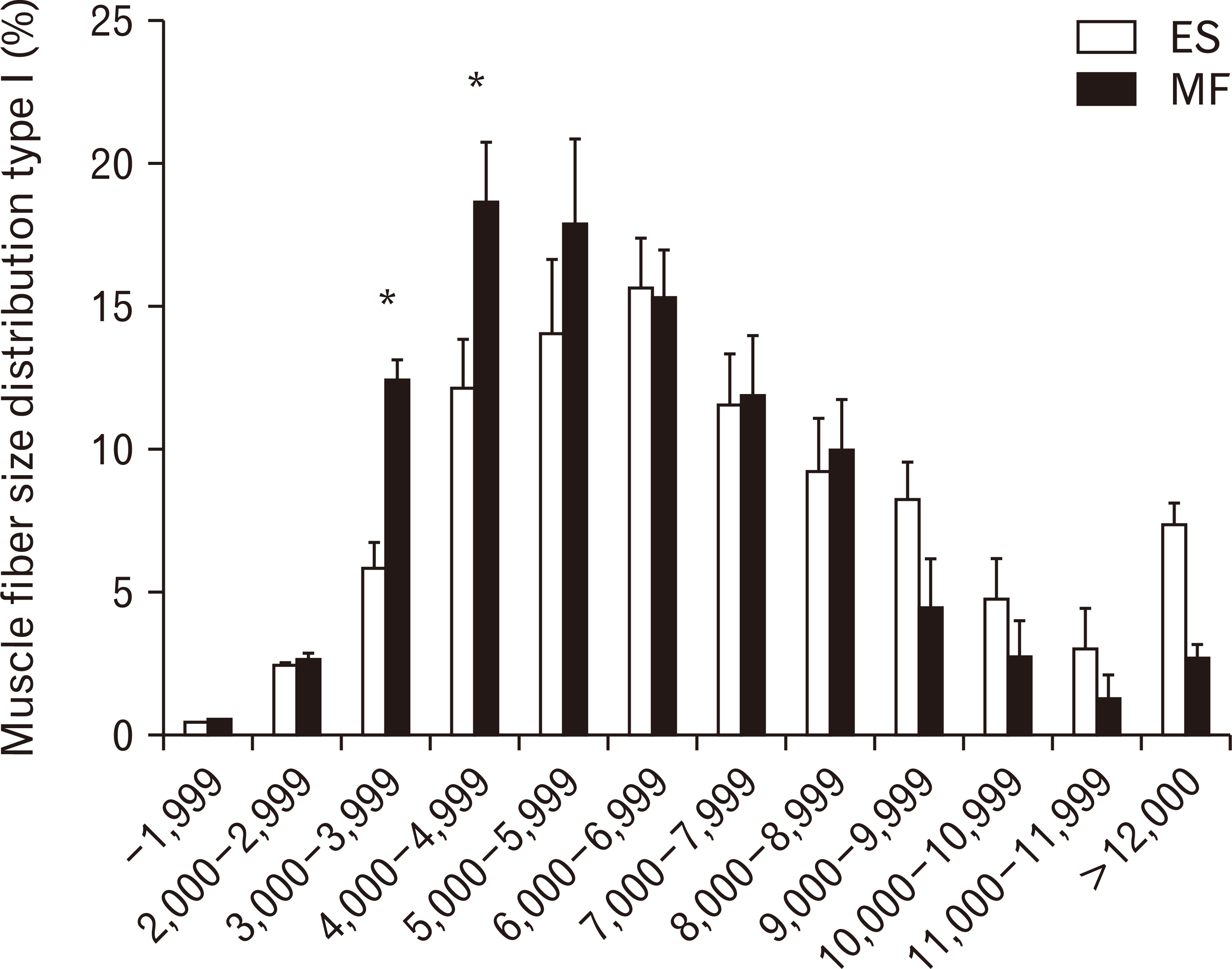
Fig. 5 Muscle fibre size distribution (in percentage) for type I muscle fibres in the MF (white) and ES (black). Values are presented as mean±SE. ES, erector spinae; MF, multifidus; SE, standard error. *P<0.05 ES vs. MF.
Reference
-
1. Gibbons SGT, Comerford MJ. 2001; Strength versus stability part I: concept and terms. Orthopaedic Division Review. 43:21–7.2. Comerford MJ, Mottram SL. 2001; Movement and stability dysfunction--contemporary developments. Man Ther. 6:15–26. DOI: 10.1054/math.2000.0388. PMID: 11243905.3. Goel VK, Kong W, Han JS, Weinstein JN, Gilbertson LG. 1993; A combined finite element and optimization investigation of lumbar spine mechanics with and without muscles. Spine (Phila Pa 1976). 18:1531–41. DOI: 10.1097/00007632-199318110-00019. PMID: 8235826.
Article4. Macintosh JE, Valencia F, Bogduk N, Munro RR. 1986; The morphology of the human lumbar multifidus. Clin Biomech (Bristol, Avon). 1:196–204. DOI: 10.1016/0268-0033(86)90146-4.
Article5. Hansen L, de Zee M, Rasmussen J, Andersen TB, Wong C, Simonsen EB. 2006; Anatomy and biomechanics of the back muscles in the lumbar spine with reference to biomechanical modeling. Spine (Phila Pa 1976). 31:1888–99. DOI: 10.1097/01.brs.0000229232.66090.58. PMID: 16924205.
Article6. Rosatelli AL, Ravichandiran K, Agur AM. 2008; Three-dimensional study of the musculotendinous architecture of lumbar multifidus and its functional implications. Clin Anat. 21:539–46. DOI: 10.1002/ca.20659. PMID: 18627104.
Article7. Bogduk N. 1980; A reappraisal of the anatomy of the human lumbar erector spinae. J Anat. 131(Pt 3):525–40.8. Bustami FM. 1986; A new description of the lumbar erector spinae muscle in man. J Anat. 144:81–91.9. Schiaffino S, Reggiani C. 1996; Molecular diversity of myofibrillar proteins: gene regulation and functional significance. Physiol Rev. 76:371–423. DOI: 10.1152/physrev.1996.76.2.371. PMID: 8618961.
Article10. Caiozzo VJ. 2002; Plasticity of skeletal muscle phenotype: mechanical consequences. Muscle Nerve. 26:740–68. DOI: 10.1002/mus.10271. PMID: 12451599.
Article11. Scott W, Stevens J, Binder-Macleod SA. 2001; Human skeletal muscle fiber type classifications. Phys Ther. 81:1810–6. DOI: 10.1093/ptj/81.11.1810. PMID: 11694174.
Article12. Jørgensen K, Nicholaisen T, Kato M. 1993; Muscle fiber distribution, capillary density, and enzymatic activities in the lumbar paravertebral muscles of young men. Significance for isometric endurance. Spine (Phila Pa 1976). 18:1439–50. DOI: 10.1097/00007632-199318110-00007.13. Rantanen J, Rissanen A, Kalimo H. 1994; Lumbar muscle fiber size and type distribution in normal subjects. Eur Spine J. 3:331–5. DOI: 10.1007/BF02200146. PMID: 7532536.
Article14. Thorstensson A, Carlson H. 1987; Fibre types in human lumbar back muscles. Acta Physiol Scand. 131:195–202. DOI: 10.1111/j.1748-1716.1987.tb08226.x. PMID: 2960128.
Article15. Agten A, Verbrugghe J, Stevens S, Boomgaert L, O Eijnde B, Timmermans A, Vandenabeele F. 2018; Feasibility, accuracy and safety of a percutaneous fine-needle biopsy technique to obtain qualitative muscle samples of the lumbar multifidus and erector spinae muscle in persons with low back pain. J Anat. 233:542–51. DOI: 10.1111/joa.12867. PMID: 30033540.
Article16. Bloemberg D, Quadrilatero J. 2012; Rapid determination of myosin heavy chain expression in rat, mouse, and human skeletal muscle using multicolor immunofluorescence analysis. PLoS One. 7:e35273. DOI: 10.1371/journal.pone.0035273. PMID: 22530000. PMCID: PMC3329435.
Article17. Mannion AF, Dumas GA, Cooper RG, Espinosa FJ, Faris MW, Stevenson JM. 1997; Muscle fibre size and type distribution in thoracic and lumbar regions of erector spinae in healthy subjects without low back pain: normal values and sex differences. J Anat. 190(Pt 4):505–13. DOI: 10.1046/j.1469-7580.1997.19040505.x. PMID: 9183674. PMCID: PMC1467636.
Article18. Ceglia L, Niramitmahapanya S, Price LL, Harris SS, Fielding RA, Dawson-Hughes B. 2013; An evaluation of the reliability of muscle fiber cross-sectional area and fiber number measurements in rat skeletal muscle. Biol Proced Online. 15:6. DOI: 10.1186/1480-9222-15-6. PMID: 23497012. PMCID: PMC3599694.
Article19. Cicchetti DV. 1994; Guidelines, criteria, and rules of thumb for evaluating normed and standardized assessment instruments in psychology. Psychol Assess. 6:284–90. DOI: 10.1037/1040-3590.6.4.284.
Article20. Hesse B, Fröber R, Fischer MS, Schilling N. 2013; Functional differentiation of the human lumbar perivertebral musculature revisited by means of muscle fibre type composition. Ann Anat. 195:570–80. DOI: 10.1016/j.aanat.2013.07.003. PMID: 24028860.
Article21. Sirca A, Kostevc V. 1985; The fibre type composition of thoracic and lumbar paravertebral muscles in man. J Anat. 141:131–7. PMID: 2934358. PMCID: PMC1166395.22. Ehrenfellner B, Zissler A, Steinbacher P, Monticelli FC, Pittner S. 2017; Are animal models predictive for human postmortem muscle protein degradation? Int J Legal Med. 131:1615–21. DOI: 10.1007/s00414-017-1643-1. PMID: 28721468. PMCID: PMC5635072.
Article23. MacDonald DA, Moseley GL, Hodges PW. 2006; The lumbar multifidus: does the evidence support clinical beliefs? Man Ther. 11:254–63. DOI: 10.1016/j.math.2006.02.004. PMID: 16716640.
Article24. Ward SR, Tomiya A, Regev GJ, Thacker BE, Benzl RC, Kim CW, Lieber RL. 2009; Passive mechanical properties of the lumbar multifidus muscle support its role as a stabilizer. J Biomech. 42:1384–9. DOI: 10.1016/j.jbiomech.2008.09.042. PMID: 19457491. PMCID: PMC2752430.
Article25. Stark H, Fröber R, Schilling N. 2013; Intramuscular architecture of the autochthonous back muscles in humans. J Anat. 222:214–22. DOI: 10.1111/joa.12005. PMID: 23121477. PMCID: PMC3632226.
Article26. Loeb EP, Giszter SF, Saltiel P, Bizzi E, Mussa-Ivaldi FA. 2000; Output units of motor behavior: an experimental and modeling study. J Cogn Neurosci. 12:78–97. DOI: 10.1162/08989290051137611. PMID: 10769307.
Article27. Amonoo-Kuofi HS. 1983; The density of muscle spindles in the medial, intermediate and lateral columns of human intrinsic postvertebral muscles. J Anat. 136(Pt 3):509–19.28. Botterman BR, Binder MD, Stuart DG. 1978; Functional anatomy of the association between motor units and muscle receptors. Am Zool. 18:135–52. DOI: 10.1093/icb/18.1.135.
Article29. Tsao H, Danneels L, Hodges PW. 2011; Individual fascicles of the paraspinal muscles are activated by discrete cortical networks in humans. Clin Neurophysiol. 122:1580–7. DOI: 10.1016/j.clinph.2011.01.048. PMID: 21377923.
Article
- Full Text Links
-
- Actions
-
Cited
- CITED
-
- Close
- Share
-
- Similar articles
-
- The Variation in the Lumbar Facet Joint Orientation in an Adult Asian Population and Its Relationship with the Cross-Sectional Area of the Multifidus and Erector Spinae
- Facilitating Effects of Fast and Slope Walking on Paraspinal Muscles
- Relationship between Lumbar Disc Degeneration and Back Muscle Degeneration
- Asymmetric Atrophy of Paraspinal Muscles in Patients With Chronic Unilateral Lumbar Radiculopathy
- Association between Cross-sectional Areas of Lumbar Muscles on Magnetic Resonance Imaging and Chronicity of Low Back Pain
