J Korean Foot Ankle Soc.
2020 Jun;24(2):94-97. 10.14193/jkfas.2020.24.2.94.
The Efficacy of Newly Designed Screw for Prevention of the Screw Breakage in Syndesmosis Fixation: Biomechanical Study
- Affiliations
-
- 1Department of Orthopedic Surgery, Myongji Hospital, Hanyang University College of Medicine, Goyang, Korea
- KMID: 2502916
- DOI: http://doi.org/10.14193/jkfas.2020.24.2.94
Abstract
- Purpose
Fatigue breakage of cortical screws sometimes occurs after syndesmosis fixation, regardless of the period of screw retention. This study compared the fatigue strength of a novel screw design to conventional cortical screws in the fixed state of syndesmosis.
Materials and Methods
Twelve sawbone models were tested mechanically to determine the fatigue strength of three screw designs. The first group was composed of cortical screws, while the second and third groups were newly-designed screws. The second group was composed of screws with a 2.4-mm diameter thread-free portion of the mid-shank while the third group had a 2.0-mm diameter threadfree mid-shank. A 400 N load was applied repetitively to a fibula model and the number of cycles until screw failure was recorded. Four screws from each group were tested, giving a total of 12 fatigue tests.
Results
The average cycles until screw failure for groups 1, 2, and 3 were 8,134, 63,186, and 2,581, respectively. The second group showed the highest fatigue strength (p=0.018). The other two screw designs showed similar fatigue strength (p=0.401).
Conclusion
New screw designs with a thread-free portion in the mid-shank could reduce the occurrence of fatigue breakage after syndesmosis fixation.
Figure
-
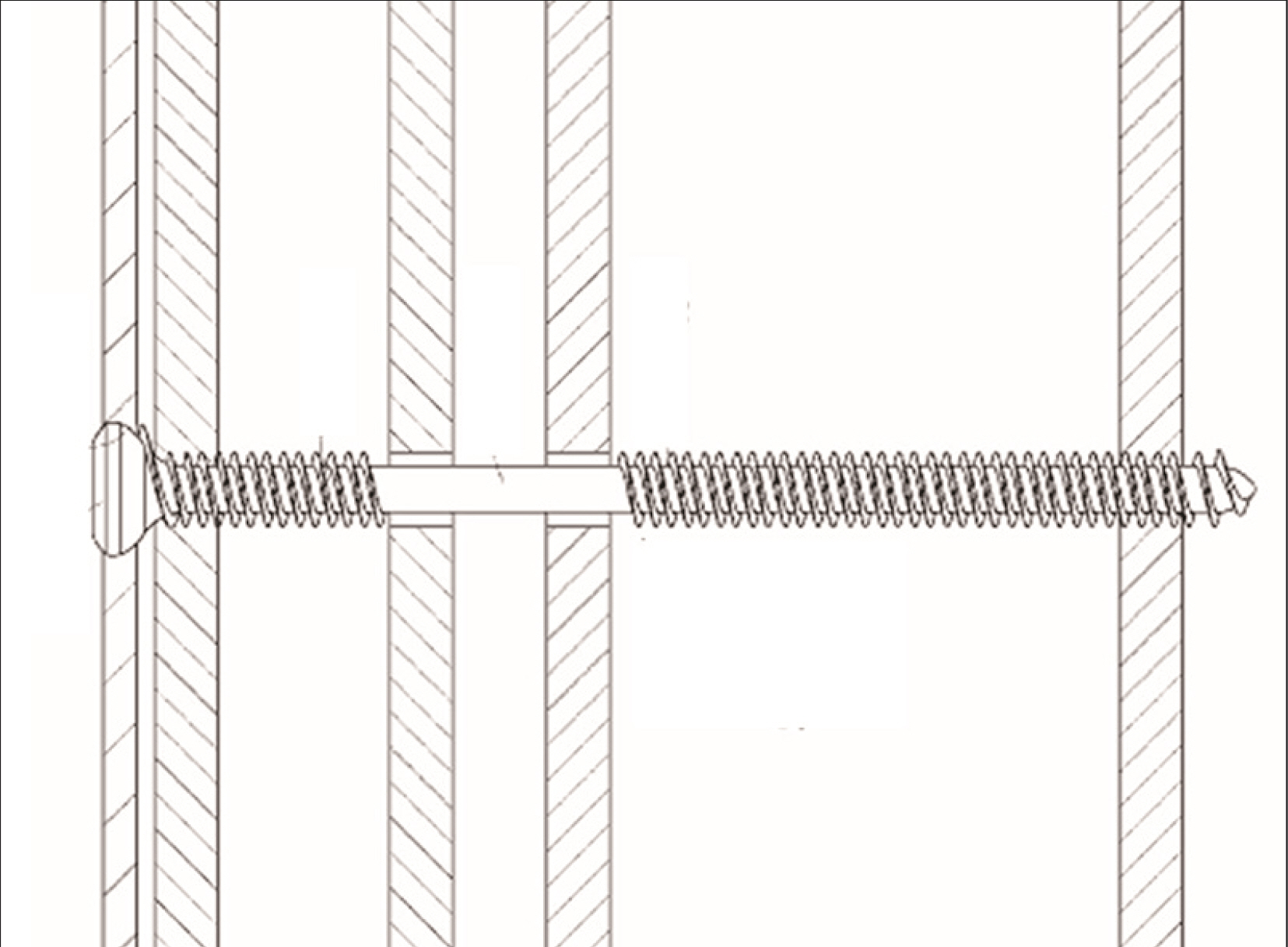
Figure. 1 Schematic drawing of a novel screw design.
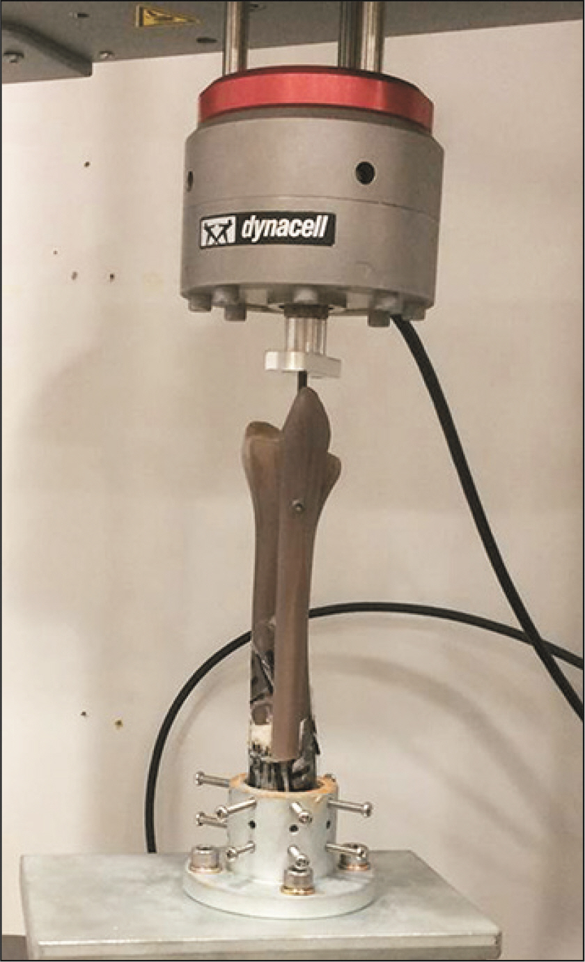
Figure. 2 Testing machine to apply repetitive load to sawbone model.
Reference
-
1. Clanton TO, Paul P. 2002; Syndesmosis injuries in athletes. Foot Ankle Clin. 7:529–49. doi: 10.1016/s1083-7515(02)00045-1. DOI: 10.1016/S1083-7515(02)00045-1. PMID: 12512408.
Article2. Teitz CC, Harrington RM. 1998; A biochemical analysis of the squeeze test for sprains of the syndesmotic ligaments of the ankle. Foot Ankle Int. 19:489–92. doi: 10.1177/107110079801900713. DOI: 10.1177/107110079801900713. PMID: 9694130.3. Peña FA, Coetzee JC. 2006; Ankle syndesmosis injuries. Foot Ankle Clin. 11:35–50. viiidoi: 10.1016/j.fcl.2005.12.007. DOI: 10.1016/j.fcl.2005.12.007. PMID: 16564452.
Article4. Bell DP, Wong MK. 2006; Syndesmotic screw fixation in Weber C ankle injuries--should the screw be removed before weight bearing? Injury. 37:891–8. doi: 10.1016/j.injury.2006.02.003. DOI: 10.1016/j.injury.2006.02.003. PMID: 16630621.
Article5. Ramsey PL, Hamilton W. 1976; Changes in tibiotalar area of contact caused by lateral talar shift. J Bone Joint Surg Am. 58:356–7. DOI: 10.2106/00004623-197658030-00010. PMID: 1262367.
Article6. Tucker A, Street J, Kealey D, McDonald S, Stevenson M. 2013; Functional outcomes following syndesmotic fixation: a comparison of screws retained in situ versus routine removal - is it really necessary? Injury. 44:1880–4. doi: 10.1016/j.injury.2013.08.011. DOI: 10.1016/j.injury.2013.08.011. PMID: 24021584.
Article7. Peek AC, Fitzgerald CE, Charalambides C. 2014; Syndesmosis screws: how many, what diameter, where and should they be removed? A literature review. Injury. 45:1262–7. doi: 10.1016/j.injury.2014.05.003. DOI: 10.1016/j.injury.2014.05.003. PMID: 24917210.
Article8. Nousiainen MT, McConnell AJ, Zdero R, McKee MD, Bhandari M, Schemitsch EH. 2008; The influence of the number of cortices of screw purchase and ankle position in Weber C ankle fracture fixation. J Orthop Trauma. 22:473–8. doi: 10.1097/BOT.0b013e31817ae635. DOI: 10.1097/BOT.0b013e31817ae635. PMID: 18670288.
Article9. Teramoto A, Suzuki D, Kamiya T, Chikenji T, Watanabe K, Yamashita T. 2011; Comparison of different fixation methods of the suture-button implant for tibiofibular syndesmosis injuries. Am J Sports Med. 39:2226–32. doi: 10.1177/0363546511413455. DOI: 10.1177/0363546511413455. PMID: 21768530.
Article10. LaMothe JM, Baxter JR, Murphy C, Gilbert S, DeSandis B, Drakos MC. 2016; Three-dimensional analysis of fibular motion after fixation of syndesmotic injuries with a screw or suture-button construct. Foot Ankle Int. 37:1350–6. doi: 10.1177/1071100716666865. DOI: 10.1177/1071100716666865. PMID: 27654046.
Article11. Storey P, Gadd RJ, Blundell C, Davies MB. 2012; Complications of suture button ankle syndesmosis stabilization with modifications of surgical technique. Foot Ankle Int. 33:717–21. doi: 10.3113/FAI.2012.0717. DOI: 10.3113/FAI.2012.0717. PMID: 22995257.
Article12. Hansen M, Le L, Wertheimer S, Meyer E, Haut R. 2006; Syndesmosis fixation: analysis of shear stress via axial load on 3.5-mm and 4.5-mm quadricortical syndesmotic screws. J Foot Ankle Surg. 45:65–9. doi: 10.1053/j.jfas.2005.12.004. DOI: 10.1053/j.jfas.2005.12.004. PMID: 16513499.
Article13. Needleman RL, Skrade DA, Stiehl JB. 1989; Effect of the syndesmotic screw on ankle motion. Foot Ankle. 10:17–24. doi: 10.1177/107110078901000104. DOI: 10.1177/107110078901000104. PMID: 2767562.
Article14. Manjoo A, Sanders DW, Tieszer C, MacLeod MD. 2010; Functional and radiographic results of patients with syndesmotic screw fixation: implications for screw removal. J Orthop Trauma. 24:2–6. doi: 10.1097/BOT.0b013e3181a9f7a5. DOI: 10.1097/BOT.0b013e3181a9f7a5. PMID: 20035170.
Article15. Hamid N, Loeffler BJ, Braddy W, Kellam JF, Cohen BE, Bosse MJ. 2009; Outcome after fixation of ankle fractures with an injury to the syndesmosis: the effect of the syndesmosis screw. J Bone Joint Surg Br. 91:1069–73. doi: 10.1302/0301-620X.91B8.22430. DOI: 10.1302/0301-620X.91B8.22430. PMID: 19651836.16. Beumer A, Campo MM, Niesing R, Day J, Kleinrensink GJ, Swierstra BA. 2005; Screw fixation of the syndesmosis: a cadaver model comparing stainless steel and titanium screws and three and four cortical fixation. Injury. 36:60–4. doi: 10.1016/j.injury.2004.05.024. DOI: 10.1016/j.injury.2004.05.024. PMID: 15589915.
Article17. Markolf KL, Jackson SR, McAllister DR. 2013; Syndesmosis fixation using dual 3.5 mm and 4.5 mm screws with tricortical and quadricortical purchase: a biomechanical study. Foot Ankle Int. 34:734–9. doi: 10.1177/1071100713478923. DOI: 10.1177/1071100713478923. PMID: 23405026.18. Welk GJ, Differding JA, Thompson RW, Blair SN, Dziura J, Hart P. 2000; The utility of the Digi-walker step counter to assess daily physical activity patterns. Med Sci Sports Exerc. 32(9 Suppl):S481–8. doi: 10.1097/00005768-200009001-00007. DOI: 10.1097/00005768-200009001-00007. PMID: 10993418.
Article
- Full Text Links
-
- Actions
-
Cited
- CITED
-
- Close
- Share
-
- Similar articles
-
- Comparison between Suture-Button Technique with Syndesmotic Repair and Screw Fixation Technique for Complete Ankle Syndesmotic Injury: Biomechanical Cadaveric Study
- Comparison of Clinical Results between Deltoid Ligament Augmentation and Syndesmosis Screw Fixation in Bimalleolar Equivalent Fracture
- Comparison of Treatment Outcomes: Screw Fixation versus Suture-Button Fixation in Distal Tibiofibular Syndesmosis Diastasis Combined with Ankle Fractures
- Comparative Analysis of Trans-syndesmotic Versus Non-syndesmotic Screw Fixation in Surgical Treatment of Ankle Fracture with Diastasis
- Segmental Breakage of Distal Interlocking Screw Complicating removal of broken nail: A Case Report
