Arch Hand Microsurg.
2020 Mar;25(1):50-54. 10.12790/ahm.19.0070.
Venous Aneurysm in Superficial Venous Palmar Arch: A Case Report
- Affiliations
-
- 1Department of Orthopedic Surgery, Hanil General Hospital, Seoul, Korea
- KMID: 2501111
- DOI: http://doi.org/10.12790/ahm.19.0070
Abstract
- Venous aneurysms in the upper limbs are a rare disease and are often misdiagnosed as benign tumors of soft tissues. Conservative treatment is considered in the absence of symptoms caused by venous aneurysms. However, if symptoms are present, surgical resection or vascular ligation of venous aneurysm may be necessary. A 56-year-old woman with no specific medical history and traumatic history of hand developed tenderness and radiation pain due to the palmar mass of her left hand and was diagnosed as venous aneurysm. After resection and ligation of venous aneurysm, patient's symptoms disappeared and there was no recurrence. Careful examination of the mass causing the symptom is necessary and the possibility of venous aneurysm should be considered.
Figure
-
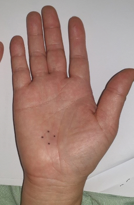
Fig. 1. Palpable mass of the palm (preoperative medical photo).
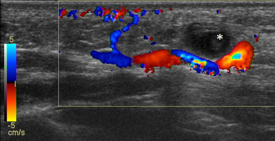
Fig. 2. Color Doppler ultrasonography shows well-delineated mass (asterisk) measuring 8 mm in diameter. The mass was predominantly isoechoic and avascular.
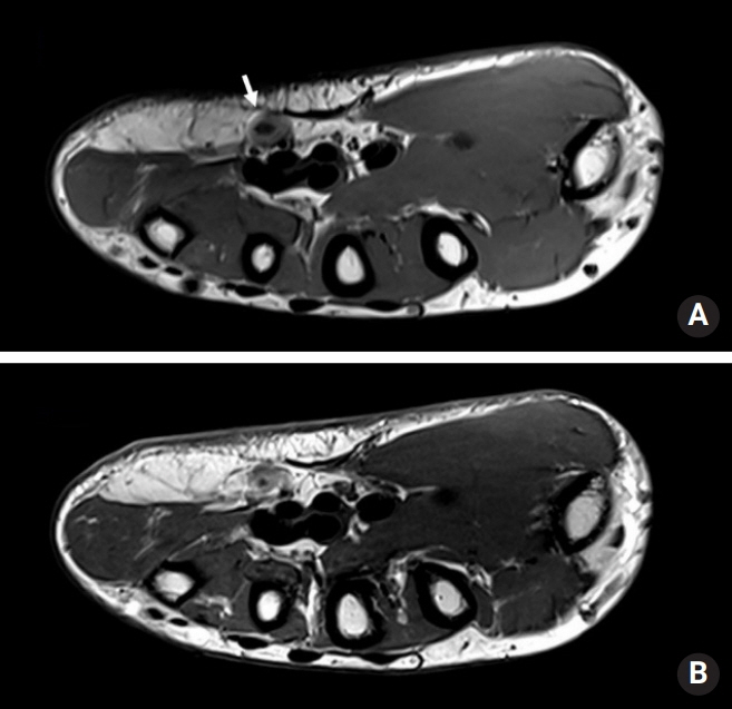
Fig. 3. Magnetic resonance (MR) imaging of the hand. (A) T2-weighted MR image in the transverse plane shows a mass in the volar and ulnar side of the palm containing a hypointense thrombus (arrow). (B) T1-weighted MR image in the transverse plane obtained after intravenous administration of a gadolinium chelate shows unenhanced mass corresponding to the thrombus.
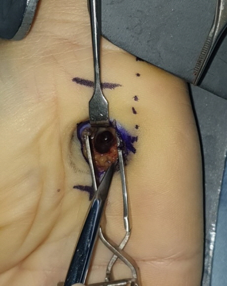
Fig. 4. Venous aneurysm in superficial venous palmar arch (intraoperative medical photo).
Reference
-
References
1. Calligaro KD, Ahmad S, Dandora R, et al. Venous aneurysms: surgical indications and review of the literature. Surgery. 1995; 117:1–6.
Article2. Gabrielli R, Rosati MS, Siani A, Irace L. Management of symptomatic venous aneurysm. The Scientific World Journal. 2012; 2012:386478.
Article3. Pascarella L, Al-Tuwaijri M, Bergan JJ, Mekenas LM. Lower extremity superficial venous aneurysms. Ann Vasc Surg. 2005; 19:69–73.
Article4. Gillespie DL, Villavicencio JL, Gallagher C, et al. Presentation and management of venous aneurysms. J Vasc Surg. 1997; 26:845–52.
Article5. Lev M, Saphir O. Endophlebohypertrophy and phlebosclerosis. I. The popliteal vein. AMA Arch Pathol. 1951; 51:154–78.6. McKesey J, Cohen PR. Spontaneous venous aneurysm: report of a non-traumatic superficial venous aneurysm on the distal arm. Cureus. 2018; 10:e2641.
Article7. Flors L, Leiva-Salinas C, Maged IM, et al. MR imaging of soft-tissue vascular malformations: diagnosis, classification, and therapy follow-up. Radiographics. 2011; 31:1321–41.
Article8. Garetier M, Moynot JC, Andro C, Vicard A, Rousset J. MR imaging findings of superficial venous aneurysm of the hand. Diagn Interv Imaging. 2016; 97:475–7.
Article
- Full Text Links
-
- Actions
-
Cited
- CITED
-
- Close
- Share
-
- Similar articles
-
- The Morphologic Study of he Superficial Palmar Arch in Korean
- Malposition of central venous catheter in the jugular venous arch via external jugular vein: a case report
- Replantation of Hand Amputated Through the Palmar Arch Level
- Surgical Reconstruction of Traumatic Pseudoaneurysm of Palmar Arch Caused by Blunt Trauma
- A Case of Arteriovenous Malformation Harboring of Large Venous Aneurysm
