Left Ventricular Systolic Dysfunction Caused by the Fistula from the Aortic Graft Pseudoaneurysm to the Left Ventricle
- Affiliations
-
- 1Kartal Kosuyolu Research and Education Hospital, İstanbul ,Turkey
- KMID: 2500958
- DOI: http://doi.org/10.4070/kcj.2019.0278
Figure
-
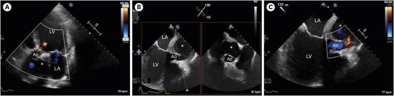
Figure 1 Transthoracic and transesophageal echocardiographic images. (A) Transthoracic echocardiography apical 5-chamber view showed pseudoaneurysm of the aortic graft (asterisk). (B) TEE mid-esophageal long axis biplane view revealed severe compression of the aortic graft by the pseudoaneurysm (asterisk). (C) TEE mid-esophageal long axis color Doppler view showed flow (arrow) towards pseudoaneurysm from the aortic graft (asterisk).Ao = aorta; LA = left atrium; LV = left ventricular; TEE = transesophageal echocardiography.
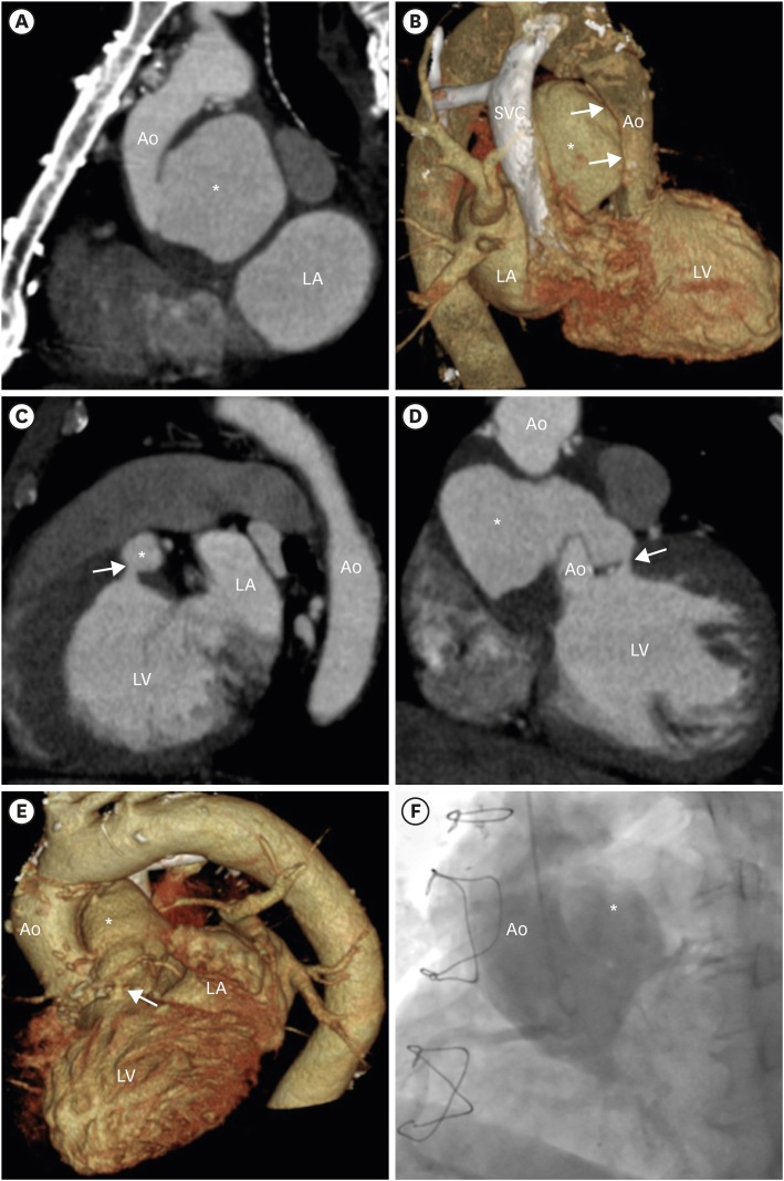
Figure 2 Computed tomographic and aortography images. (A) CT showed pseudoaneurysm of the aortic graft (asterisk). (B) CT 3D view showed compression (arrow) of the aortic graft by the pseudoaneurysm (asterisk). (C) CT sagittal view, (D) CT coronal view, and (E) CT 3D view revealed fistulization from the pseudoaneurysm of the aortic graft (asterisk) to the left ventricle (arrow), respectively. (F) Aortography showed contrast flow from the aorta to the pseudoaneurysm of the aortic graft (asterisk).Ao = aorta; CT = computed tomography; LA = left atrium; LV = left ventricular.
- Full Text Links
-
- Actions
-
Cited
- CITED
-
- Close
- Share
-
- Similar articles
-
- A Case of Left Ventricular Pseudoaneurysm in the Left Atrioventricular Groove after Mitral Valve Replacement
- Left Ventricular Diastolic Functions by M-Mode Echocardiogram in Essential Hypertensive Patients
- A Case of Left Ventricular Pseudoaneurysm Detected by Transesophageal Echocardiography
- Multiple Fistula Emptying into the Left Ventricle through the Entire Left Ventricular Wall
- Transient Left Ventricular Systolic Dysfunction Associated with Carbon Monoxide Toxicity
