Clin Endosc.
2020 Jan;53(1):60-64. 10.5946/ce.2019.081.
Utility of Forward-View Echoendoscopy for Transcolonic Fine-Needle Aspiration of Extracolonic Lesions: An Institutional Experience
- Affiliations
-
- 1Department of Gastroenterology, Aichi Cancer Center Hospital, Nagoya, Japan
- 2Department of Internal Medicine, Faculty of Medicine, Chiang Mai University, Chiang Mai, Thailand
- KMID: 2500906
- DOI: http://doi.org/10.5946/ce.2019.081
Abstract
- Background/Aims
Non-invasive tissue sampling from the lower intra-abdominal and pelvic cavity is challenging. The role of endoscopic ultrasound-guided fine-needle aspiration (EUS-FNA) in this situation is not well-established because of the limitations of the curved linear-array echoendoscopy-EUS for colonic insertion. The aim of this study was to report our institutional experience of transcolonic EUS-FNA using forward-viewing therapeutic linear echoendoscopy-EUS (FV-EUS) in combination with fluoroscopic guidance.
Methods
Medical records of 13 patients who underwent transcolonic EUS-FNA of extracolonic lesions using FV-EUS in combination with fluoroscopic guidance at Aichi Cancer Center Hospital, Nagoya, Japan from June 2015 to November 2018 were retrospectively reviewed.
Results
Using FV-EUS under fluoroscopic guidance, the FNA procedure could be performed successfully in all patients (100% technical success), with a median procedure time of 31 minutes. The sensitivity, specificity, and accuracy of EUS-FNA for detecting malignant lesions in this study were 91%, 100%, and 92%, respectively. There were no adverse events associated with the EUS-FNA procedure.
Conclusions
FV-EUS in combination with fluoroscopic guidance is an easy, safe, and effective technique for FNA of extracolonic lesions in the lower abdomen.
Keyword
Figure
-
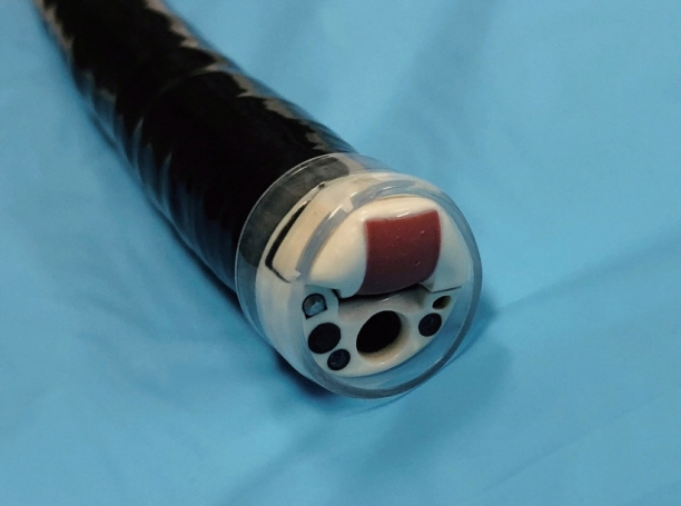
Fig. 1. Forward-view echoendoscope (TGF-UC260J; Olympus Medical Systems, Tokyo, Japan) with a transparent cap.

Fig. 2. The target lesion (green arrow) on positron emission tomography/computed tomography (PET/CT) (A), and the scope tip position on fluoroscopy (B) in patient 9. The target lesion (green arrow) on PET/CT (C), and the scope tip position on fluoroscopy (D) in patient 11. The target lesion (green arrow) on PET/CT (E), and the scope tip position on fluoroscopy (F) in patient 13.
Cited by 1 articles
-
Transcolonic Endoscopic Ultrasound-Guided Fine-Needle Aspiration Has a Promising Future
Sharmila Sachithanandan
Clin Endosc. 2020;53(1):3-4. doi: 10.5946/ce.2020.012.
Reference
-
1. Dumonceau JM, Polkowski M, Larghi A, et al. Indications, results, and clinical impact of endoscopic ultrasound (EUS)-guided sampling in gastroenterology: European Society of Gastrointestinal Endoscopy (ESGE) clinical guideline. Endoscopy. 2011; 43:897–912.
Article2. Nishida T, Kawai N, Yamaguchi S, Nishida Y. Submucosal tumors: comprehensive guide for the diagnosis and therapy of gastrointestinal submucosal tumors. Dig Endosc. 2013; 25:479–489.
Article3. Yoshinaga S, Suzuki H, Oda I, Saito Y. Role of endoscopic ultrasound-guided fine needle aspiration (EUS-FNA) for diagnosis of solid pancreatic masses. Dig Endosc. 2011; 23 Suppl 1:29–33.
Article4. Mohammad Alizadeh AH, Shahrokh S, Hadizadeh M, Padashi M, Zali MR. Diagnostic potency of EUS-guided FNA for the evaluation of pancreatic mass lesions. Endosc Ultrasound. 2016; 5:30–34.
Article5. Chen VK, Eloubeidi MA. Endoscopic ultrasound-guided fine needle aspiration is superior to lymph node echofeatures: a prospective evaluation of mediastinal and peri-intestinal lymphadenopathy. Am J Gastroenterol. 2004; 99:628–633.
Article6. Chin YK, Iglesias-Garcia J, de la Iglesia D, et al. Accuracy of endoscopic ultrasound-guided tissue acquisition in the evaluation of lymph nodes enlargement in the absence of on-site pathologist. World J Gastroenterol. 2017; 23:5755–5763.
Article7. Edelman BR, Weiser MR. Endorectal ultrasound: its role in the diagnosis and treatment of rectal cancer. Clin Colon Rectal Surg. 2008; 21:167–177.
Article8. Hünerbein M, Totkas S, Moesta KT, Ulmer C, Handke T, Schlag PM. The role of transrectal ultrasound-guided biopsy in the postoperative follow-up of patients with rectal cancer. Surgery. 2001; 129:164–169.
Article9. Pishvaian AC, Ahlawat SK, Garvin D, Haddad NG. Role of EUS and EUS-guided FNA in the diagnosis of symptomatic rectosigmoid endometriosis. Gastrointest Endosc. 2006; 63:331–335.
Article10. Shami VM, Parmar KS, Waxman I. Clinical impact of endoscopic ultrasound and endoscopic ultrasound-guided fine-needle aspiration in the management of rectal carcinoma. Dis Colon Rectum. 2004; 47:59–65.
Article11. Sailer M, Bussen D, Fein M, et al. Endoscopic ultrasound-guided transrectal biopsies of pelvic tumors. J Gastrointest Surg. 2002; 6:342–346.12. Fernández-Esparrach G, Alberghina N, Subtil JC, et al. Endoscopic ultrasound-guided fine needle aspiration is highly accurate for the diagnosis of perirectal recurrence of colorectal cancer. Dis Colon Rectum. 2015; 58:469–473.
Article13. Amin K, Olyaee M, Tawfik O, Fan F. Endoscopic ultrasound-guided fine needle aspiration as a diagnostic and staging tool for rectal and perirectal lesions-an institutional experience. Ann Diagn Pathol. 2013; 17:494–497.
Article14. Rotondano G, Esposito P, Pellecchia L, Novi A, Romano G. Early detection of locally recurrent rectal cancer by endosonography. Br J Radiol. 1997; 70:567–571.
Article15. Sasaki Y, Niwa Y, Hirooka Y, et al. The use of endoscopic ultrasound-guided fine-needle aspiration for investigation of submucosal and extrinsic masses of the colon and rectum. Endoscopy. 2005; 37:154–160.
Article16. Nguyen-Tang T, Shah JN, Sanchez-Yague A, Binmoeller KF. Use of the front-view forward-array echoendoscope to evaluate right colonic subepithelial lesions. Gastrointest Endosc. 2010; 72:606–610.
Article17. Uchida N, Galasso D, Seerden TC, et al. EUS-FNA of extracolonic lesions by using the forward-viewing linear echoendoscope. Gastrointest Endosc. 2010; 72:1321–1323.
Article
- Full Text Links
-
- Actions
-
Cited
- CITED
-
- Close
- Share
-
- Similar articles
-
- The Usefulness of Ultrasound-Guided Fine Needle Aspiration in Breast Lesions
- Fine Needle Aspiration Cytology of Thyroid Follicular Proliferative Lesions
- Transcolonic Endoscopic Ultrasound-Guided Fine-Needle Aspiration Has a Promising Future
- Fine Needle Aspiration Biopsy of Posterior Orbital Tumors
- Fine Needle Aspiration Biopsy with a Scalp Needle
