A case of retinopathy with antirecoverinantibody preceding the diagnosis ofcancer
- Affiliations
-
- 1Department of Neurology, Haeundae Paik Hospital, Inje University College of Medicine, Busan, Korea
- KMID: 2500306
- DOI: http://doi.org/10.14253/acn.2020.22.1.48
Figure
-
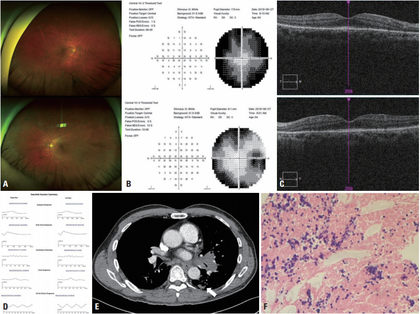
Fig. 1. . Findings of ophthalmology, imaging, and pathology examinations. (A) Fundoscopy produced normal findings in both eyes (upper: right eye, lower: left eye). (B) Humphrey visual field demonstrating generalized field constriction with a sparing of the central vision in both eyes (upper: right eye, lower: left eye). (C) Optical coherence tomography shows loss of the outer limiting membrane at the junction of the inner and outer segments with sparing of the central region (upper: right eye, lower: left eye). (D) Electroretinogram reveals diffused decreased wave potentials for all stimuli. (E) Chest computed tomography reveals a 1.8 cm nodule with adjacent tiny nodules in a superior segment of the left lower lobe (arrow). (F) Immunohistochemistry of a lymph node demonstrates metastatic small-cell carcinoma. Hematoxylin and eosin staining (×200) of the biopsy specimen revealed proliferation comprising small cells with hyperchromatic nuclei infiltrating the lymph node.
Reference
-
1. Grewal DS, Fishman GA, Jampol LM. Autoimmune retinopathy and antiretinal antibodies: a review. Retina. 2014; 34:827–845.2. Hoogewoud F, Butori P, Blanche P, Brézin AP. Cancer-associated retinopathy preceding the diagnosis of cancer. BMC Ophthalmol. 2018; 18:285.
Article
- Full Text Links
-
- Actions
-
Cited
- CITED
-
- Close
- Share
-
- Similar articles
-
- New Modalities for the Diagnosis and Treatment of Diabetic Retinopathy
- Retinopathy of prematurity-mimicking retinopathy in full-term babies
- Diabetic Retinopathy
- Incidence and Time of Onset of Retinopathy in Premature Infants in Korea
- The Influences of Arteriosclerosis on the Development and Progression of Diabetic Retinopathy
