Formaldehyde-induced Arthropathy in Rabbits
- Affiliations
-
- 1Department of Orthopaedics, Catholic Medical College, Seoul, Korea.
- KMID: 2471445
- DOI: http://doi.org/10.4055/jkoa.1975.10.1.51
Abstract
- Of the several methods available for producing an experimental arthritis, authors adopted a relatively simple and constant method for the production of an experimental arthritis by intra-articular injection of formaldehyde, and gave a detailed description for its sequential change of articular cartilage as observed by gross examination and by light microscopy. A total of 15 growing white rabbits, weighing 1.5-1.6 kg. were used. This animal were divided into two main groups, one designated as a control and the other as experimental group. Experimental group was subdivided into three groups to produce three form of arthritis simulating clinical forms of rheumatoid arthritis. Both knees were selected for the site of injection. Four rabbits received a single intra-articular injection of formaldehyde, while the other eight received 5 doses, halves at daily interval and the other halves at one week interval The rabbits observed clinically during the course of the experiment. They were sacrificed at 1 month after last intra-articular injection. The knee joints were then opened and examined grossly. The proximal end of tibia with the adjacent synovium of the infrapatellar region was excised ex Wec and examined. Sections were stained with hematoxylin and eosin. Those joints were normal in control group, but formaldehyde induced arthritic joint had mild to severe change of articular cartilage, A single intra-articular injection could cause arthritis transiently but minimum cartilage destruction by pannus invasion at periphery. Flve daily successive injection could cause most severe progressive form of arthritis which persisted over 6 months and caused most marked cartilage destruction in entire area but 5 successive injection at 1 week interval could cause intermediate form of arthritis persisting about 2 months after last injection. Cartilage destruction were moderate at periphery. Authors do not, however, as yet have a complete explanation for the longer perpetuation of the arthritis in the 5 doses of successive daily injection group. Severity of the joint destruction including cartilage seems to be responsible for the severity of initial insult. The diseased synovium itself could be responsible for the continued joint damage and it's severity including cartilage, perhaps by the release of proteolytic enzymes and the perpetuation of a chronic inflammatory response. Also it is postulated that many of the late changes in the cartilage are simply the end result of the initial Injury to synovium and articular cartilage by formaldehyde.
MeSH Terms
Figure
-
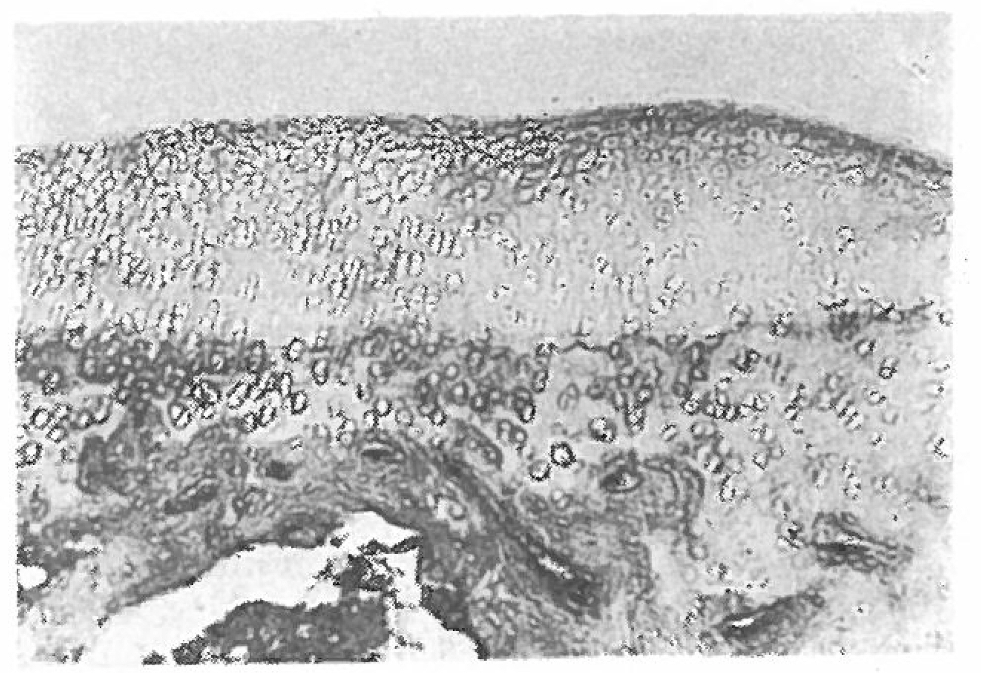
Fig. 1. Articular cartilage are well arranged in well defined linear arcades (Hematoxylinᅳeosin stain: one month after intraarticular injection of normal saline X100).

Fig. 2. There is mild synovial reaction with a superficial marginal cartilage change(Hematoxylin- eosin stainæ one month after a single intraarticular formaldehyde injection X100 (A), X450 (B)).
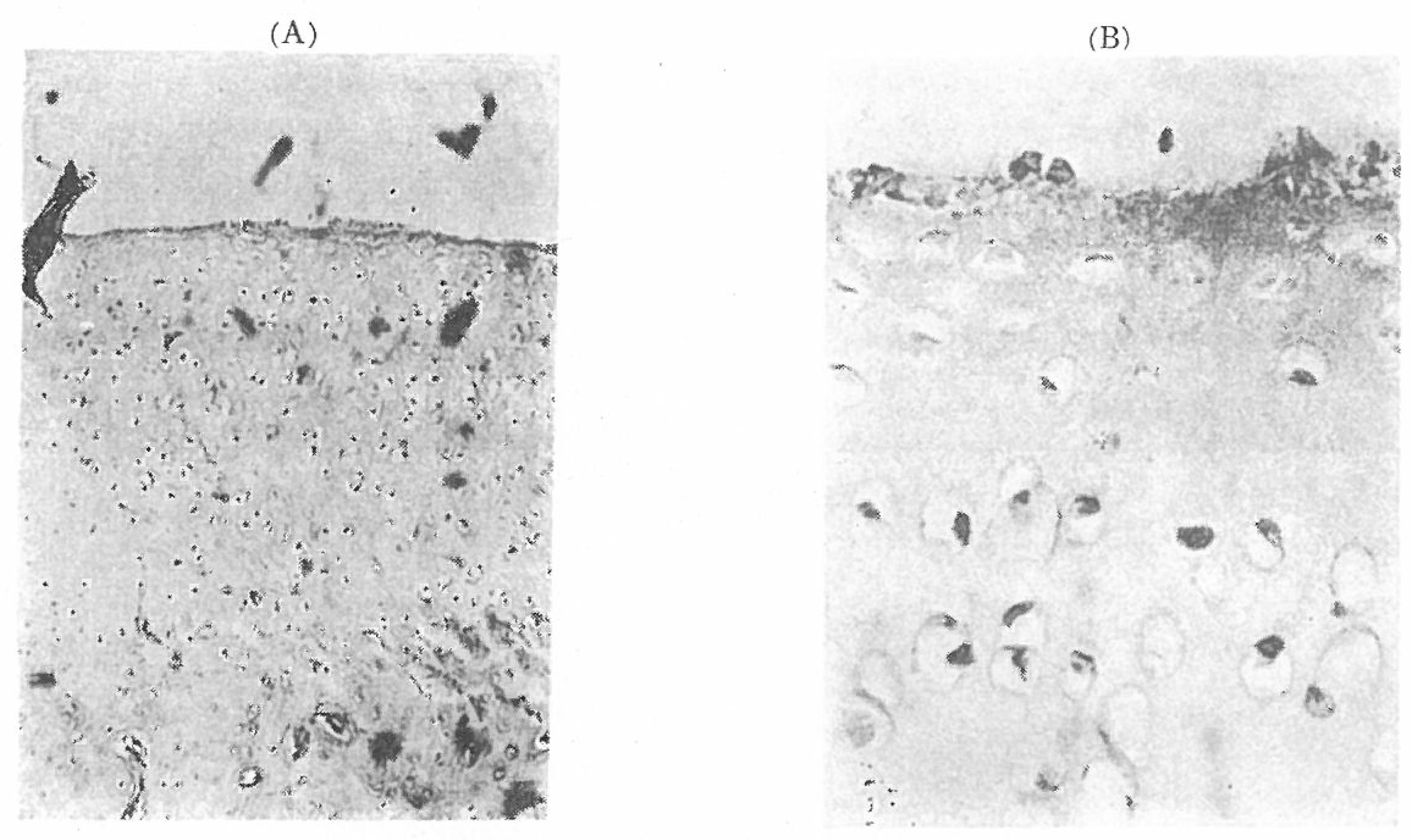
Fig. 3. There is mild fraying of marginal articular surface, superficial ulceration and early chondrocyte degeneration in the tangential and transitional layer (Hematoxylin-eosiη stain: one month after 5 successive intraarticular formaldehyde injection at a week interval X100 (A), X450 (B)).

Fig. 4. There is a moderate fibrillation-fraying of the cartilage surface, ulceration at the pariphery, fibrillary appearance of matrix from tangential zone to upper radial zone. Also there is nuclear degeneration and empty lacuna(Hematoxylin-eosin stain: one month after 5 successive intraarticular injection at daily interval X100 (A), X450 (B)).
Reference
-
Ball J.1969. Pathological Aspects of Rheumatoid Arthritis. In Early Synovectomy in Rheumatoid Arthritis. Excerpta Medica.Bourne G. H.1951. Some histological aspects of formalin “arthritis” in rats. Brit. J. Exp. Path. 32:377.Chaplin D. M.1971. The pattern of bone and cartilage damage in the rheumatoid knee. f. Bone and Joint Surg. 53:404.
Article최기홍(1966). 실험적만성관절염에미치는Corticosteroid 와영향. 대한외과학회잡지제권제1호57-72,.Harris E. D. Jr.1974. Degradation of cartilage collagen by rheumatoid synovial collagenase. f. New Eng. Med. 290:1.Kulka J. P.1959. The pathogenesis of rheumatoid arthritis. Journal of Chronic Disease. 10:388.
ArticleParratt J. R.., West G. B.{. 1958. Inhibition by various substances of oedema formation in the hind- paw of the rat induced by 5-hydroxy-tryptamine, histamine, egg white and compound 48¡80. Brit. J. J. Pharmacol. 14:484.Pemberton R.., Eiman J.., Patterson S.., Stack- jous E. A.1947. Attempts at the experimental production of arthritis, f. Lab. Med. 32:1121.Policard A.1936. In physiologie generaledes articulations a Vetat normal et pathologique, Paris, cited from Masson et Cie:. J. J. Bone and Joint Jur g. 53-B:732–750.Selye H.1949. Further studies in the participation of the adrenal cortex in the pathogenesis of arthritis. Brit. Med. J. 2:1129.Steinberg M. E.., McCrae C. R.., Cohon L. D.., Schumaches H. R.1973. Pathothogenesis of an- tigeninduced arthritis. Clinical Orthopaedic and Related Research. 97:248.
- Full Text Links
-
- Actions
-
Cited
- CITED
-
- Close
- Share
-
- Similar articles
-
- Sources of Formalin: III. Formalin Concentration in Adhesives
- Detection of formaldehyde in textiles
- Neuropathic Arthropathy Induced by Syringomyelia due to Arnold-Chiari I Malformation: A case report
- Sources of Formalin: I. Formalin Concentration in Shampoos
- Histological Studies of Endocrine Organs after the Injection of Garlic Extract (Rocambole)
