J Korean Orthop Assoc.
1975 Sep;10(3):265-271. 10.4055/jkoa.1975.10.3.265.
Treatment of Plantar Fascitis and Calcaneal Spur with UC-BL shoe insert
- Affiliations
-
- 1Department of Orthopaedics, Catholic Medical College, Seoul, Korea.
- 2Department of Internal Medicine, Catholic Medical College, Seoul, Korea.
- KMID: 2471425
- DOI: http://doi.org/10.4055/jkoa.1975.10.3.265
Abstract
- The ideal arch support should satisfy the following conditions; that is, it should reduce the tension of plantar fascia during weight bearing and make the high longitudinal arch. According to Campbell and Inman (1994). the defect of a conventional arch support is increment of tension of plantar fascia with bow-string effects, and therefore it cannot relieve the tension and plantar pain in flat foot though it can effectively make the high arch. Authors pointed out the restriction of dorsiflexion of toes during push off phase as a another basic important defect of a conventional arch support in this report. New arch support which satisfy these defects came to the front by Campbell and Inman (1974) recently. Yet, there are still technical difficulties to make it. Lastly, authors introduced clinical experiences of the UC-BL shoe insert which effectively relieved the obstinate painful flat foot, fasciitis and calcaneal spur.
Figure
-

Fig. 1. Bowstring effect resulting from upward force on plantar fascia exerted by an arch support. (Campbell and Inman. 1974).

Fig. 2. Axis of the subtalar joint. (Manter and Hicks. 1941.).
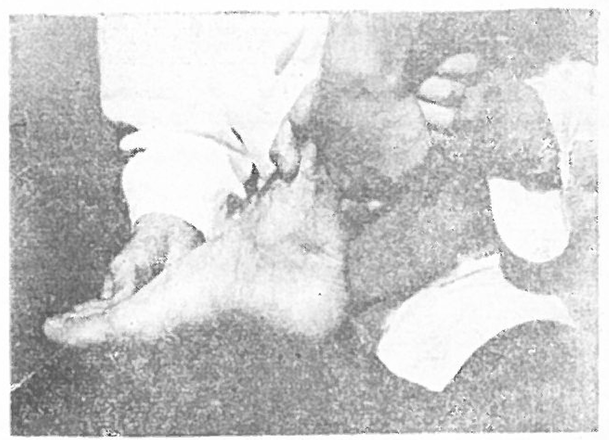
Fig. 3. Position of foot for taking plaster cast for shoe insert.
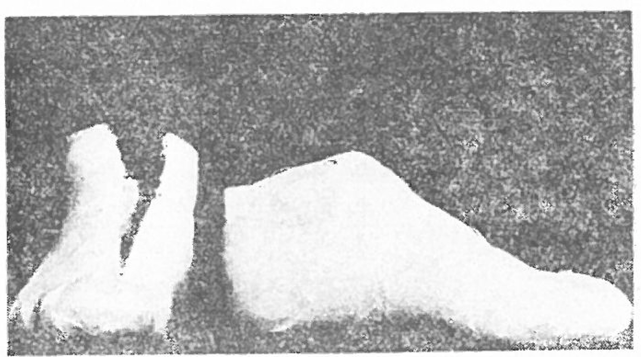
Fig. 4. Plaster-negative mould.
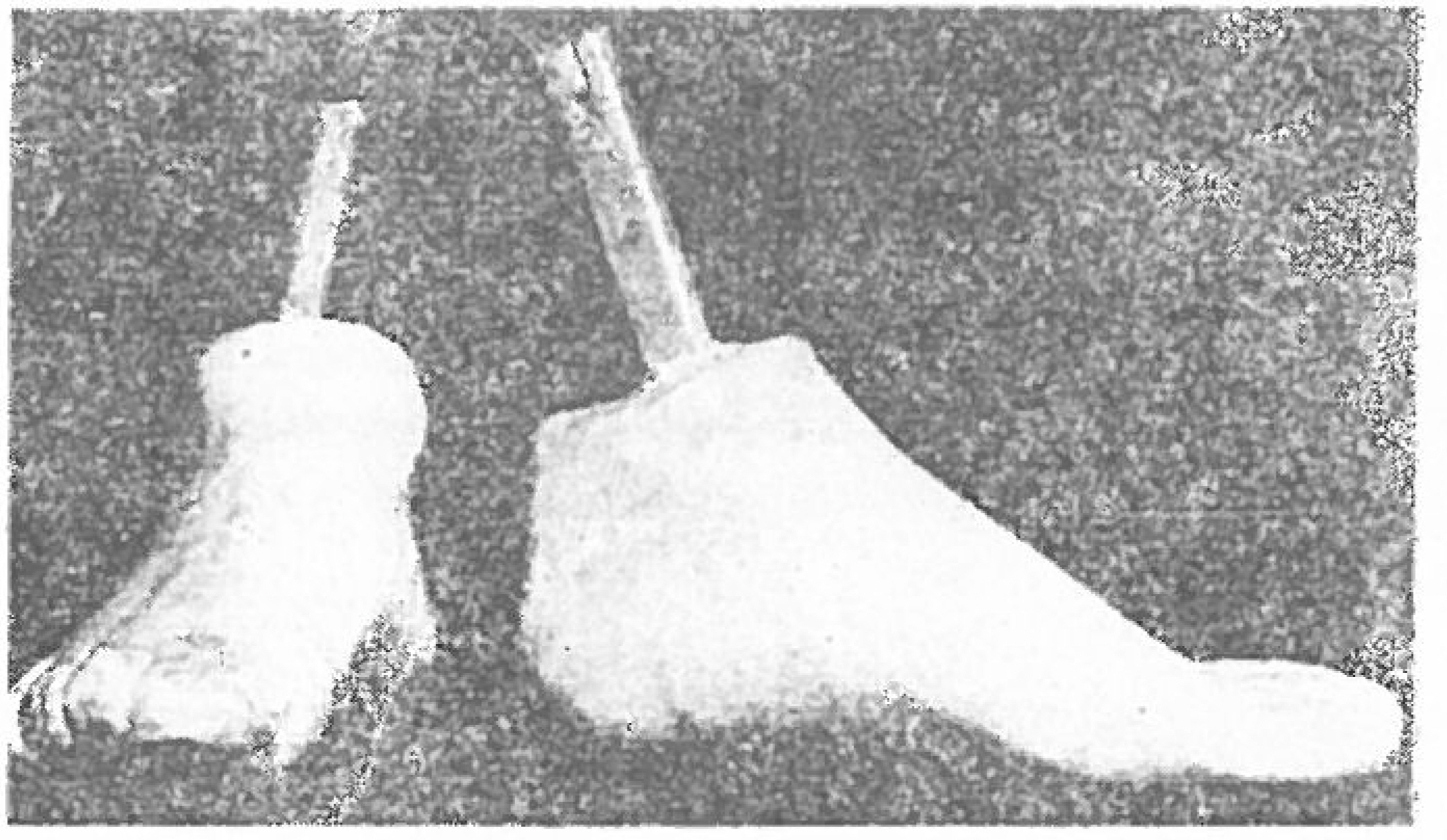
Fig. 5. Plaster-positive mould

Fig. 6. Cilastic UC-BL shoe insert.
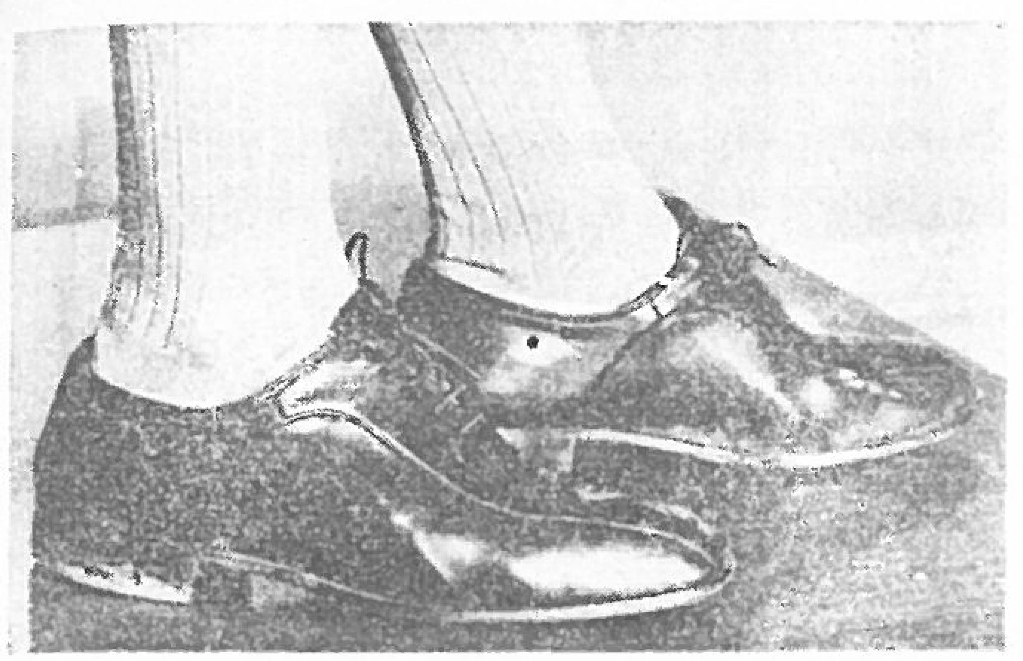
Fig. 7. Outlook of a patient's shoes with the UC- BL shoe insert in his shoes

Fig. 8. Arch development. Engel and Staheli, (1974)

Fig. 9. Arch makers responsible to each phase of a walking cycle (Mann and Inman. 1964.)

Fig. 10. The windlass mechanism. (Hicks 1954.)
Reference
-
1.Basmajian J.V.., Stecko G.1963. The role of muscles in arch support of the foot: an electromyographic study, f. Bone Joint Surg. 45A:1184.2.Bruce. J.., Walmsley R.1938. Some observations on the arches of the foot and flat foot. The lancet sept. 17:656–659.3.Campbell J. W.., Inman V. T.1974. Treatment of plantar fasciitis and calcaneal spurs with the UC-BL shoe insert, eli. orth. No. 103. 57-62.4.Elftman ., Herbert . 1960. The Transverse tarsal Joint and its control, clin, orthop. 16:41–46.5.Engel G. M.., Staheli L. T.1974. The natural history of torsion and other factors gait in casting. A study of the angle of gait influencing tibial torsion, knee angle, hip rotation, and developinent of the arch in normal children, cli. orth. 99:1217.6.Giannestras N.J.1973. Foot disorders. Medical and surgical management 2np ed 17-23.7.Henderson W. H.., Campbell J.W.1967. UC-BJ shoe insert; Casting and fabrication. Biomechanics Laboratory, University of California, San Francisco and Berkeley. Technical report 53. Reprinted in Bull. Prosthet. Res. 10-11: 215.8.Hick J. H.1954. The mechanics of the foot. The plantar aponeurosis and the arch. J. Anat. 88;25.9.Mann R.., Inman V. T.1964. Phasic activity of intrinsic nuscles of the foot. J. Bone and Joint Sur g. 46 A; 469.10.Manter J. T.., Hicks J. H.1941. Movements of the subtalar and transverse tarsal Joints. Anat, Res. 80:397.
Article11.Snook G. A.., Chrisman O. D.1972. The management of subcalcaneal pain. Cli orth. and Related research. No. 82:163–168.
Article12.Viladot A.1973. Metatarsalgia due to Biomechanical alterations of the forefoot. Orth. Cli. of North Arne. 4;1, Jan. 165-178.13.Wright D. G.., Desal S. M.1964. Action of the subtalar and ankle Joint complex During the stance phase of walking. J. Bone and Joint Surg. 46-A:361–382.14.Wright D. C.., Rennels D. C.1964. A study of the elastic properties of plantar fascia. J. Bone and Joint Surg. 46-A:No.(3):Apr.482–492.
Article
- Full Text Links
-
- Actions
-
Cited
- CITED
-
- Close
- Share
-
- Similar articles
-
- Ultrasonographic Appearances of the Plantar Fasciitis
- Biomechanical Factors Associated with Plantar Fasciitis in Non-obese Patients
- A Study of Prognostic Factors of Conservative Treatment in Plantar Fasciitis
- Clinical Characteristics of the Causes of Plantar Heel Pain
- Age, Body Mass Index, and Spur Size Associated with Patients’ Symptoms in Plantar Fasciitis
