A Canine Model for Lymphangiography and Thoracic Duct Access
- Affiliations
-
- 1Department of Radiology and Research Institute of Clinical Medicine of Chonbuk National University-Biomedical Research Institute of Chonbuk National University Hospital, Jeonju, Korea.
- 2Biomedical Engineering Research Center, Asan Medical Center, University of Ulsan College of Medicine, Seoul, Korea.
- 3Department of Interventional Therapy, National Cancer Center/National Clinical Research Center for Cancer/Cancer Hospital, Chinese Academy of Medical Sciences and Peking Union Medical College, Beijing, China.
- 4Department of Radiology and Research Institute of Radiology, Asan Medical Center, University of Ulsan College of Medicine, Seoul, Korea. jhshin@amc.seoul.kr
- 5Department of Radiology, The Affiliated Cancer Hospital of Zhengzhou University, Zhengzhou, China.
- KMID: 2470753
- DOI: http://doi.org/10.3348/kjr.2019.0313
Abstract
OBJECTIVE
To evaluate the technical feasibility of intranodal lymphangiography and thoracic duct (TD) access in a canine model.
MATERIALS AND METHODS
Five male mongrel dogs were studied. The dog was placed in the supine position, and the most prominent lymph node in the groin was accessed using a 26-gauge spinal needle under ultrasonography (US) guidance. If the cisterna chyli (CC) was not opacified by bilateral lymphangiography, the medial iliac lymph nodes were directly punctured and Lipiodol was injected. After opacification, the CC was directly punctured with a 22-gauge needle. A 0.018-in microguidewire was advanced through the CC and TD. A 4-Fr introducer and dilator were then advanced over the wire. The microguidewire was changed to a 0.035-in guidewire, and this was advanced into the left subclavian vein through the terminal valve of the TD. Retrograde TD access was performed using a snare kit.
RESULTS
US-guided lymphangiography (including intranodal injection of Lipiodol [Guerbet]) was successful in all five dogs. However, in three of the five dogs (60%), the medial iliac lymph nodes were not fully opacified due to overt Lipiodol extravasation at the initial injection site. In these dogs, contralateral superficial inguinal intranodal injection was performed. However, two of these three dogs subsequently underwent direct medial iliac lymph node puncture under fluoroscopy guidance to deliver additional Lipiodol into the lymphatic system. Transabdominal CC puncture and cannulation with a 4-Fr introducer was successful in all five dogs. Transvenous retrograde catheterization of the TD (performed using a snare kit) was also successful in all five dogs.
CONCLUSION
A canine model may be appropriate for intranodal lymphangiography and TD access. Most lymphatic intervention techniques can be performed in a canine using the same instruments that are employed in a clinical setting.
MeSH Terms
Figure
-
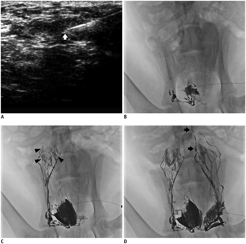
Fig. 1 Inguinal lymph node puncture. A. Ultrasound image shows needle tip (arrow) located in cortex of lymph node. B. Lymphangiography via intranodal injection of Lipiodol (Guerbet) shows tortuous lymphatic collectors connected to superficial inguinal lymph node. C. Lipiodol drains via pelvic retroperitoneal wall to right medial iliac lymph nodes (arrowheads). D. Contralateral access was re-tried due to overt extravasation at injection site on right-hand side. Lipiodol traversing left medial iliac lymph nodes to distal thoracic duct (arrows).

Fig. 2 Thoracic duct lymphangiography. A. Distal thoracic duct (arrowheads) and cisterna chyli (arrow) are visible. Either thoracic duct or cisterna chyli could be punctured. B. In another canine, lymphangiography shows that distal thoracic duct consists of two thin ducts (arrowheads). Cisterna chyli (arrow) is also visible.
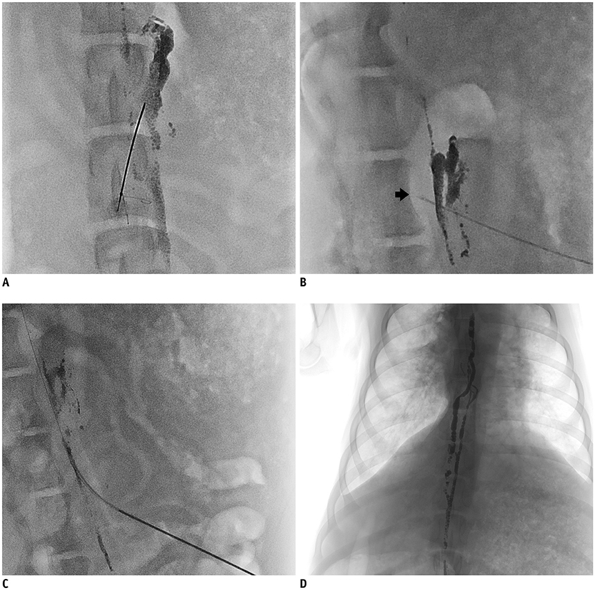
Fig. 3 Transabdominal puncture of cisterna chyli. A. Anterior/posterior view shows seemingly successful transabdominal puncture of cisterna chyli. B. Lateral-oblique view shows that needle tip is penetrating cisterna chyli (arrow). C. Microguidewire and 4-Fr introducer catheter are advanced through cisterna chyli. D. Entire thoracic duct is visible by lymphangiography via 4-Fr introducer catheter.
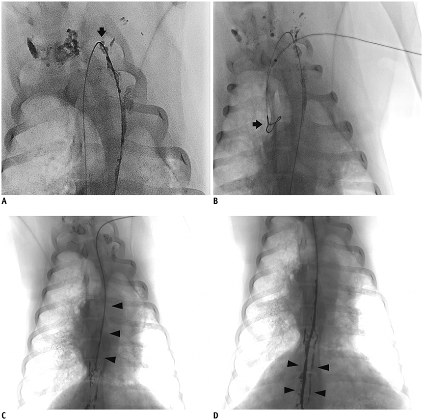
Fig. 4 Retrograde thoracic duct access technique. A. 0.035-in guidewire is passed through terminal thoracic duct (arrow). B. 4-Fr goose neck snare kit is inserted through left subclavian vein, and is used to capture 0.035-in guidewire tip (arrow). C. 5-Fr catheter is advanced over guidewire (arrowheads). D. Final lymphangiography via retrograde access shows distal thoracic duct (arrowheads).
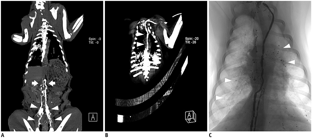
Fig. 5 Post-procedural CT images. A. Coronal reconstructed CT image shows pelvic retroperitoneal lymphatic tract (arrowheads) and intraabdominal portion of thoracic duct (arrows). B. Coronal reconstructed CT image shows intrathoracic portion of thoracic duct (arrowheads). C. Fluoroscopy image after lymphangiography shows multiple Lipiodol droplets distributed along both pulmonary arteries (arrowheads).
Reference
-
1. Alejandre-Lafont E, Krompiec C, Rau WS, Krombach GA. Effectiveness of therapeutic lymphography on lymphatic leakage. Acta Radiol. 2011; 52:305–311.
Article2. Inoue M, Nakatsuka S, Yashiro H, Tamura M, Suyama Y, Tsukada J, et al. Lymphatic intervention for various types of lymphorrhea: access and treatment. Radiographics. 2016; 36:2199–2211.
Article3. Johnson OW, Chick JF, Chauhan NR, Fairchild AH, Fan CM, Stecker MS, et al. The thoracic duct: clinical importance, anatomic variation, imaging, and embolization. Eur Radiol. 2016; 26:2482–2493.
Article4. Matsumoto T, Kudo T, Endo J, Hashida K, Tachibana N, Murakoshi T, et al. Transnodal lymphangiography and post-CT for protein-losing enteropathy in Noonan syndrome. Minim Invasive Ther Allied Technol. 2015; 24:246–249.
Article5. Nadolski GJ, Chauhan NR, Itkin M. Lymphangiography and lymphatic embolization for the treatment of refractory chylous ascites. Cardiovasc Intervent Radiol. 2018; 41:415–423.
Article6. Toliyat M, Singh K, Sibley RC, Chamarthy M, Kalva SP, Pillai AK. Interventional radiology in the management of thoracic duct injuries: anatomy, techniques and results. Clin Imaging. 2017; 42:183–192.
Article7. Yamagami T, Masunami T, Kato T, Tanaka O, Hirota T, Nomoto T, et al. Spontaneous healing of chyle leakage after lymphangiography. Br J Radiol. 2005; 78:854–857.
Article8. Kim PH, Tsauo J, Shin JH. Lymphatic interventions for chylothorax: a systematic review and meta-analysis. J Vasc Interv Radiol. 2018; 29:194–202.e4.9. Koike Y, Hirai C, Nishimura J, Moriya N, Katsumata Y. Percutaneous transvenous embolization of the thoracic duct in the treatment of chylothorax in two patients. J Vasc Interv Radiol. 2013; 24:135–137.
Article10. Mittleider D, Dykes TA, Cicuto KP, Amberson SM, Leusner CR. Retrograde cannulation of the thoracic duct and embolization of the cisterna chyli in the treatment of chylous ascites. J Vasc Interv Radiol. 2008; 19:285–290.
Article11. Ito R, Suami H. Lymphatic territories (lymphosomes) in swine: an animal model for future lymphatic research. Plast Reconstr Surg. 2015; 136:297–304.12. Suami H, Yamashita S, Soto-Miranda MA, Chang DW. Lymphatic territories (lymphosomes) in a canine: an animal model for investigation of postoperative lymphatic alterations. PLoS One. 2013; 8:e69222.
Article13. Cope C. Percutaneous thoracic duct cannulation: feasibility study in swine. J Vasc Interv Radiol. 1995; 6:559–564.
Article14. Kraitchman D, Kamel I, Weiss C, Georgiades C. Elucidation of percutaneously accessible lymph nodes in swine: a large animal model for interventional lymphatic research. J Vasc Interv Radiol. 2017; 28:451–456.
Article15. Mayer MN, Silver TI, Lowe CK, Anthony JM. Radiographic lymphangiography in the dog using iodized oil. Vet Comp Oncol. 2013; 11:151–161.16. Naganobu K, Ohigashi Y, Akiyoshi T, Hagio M, Miyamoto T, Yamaguchi R. Lymphography of the thoracic duct by percutaneous injection of iohexol into the popliteal lymph node of dogs: experimental study and clinical application. Vet Surg. 2006; 35:377–381.
Article17. Suami H. Lymphosome concept: anatomical study of the lymphatic system. J Surg Oncol. 2017; 115:13–17.
Article
- Full Text Links
-
- Actions
-
Cited
- CITED
-
- Close
- Share
-
- Similar articles
-
- Postoperative Chylothorax: the Use of Dynamic Magnetic Resonance Lymphangiography and Thoracic Duct Embolization
- Lymphangiography to Treat Postoperative Lymphatic Leakage: A Technical Review
- Thoracic Duct Embolization for Treatment of Chyle Leakage After Thyroidectomy and Neck Dissection
- Novel Interventional Radiology for the Treatment of Various Lymphatic Leakages: Lymphatic Intervention and Embolization
- Retrograde Distal Thoracic Duct Leak Embolization via Access Through Lymphocele After Thyroidectomy and Neck Dissection
