Restor Dent Endod.
2020 Feb;45(1):e7. 10.5395/rde.2020.45.e7.
Isthmuses, accessory canals, and the direction of root curvature in permanent mandibular first molars: an in vivo computed tomography study
- Affiliations
-
- 1Department of Dental Morphology, Dental Branch, Islamic Azad University, Tehran, Iran.
- 2Department of Removable Prosthodontics, Dental Faculty, Tehran Medical Sciences, Islamic Azad University, Tehran, Iran. dr.ma.navabi@gmail.com
- KMID: 2470342
- DOI: http://doi.org/10.5395/rde.2020.45.e7
Abstract
OBJECTIVES
This study was performed to assess the anatomy of mandibular first molars.
MATERIALS AND METHODS
In this in vivo study, cone-beam computed tomography (CBCT) volumes of 312 bilateral intact first mandibular molars from 156 patients (79 men and 77 women; average age, 35.6 ± 11.2 years) were investigated in terms of the direction of each canal's curvature in the buccolingual and mesiodistal dimensions (direction of the position of the apex in relation to the longitudinal axis of the root), the presence of an isthmus (a narrow, ribbon-shaped communication between 2 root canals) in 3 segments (0-2, 2-4, and 4-6 mm) from the apex), and the presence and number of accessory canals (smaller canals besides the main root canals, connecting the pulp to the periodontium). Data were analyzed statistically (α = 0.05).
RESULTS
Mesiolingual canals were mostly buccally and distally inclined, while mesiobuccal and distolingual canals were mostly distally curved. Isthmuses were more common in younger patients (χ2 test, p < 0.05). The average numbers of accessory canals in the apical, middle, and coronal segments were 9.9 ± 4.2, 6.9 ± 2.9, and 9.3 ± 3.0 canals per segment, respectively (analysis of variance, p < 0.001). Age and sex were not associated with the number of accessory canals (p > 0.05).
CONCLUSIONS
The complex anatomy of these teeth deserves attention during non-surgical or surgical endodontic treatment. Around the apex, isthmuses might be more prevalent in younger and female individuals.
Keyword
Figure
-
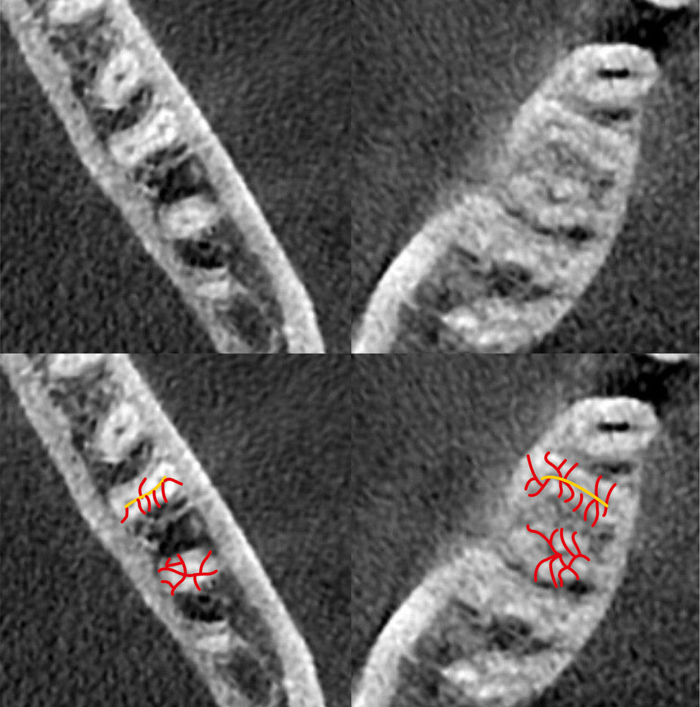
Figure 1. An example of accessory canals. In the bottom image, the main (yellow) and accessory (red) canals are marked.
Reference
-
References
1. Akhlaghi NM, Abbas FM, Mohammadi M, Shamloo MR, Radmehr O, Kaviani R, Rakhshan V. Radicular anatomy of permanent mandibular second molars in an Iranian population: a preliminary study. Dent Res J (Isfahan). 2016; 13:362–366.2. Mohammadzadeh Akhlaghi N, Khalilak Z, Vatanpour M, Mohammadi S, Pirmoradi S, Fazlyab M, Safavi K. Root canal anatomy and morphology of mandibular first molars in a selected Iranian population: an in vitro study. Iran Endod J. 2017; 12:87–91.3. Madani ZS, Mehraban N, Moudi E, Bijani A. Root and canal morphology of mandibular molars in a selected Iranian population using cone-beam computed tomography. Iran Endod J. 2017; 12:143–148.4. Mehrvarzfar P, Akhlagi NM, Khodaei F, Shojaee G, Shirazi S. Evaluation of isthmus prevalence, location, and types in mesial roots of mandibular molars in the Iranian population. Dent Res J (Isfahan). 2014; 11:251–256.5. Weller RN, Niemczyk SP, Kim S. Incidence and position of the canal isthmus. Part 1. Mesiobuccal root of the maxillary first molar. J Endod. 1995; 21:380–383.6. von Arx T. Frequency and type of canal isthmuses in first molars detected by endoscopic inspection during periradicular surgery. Int Endod J. 2005; 38:160–168.
Article7. Al-Qudah AA, Awawdeh LA. Root and canal morphology of mandibular first and second molar teeth in a Jordanian population. Int Endod J. 2009; 42:775–784.
Article8. Ahmed HA, Abu-bakr NH, Yahia NA, Ibrahim YE. Root and canal morphology of permanent mandibular molars in a Sudanese population. Int Endod J. 2007; 40:766–771.
Article9. Gulabivala K, Aung TH, Alavi A, Ng YL. Root and canal morphology of Burmese mandibular molars. Int Endod J. 2001; 34:359–370.
Article10. Peiris HR, Pitakotuwage TN, Takahashi M, Sasaki K, Kanazawa E. Root canal morphology of mandibular permanent molars at different ages. Int Endod J. 2008; 41:828–835.
Article11. Razmi H, Shokouhinejad N, Hooshyar M. An in vitro study of the number of distal roots and canals in mandibular first molars in Iranian population. Iran Endod J. 2008; 2:126–130.12. Choupani Dastgerdi A, Navabi M, Hafezi L, Khalilak Z, Rakhshan V. Anatomy of permanent mandibular first molars in a selected Iranian population using cone-beam computed tomography. Iran Endod J. 2018; 13:251–256.13. Lee KW, Kim Y, Perinpanayagam H, Lee JK, Yoo YJ, Lim SM, Chang SW, Ha BH, Zhu Q, Kum KY. Comparison of alternative image reformatting techniques in microcomputed tomography and tooth clearing for detailed canal morphology. J Endod. 2014; 40:417–422.
Article14. Dalili Kajan Z, Taramsari M, Khosravi Fard N, Kanani M. Accuracy of cone-beam computed tomography in comparison with standard method in evaluating root canal morphology: an in vitro study. Iran Endod J. 2018; 13:181–187.15. Faramarzi F, Vossoghi M, Shokri A, Shams B, Vossoghi M, Khoshbin E. Cone beam computed tomography study of root and canal morphology of maxillary first molar in an Iranian population. Avicenna J Dent Res. 2015; 7:e24038.
Article16. Hsu YY, Kim S. The resected root surface. The issue of canal isthmuses. Dent Clin North Am. 1997; 41:529–540.17. Estrela C, Bueno MR, Barletta FB, Guedes OA, Porto OC, Estrela CR, Pécora JD. Identification of apical and cervical curvature radius of human molars. Braz Dent J. 2015; 26:351–356.
Article18. Schäfer E, Diez C, Hoppe W, Tepel J. Roentgenographic investigation of frequency and degree of canal curvatures in human permanent teeth. J Endod. 2002; 28:211–216.19. Rocha LF, Sousa Neto MD, Fidel SR, da Costa WF, Pécora JD. External and internal anatomy of mandibular molars. Braz Dent J. 1996; 7:33–40.20. de Pablo ÓV, Estevez R, Péix Sánchez M, Heilborn C, Cohenca N. Root anatomy and canal configuration of the permanent mandibular first molar: a systematic review. J Endod. 2010; 36:1919–1931.
Article21. Zafar M, Alrahabi M. Cone beam computed tomography for exploring morphology of mandibular first molar. Br J Med Med Res. 2015; 6:514–521.
Article22. Arjmand N, Kolahdouzan A, Rouhi N. Evaluation of root and canal morphology of first and second mandibular molars according to Vertucci and Weine classification by using cone-beam computed tomography archive in Partow Radiology Center in Qazvin in 1391 [dissertation]. Qazvin: Qazvin University of Medical Sciences;2014.23. Masoudi SM, Rouhi N. Evaluation of root canal number and anatomy of first and second mandibular molars with available CBCT in Qazvin in 2013 [dissertation]. Qazvin: Qazvin University of Medical Sciences;2014.24. Gu L, Wei X, Ling J, Huang X. A microcomputed tomographic study of canal isthmuses in the mesial root of mandibular first molars in a Chinese population. J Endod. 2009; 35:353–356.
Article25. Villas-Bôas MH, Bernardineli N, Cavenago BC, Marciano M, Del Carpio-Perochena A, de Moraes IG, Duarte MH, Bramante CM, Ordinola-Zapata R. Micro-computed tomography study of the internal anatomy of mesial root canals of mandibular molars. J Endod. 2011; 37:1682–1686.
Article26. Fan B, Pan Y, Gao Y, Fang F, Wu Q, Gutmann JL. Three-dimensional morphologic analysis of isthmuses in the mesial roots of mandibular molars. J Endod. 2010; 36:1866–1869.
Article27. Demirbuga S, Sekerci AE, Dinçer AN, Cayabatmaz M, Zorba YO. Use of cone-beam computed tomography to evaluate root and canal morphology of mandibular first and second molars in Turkish individuals. Med Oral Patol Oral Cir Bucal. 2013; 18:e737–e744.
Article28. Faraz SA, Tariq A, Jameel A. Root canal morphology of mandibular first permanent molars-Karachi sample. Pak Oral Dent J. 2015; 35:294–298.29. Chourasia HR, Meshram GK, Warhadpande M, Dakshindas D. Root canal morphology of mandibular first permanent molars in an Indian population. Int J Dent. 2012; 2012:745152.
Article30. Wu DM, Wu YN, Guo W, Sameer S. Accuracy of direct digital radiography in the study of the root canal type. Dentomaxillofac Radiol. 2006; 35:263–265.
Article31. Neelakantan P, Subbarao C, Subbarao CV. Comparative evaluation of modified canal staining and clearing technique, cone-beam computed tomography, peripheral quantitative computed tomography, spiral computed tomography, and plain and contrast medium-enhanced digital radiography in studying root canal morphology. J Endod. 2010; 36:1547–1551.
Article
- Full Text Links
-
- Actions
-
Cited
- CITED
-
- Close
- Share
-
- Similar articles
-
- Asymmetry in mesial root number and morphology in mandibular second molars: a case report
- A Study of Root Canals Morphology in Primary Molars using Computerized Tomography
- Assessment of Root and Root Canal Morphology of Human Primary Molars using CBCT
- An evaluation of canal curvature at the apical one third in type II mesial canals of mandibular molars
- Prevalence and features of distolingual roots in mandibular molars analyzed by cone-beam computed tomography
