J Surg Ultrasound.
2019 Nov;6(2):58-63. 10.0000/jsu.2019.6.2.58.
A Comparative Study of the Diagnostic Performance of Evaluating Breast Masses for Breast Surgeons versus S-Detectâ„¢ (Samsung Medison Co., Ltd, Seoul, Korea)
- Affiliations
-
- 1Department of Surgery, Presbyterian Medical Center, Jeonju, Korea. cskimmd@hotmail.com
- KMID: 2469750
- DOI: http://doi.org/10.0000/jsu.2019.6.2.58
Abstract
- PURPOSE
Ultrasonography is widely used for examining breast mass. We used the Breast Imaging-Reporting and Data System (BI-RADS) to characterize breast lesions found on ultrasonography. Among various ultrasound techniques, we used S-Detectâ„¢ (Samsung Medison Co., Ltd, Seoul, Korea), which supports the morphological analysis of breast masses found according to BI-RADS. In addition, we compared the breast surgeons' categorization of breast masses with that by S-Detectâ„¢.
METHODS
Breast surgeons evaluated the breast masses found using ultrasonography between April 2016 and December 2016. A total of 139 masses, which were categorized as BI-RADS 3 or 4, from 112 patients were reevaluated by S-Detectâ„¢ before performing vacuum-assisted resection or surgical excision.
RESULTS
Of the 139 masses, 118 were benign tumors and 21 were malignant tumors. With regard to the diagnostic performance, the sensitivity of categorization was 95% for breast surgeons, but the sensitivity was relatively lower for S-detectâ„¢ (85%). However, the specificity and accuracy of S-detectâ„¢ were 70.6% and 74.1%, respectively, which were higher than those values obtained from breast surgeons (18.5% and 30.9%, respectively).
CONCLUSION
S-detectâ„¢ can be used by breast surgeons as a diagnostic aid when evaluating and diagnosing breast masses found on ultrasonography.
MeSH Terms
Figure
-
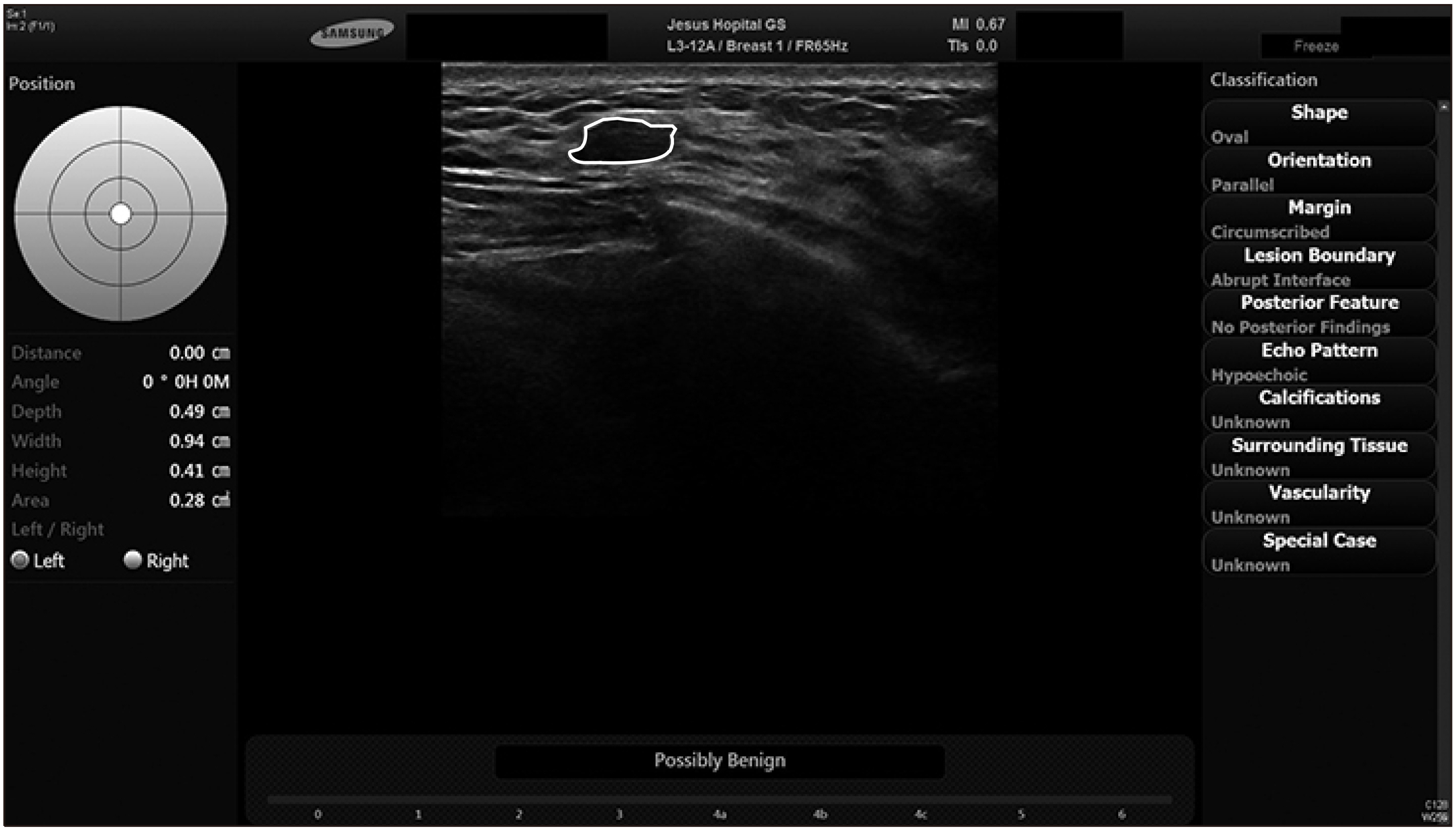
Fig. 1 A 27-year-old woman with left breast mass on B-mode ultrasound. S-DetectTM concluded that it is “Possibly Benign” based on features of the lesion listed on the right column.

Fig. 2 A 65-year-old woman with left breast mass on B-mode ultrasound. S-DetectTM concluded that it is “Possibly Malignant” based on features of the lesion listed on the right column.
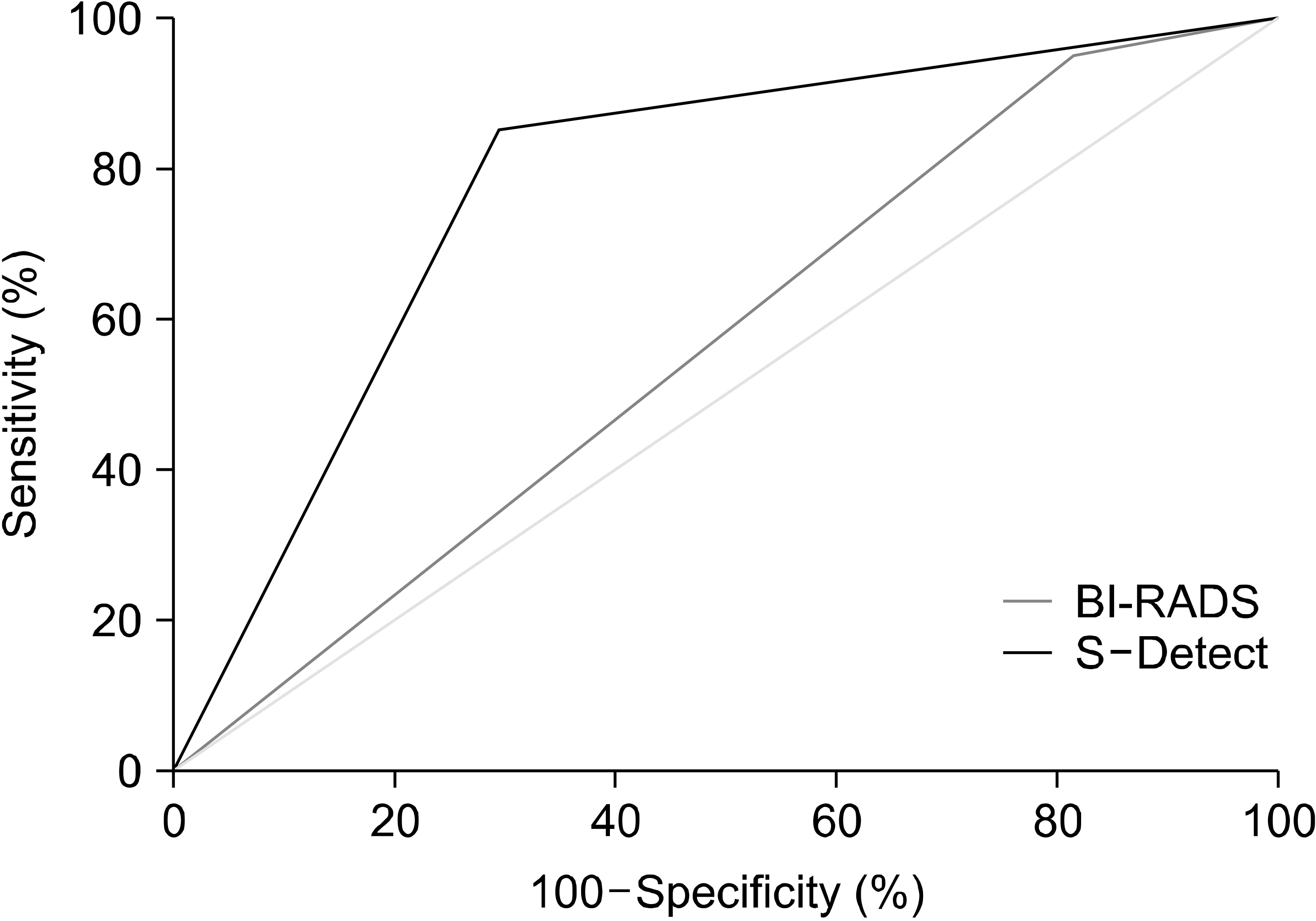
Fig. 3 Receiver operating characteristic curve for breast surgeons (BI-RADS) and S-DetectTM.
Reference
-
Korean Breast Cancer Society. Breast Cancer Facts & Figures 2018. Korean Breast Cancer Society;Seoul:Youn I., Choi S., Kook SH., Choi YJ. 2016. Mammographic breast density evaluation in korean women using fully automated volumetric assessment. J Korean Med Sci. 31:457–62. DOI: 10.3346/jkms.2016.31.3.457. PMID: 26955249. PMCID: PMC4779873.
ArticleStavros AT., Thickman D., Rapp CL., Dennis MA., Parker SH., Sisney GA. 1995. Solid breast nodules: use of sonography to distinguish between benign and malignant lesions. Radiology. 196:123–34. DOI: 10.1148/radiology.196.1.7784555. PMID: 7784555.
ArticleKim YJ., Choi HY., Moon BI., Lee SN. 2006. Categorization and evaluation of usefulness of breast lesions with using ultrasound BI-RADS (Breast Imaging Reporting and Data system). J Korean Radiol Soc. 54:313–8. DOI: 10.3348/jkrs.2006.54.4.313.
ArticlePan J., Dogan BE., Carkaci S., Santiago L., Arribas E., Cantor SB, et al. 2013. Comparing performance of the CADstream and the DynaCAD breast MRI CAD systems : CADstream vs. DynaCAD in breast MRI. J Digit Imaging. 26:971–6. DOI: 10.1007/s10278-013-9602-y. PMID: 23589186. PMCID: PMC3782607.
ArticleKolb TM., Lichy J., Newhouse JH. 2002. Comparison of the performance of screening mammography, physical examination, and breast US and evaluation of factors that influence them: an analysis of 27,825 patient evaluations. Radiology. 225:165–75. DOI: 10.1148/radiol.2251011667. PMID: 12355001.
ArticleKaplan SS. 2001. Clinical utility of bilateral whole-breast US in the evaluation of women with dense breast tissue. Radiology. 221:641–9. DOI: 10.1148/radiol.2213010364. PMID: 11719658.
ArticleBrem RF., Lenihan MJ., Lieberman J., Torrente J. 2015. Screening breast ultrasound: past, present, and future. AJR Am J Roentgenol. 204:234–40. DOI: 10.2214/AJR.13.12072. PMID: 25615743.
ArticleKopans DB. 2004. Sonography should not be used for breast cancer screening until its efficacy has been proven scientifically. AJR Am J Roentgenol. 182:489–91. DOI: 10.2214/ajr.182.2.1820489. PMID: 14736687.
ArticleGalperin M., Andre MP., Barker CH., Olson LK., O'Boyle M., Richman K, et al. 2009. Reproducibility of image analysis for breast ultrasound computer-aided diagnosis. Acoustical Imaging. 29:397–402. DOI: 10.1007/978-1-4020-8823-0_55.
ArticleSonka M., Hlavac V., Boyle R. 2014. Image Processing, Analysis, and Machine Vision. 4th ed. Cengage Learning;Boston:
ArticleChen CM., Chou YH., Han KC., Hung GS., Tiu CM., Chiou HJ, et al. 2003. Breast lesions on sonograms: computer-aided diagnosis with nearly setting-independent features and artificial neural networks. Radiology. 226:504–14. DOI: 10.1148/radiol.2262011843. PMID: 12563146.
ArticleFujita H., Uchiyama Y., Nakagawa T., Fukuoka D., Hatanaka Y., Hara T, et al. 2008. Computer-aided diagnosis: the emerging of three CAD systems induced by Japanese health care needs. Comput Methods Programs Biomed. 92:238–48. DOI: 10.1016/j.cmpb.2008.04.003. PMID: 18514362.
ArticleChoi JH., Kang BJ., Baek JE., Lee HS., Kim SH. 2018. Application of computer-aided diagnosis in breast ultrasound interpretation: improvements in diagnostic performance according to reader experience. Ultrasonography. 37:217–25. DOI: 10.14366/usg.17046. PMID: 28992680. PMCID: PMC6044219.
ArticleKim K., Song MK., Kim EK., Yoon JH. 2017. Clinical application of S-Detect to breast masses on ultrasonography: a study evaluating the diagnostic performance and agreement with a dedicated breast radiologist. Ultrasonography. 36:3–9. DOI: 10.14366/usg.16012. PMID: 27184656. PMCID: PMC5207353.
ArticleShin HJ., Kim HH., Cha JH., Park JH., Lee KE., Kim JH. 2011. Automated ultrasound of the breast for diagnosis: interobserver agreement on lesion detection and characterization. AJR Am J Roentgenol. 197:747–54. DOI: 10.2214/AJR.10.5841. PMID: 21862820.
Article
- Full Text Links
-
- Actions
-
Cited
- CITED
-
- Close
- Share
-
- Similar articles
-
- Reproducibility and diagnostic performance of the vascular index of superb microvascular imaging in real-time breast ultrasonography for evaluating breast masses
- Clinical application of S-Detect to breast masses on ultrasonography: a study evaluating the diagnostic performance and agreement with a dedicated breast radiologist
- Detection Rate of Breast Lesion on Mammogram Shown on Breast Sonogram
- Characterization of Cystic Breast Masses on Ultrasound: Comparative Study among Conventional, Tissue Harmonic, Compound, and a Combination of Tissue Harmonic and Compound Imaging
- Combination of Quantitative Parameters of Shear WaveElastography and Superb Microvascular Imaging toEvaluate Breast Masses
