J Korean Fract Soc.
2019 Oct;32(4):181-187. 10.12671/jkfs.2019.32.4.181.
Clinical Outcomes of Minimally Invasive Surgery in Sanders Type IV Intra-Articular Calcaneal Fractures
- Affiliations
-
- 1Department of Orthopaedic Surgery, School of Medicine, Chosun University, Gwangju, Korea. leejy88@chosun.ac.kr
- KMID: 2468680
- DOI: http://doi.org/10.12671/jkfs.2019.32.4.181
Abstract
- PURPOSE
This study evaluated the radiologic and clinical results in patients who underwent minimal invasive surgery using sinus tarsi approach in Sanders type IV calcaneal fracture.
MATERIALS AND METHODS
This retrospective study evaluated 13 cases of Sanders type IV calcaneus fractures that were treated by minimal invasive surgery using the sinus tarsi approach from July 2012 to April 2017. Further, these cases could be followed up for more than 12 months. Bone union, radiologic parameters such as Böhler's angle, Gissane's angle, calcaneal height, length, and width, the American Orthopaedic Foot and Ankle Society (AOFAS) ankle-hindfoot score, and the postoperative complications were evaluated.
RESULTS
Bony union was achieved in all the cases at the final follow up, and the mean union time was 5.5 months. One patient underwent reoperation for a surgical site infection, six patients had post traumatic arthritis, and two of them underwent subtalar joint fusion. The mean AOFAS ankle-hindfoot score was 81.2. At the final follow-up, the mean values of Böhler's angle and Gissane's angle were 20° and 119.8°, respectively, and the mean values of the calcaneus height, length, and width were 46.8 mm, 81.8 mm, and 45.6 mm, respectively.
CONCLUSION
Minimal invasive surgery using the sinus tarsi approach for Sanders type IV calcaneal fracture resulted in satisfactory anatomic reduction and stable fixation, and satisfactory clinical and radiologic results were obtained in most of the patients. Minimal invasive surgery is thought to reduce the soft tissue-related complications as compared to surgery using the extensile lateral approach.
MeSH Terms
Figure
-
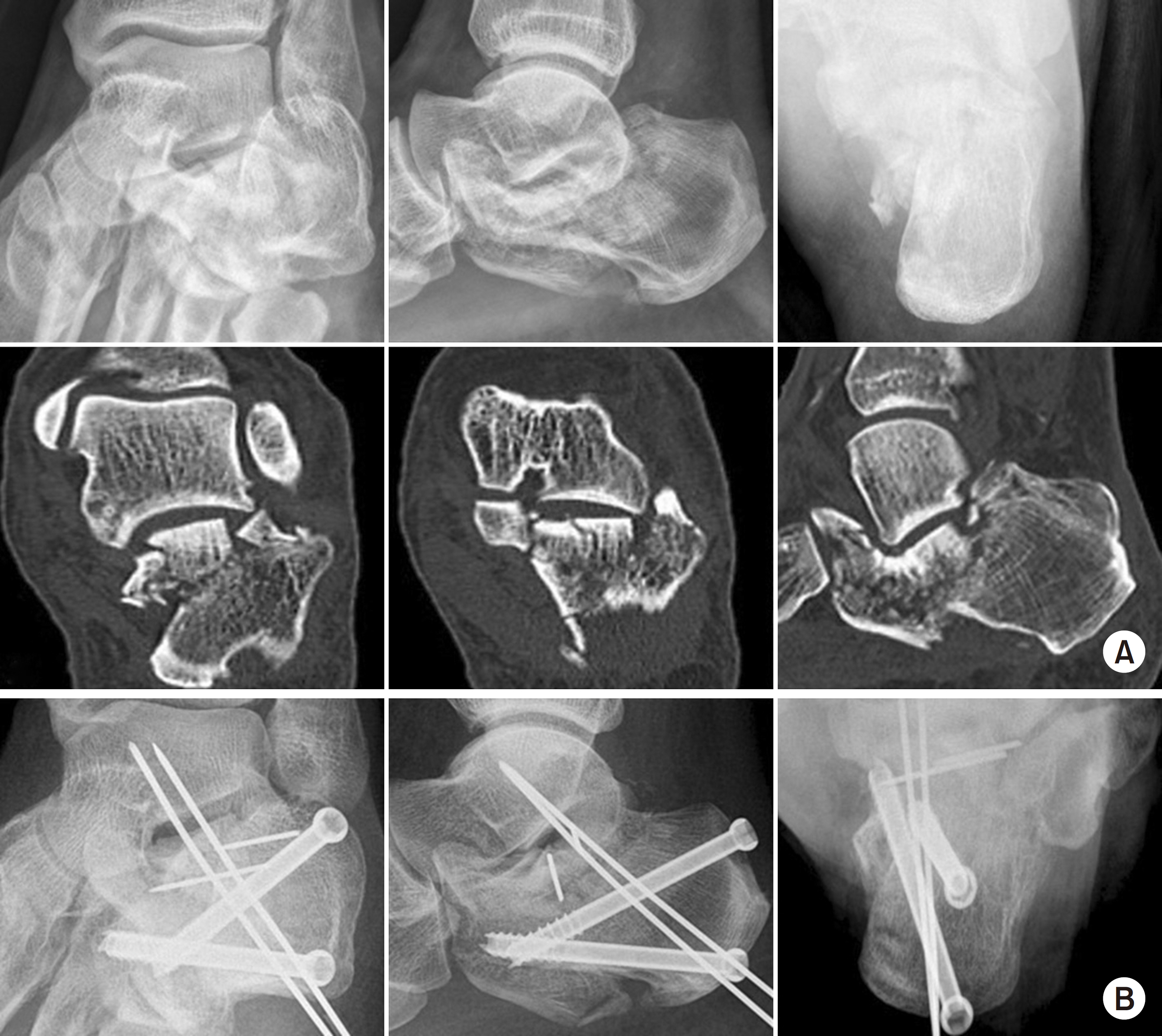
Fig. 1. (A) Preoperative X-ray and computed tomography showing a Sanders type IV and joint depressive type calcaneal fracture. (B) The postoperative X-ray showing restoration of the of Böhler's angle, Gissane's angle, and the calcaneus height, length, and width.
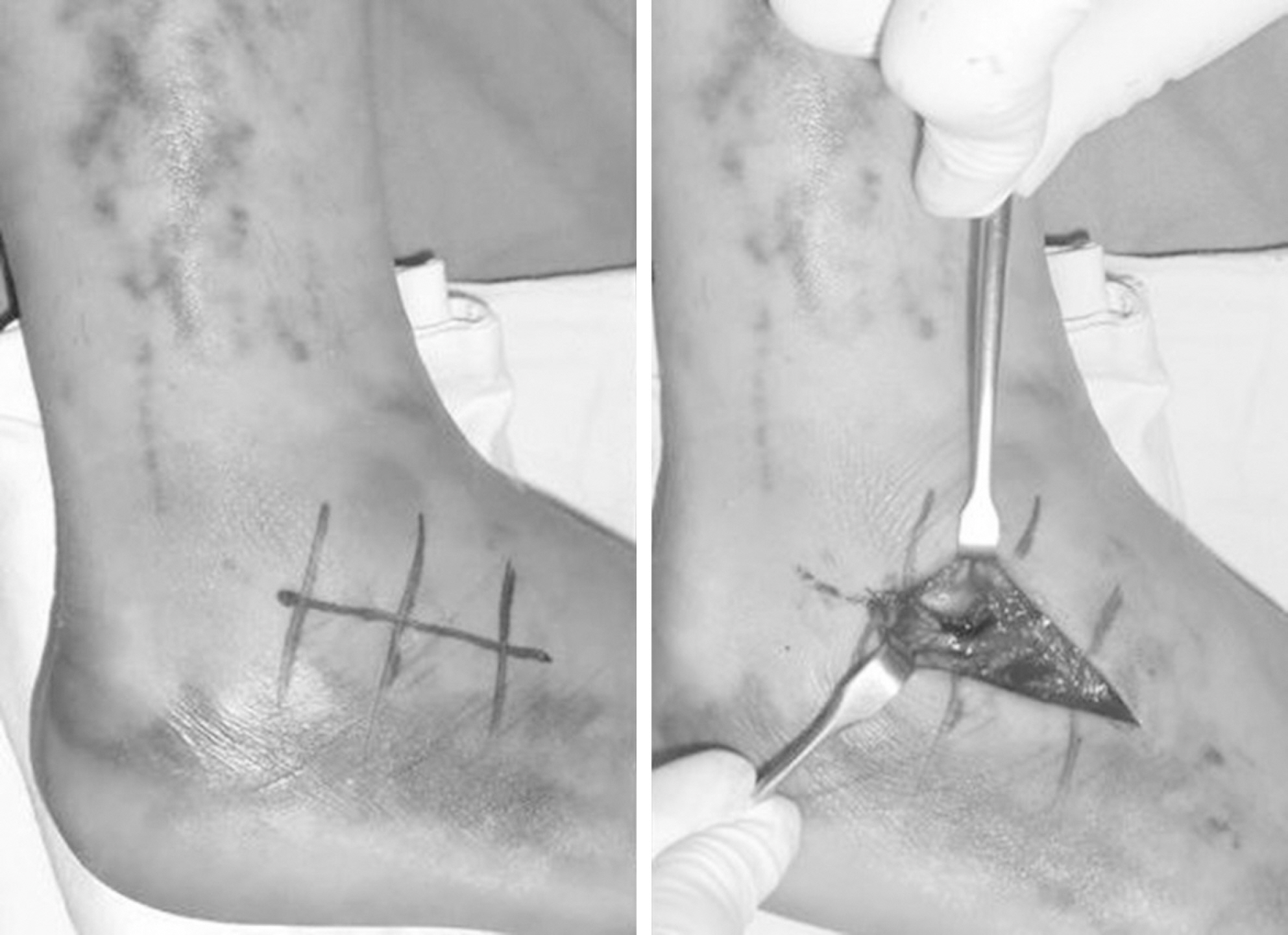
Fig. 2. Photograph showing the sinus tarsi approach.
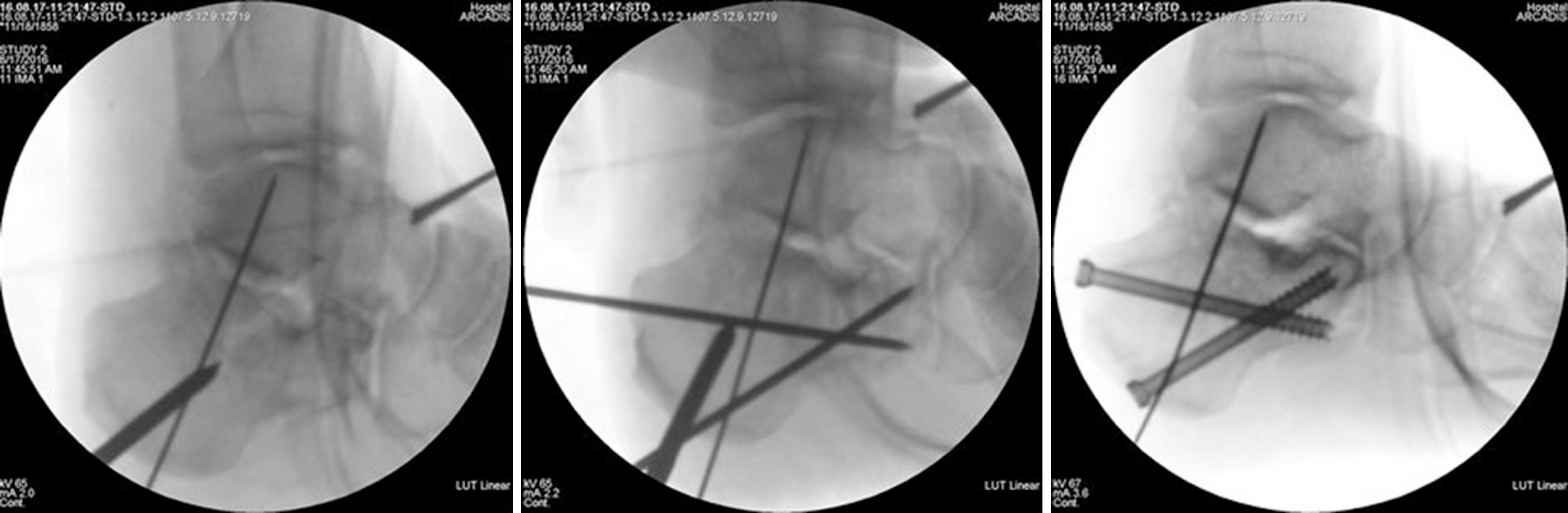
Fig. 3. Intraoperative fluoroscope images showing posterior facet reduction using Schantz pin and fixation with 6.5 mm cannulated screws and K-wire.
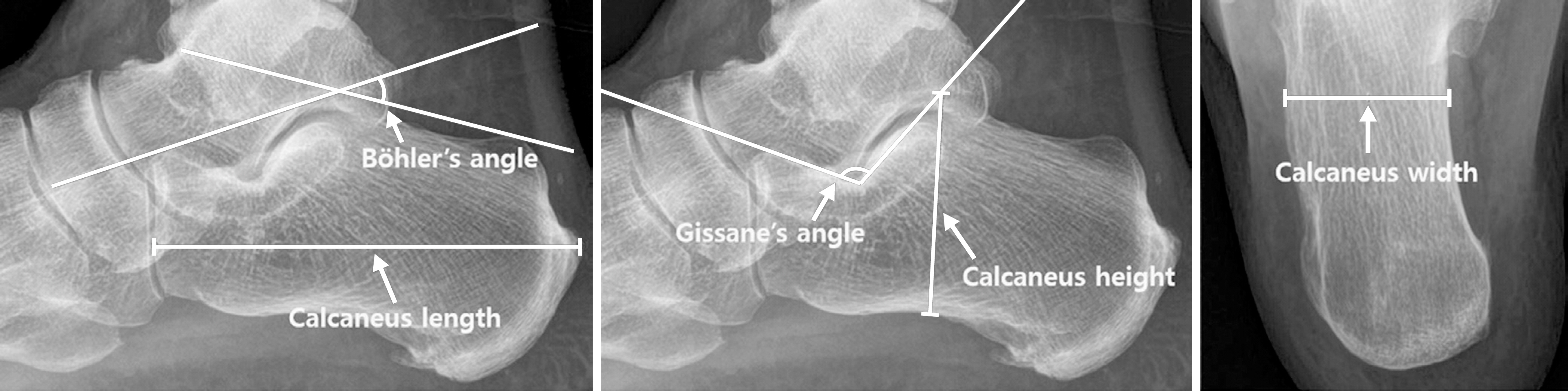
Fig. 4. Method of measurement of the calcaneal parameters.
Reference
-
References
1. Freund M, Thomsen M, Hohendorf B, Zenker W, Heller M. Optimized preoperative planning of calcaneal fractures using spiral computed tomography. Eur Radiol. 9:901–906. 1999.
Article2. Harvey EJ, Grujic L, Early JS, Benirschke SK, Sangeorzan BJ. Clinical Outcomes of Minimally Invasive Surgery in Sanders Type IV Intra-Articular Calcaneal Fractures Jun Young Lee, et al. Morbidity associated with ORIF of intra-articular calcaneus fractures using a lateral approach. Foot Ankle Int. 22:868–873. 2001.3. Essex-Lopresti P. The mechanism, reduction technique, and results in fractures of the os calcis. Br J Surg. 39:395–419. 1952.
Article4. Sanders R, Gregory P. Operative treatment of intra-articular fractures of the calcaneus. Orthop Clin North Am. 26:203–214. 1995.
Article5. Folk JW, Starr AJ, Early JS. Early wound complications of operative treatment of calcaneus fractures: analysis of 190 fractures. J Orthop Trauma. 13:369–372. 1999.
Article6. O'Farrell DA, O'Byrne JM, McCabe JP, Stephens MM. Fractures of the os calcis: improved results with internal fixation. Injury. 24:263–265. 1993.7. Paley D, Hall H. Intra-articular fractures of the calcaneus. A critical analysis of results and prognostic factors. J Bone Joint Surg Am. 75:342–354. 1993.
Article8. Sangeorzan BJ, Benirschke SK, Carr JB. Surgical management of fractures of the os calcis. Instr Course Lect. 44:359–370. 1995.9. Weber M, Lehmann O, Sägesser D, Krause F. Limited open reduction and internal fixation of displaced intra-articular fractures of the calcaneum. J Bone Joint Surg Br. 90:1608–1616. 2008.
Article10. Li LH, Guo YZ, Wang H, et al. Less wound complications of a sinus tarsi approach compared to an extended lateral approach for the treatment of displaced intraarticular calcaneal fracture: a randomized clinical trial in 64 patients. Medicine (Baltimore). 95:e4628. 2016.11. Ebraheim NA, Elgafy H, Sabry FF, Freih M, Abou-Chakra IS. Sinus tarsi approach with trans-articular fixation for displaced intra-articular fractures of the calcaneus. Foot Ankle Int. 21:105–113. 2000.
Article12. Mostafa MF, El-Adl G, Hassanin EY, Abdellatif MS. Surgical treatment of displaced intra-articular calcaneal fracture using a single small lateral approach. Strategies Trauma Limb Reconstr. 5:87–95. 2010.
Article13. Jia B, Li W, Liu Y, et al. Extensive lateral versus tarsal sinus approach to internal fixation for intra-articular calcaneal fractures. Int J Clin Exp Med. 10:15782–15788. 2017.14. SooHoo NF, Shuler M, Fleming LL. American Orthopaedic Foot and Ankle Society: Evaluation of the validity of the AOFAS Clinical Rating Systems by correlation to the SF-36. Foot Ankle Int. 24:50–55. 2003.
- Full Text Links
-
- Actions
-
Cited
- CITED
-
- Close
- Share
-
- Similar articles
-
- Outcomes of Minimally Invasive Surgery in Intra-Articular Calcaneal Fractures: Sanders Type III, Joint Depressive Type Calcaneal Fracture
- Minimally-invasive Percutaneous Screw Fixation of Displaced Intra-articular Calcaneal Fractures
- Clinical Results of Surgical Treatment with Minimally Invasive Percutaneous Plate Osteosynthesis for Displaced Intra-articular Fractures of Calcaneus
- Surgical Outcomes of Intra-articular Fractures of Calcaneus using AO Calcaneal Plate
- The Result Treated by Open Reduction and Internal Fixation with Minimally Invasive Technique in Joint Depressive Calcaneal Fracture
