Chronic Eosinophilic Pneumonia Masquerading As Bronchogenic Malignancy
- Affiliations
-
- 1Division of Pulmonary Medicine, Department of Internal Medicine, Wonkwang University School of Medicine, Iksan, Korea. eyesmile@wku.ac.kr
- KMID: 2468154
- DOI: http://doi.org/10.4068/cmj.2020.56.1.83
Abstract
- No abstract available.
MeSH Terms
Figure
-

FIG. 1 Chest X-ray indicates an ill-defined increased opacity at left upper lung (LUL) (A). Chest CT was revealed that air space consolidation and ground-glass opacity (GGO) at LUL. Furthermore, LUL lobar bronchus wall thickening and obliteration can be seen (B). Bronchoscopy revealed luminal impaction of the anterior segment of the LUL upper-division due to a mass forming lesion (C). Biopsy tissue sections featured diffuse eosinophilic infiltration (D). The biopsy sections were negative for CD56 staining (E). All biopsy sections underwent hematoxylin and eosin (H&E) staining, and photographs were taken at a magnification of 400×.
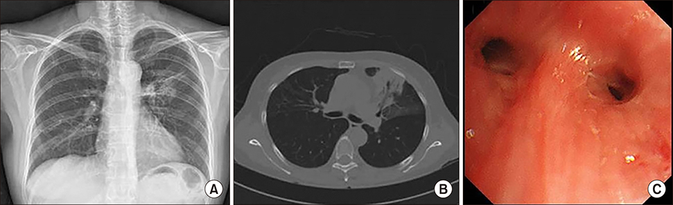
FIG. 2 Follow up chest X-ray showed a marked decrease in the extent of consolidation in the LUL (A). The post-treatment chest CT indicated a decrease in the extent of air space consolidation and GGO in the LUL and reduction of the endobronchial narrowing of the LUL lobar bronchus (B). Follow up bronchoscopy showed complete resolution of the bronchial obstruction, which was associated with the total luminal opening (C).
Reference
-
1. Marchand E, Cordier JF. Idiopathic chronic eosinophilic pneumonia. Semin Respir Crit Care Med. 2006; 27:134–141.
Article2. Kim NH, Lee KH, Kim JH, Cho JH, Kim L, Kim E. Bronchial involvement in chronic eosinophilic pneumonia: a case report. J Thorac Dis. 2015; 7:E97–E101.
