Schwannoma of the Tongue Base with Imaging Features and Differential Diagnosis: a Rare Case Report and Literature Review
- Affiliations
-
- 1Department of Radiology, Eulji University Hospital, Daejeon, Korea. mayon2@naver.com
- 2Department of Pathology, Eulji University Hospital, Daejeon, Korea.
- 3Department of Otolaryngology, Eulji University Hospital, Daejeon, Korea.
- 4Department of Otolaryngology, Bon ENT Clinic, Daejeon, Korea.
- KMID: 2468057
- DOI: http://doi.org/10.13104/imri.2019.23.4.385
Abstract
- Schwannoma or neurilemmoma is a benign peripheral nerve sheath tumor that arises from Schwann cells. Approximately 25-45% of all schwannomas occur in the head and neck regions, and the intraoral presentation of these is only 1%. We report a rare case of a patient presenting tongue base schwannoma with characteristic imaging features on computed tomography and magnetic resonance imaging.
MeSH Terms
Figure
-
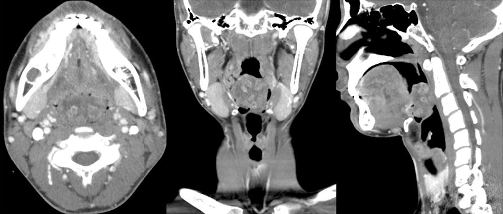
Fig. 1 Axial, coronal and sagittal contrast-enhanced computed tomography images reveal heterogeneously enhanced mass with internal multiple nodular rim enhancing foci, arising from the tongue base.
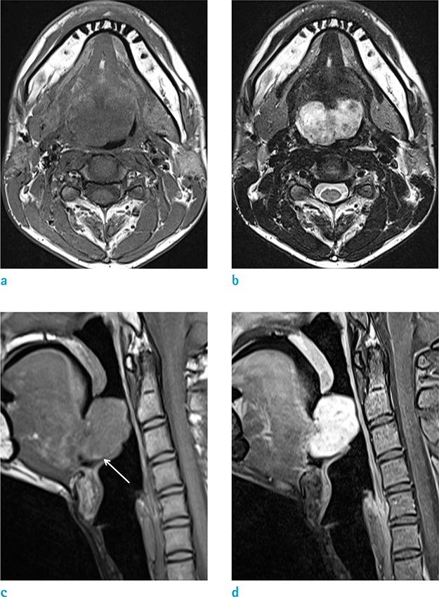
Fig. 2 The mass reveals generally iso signal intensities on T1WI (a) and heterogeneously high signal intensities with multiple internal ring-like lesions on T2WI (b). On sagittal T1WI (c), slightly high signal intensity at the peripheral portion of the mass (white arrow) is also noted. On the gadolinium-enhanced fat-suppressed T1WI (d), it shows bright and heterogeneous enhancement. T1WI = T1-weighted image; T2WI = T2-weighted image.

Fig. 3 Gross pathologic examination (a) reveals a well circumscribed brownish tan color mass. In the section, the mass was 42 × 40 × 30 mm sized in dimension. Hematoxylin and Eosin (H&E, × 100) staining shows that the tumor was composed of spindle cells and loose myxoid stroma (b). In immunostaining (S-100, × 200), the tumor cells revealed diffuse positivity (c).
Reference
-
1. Harada H, Omura K, Maeda A. A massive pleomorphic adenoma of the submandibular salivary gland accompanied by neurilemomas of the neck misdiagnosed as a malignant tumor: report of case. J Oral Maxillofac Surg. 2001; 59:931–935.2. Lee EY, Kim JJ, Seok H, Lee JY. Schwannoma of the tongue: a case report with review of literature. Maxillofac Plast Reconstr Surg. 2017; 39:17.3. Aref H, Abizeid GA. Axillary schwannoma, preoperative diagnosis on a tru-cut biopsy: case report and literature review. Int J Surg Case Rep. 2018; 52:49–53.4. Bhola N, Jadhav A, Borle R, Khemka G, Bhutekar U, Kumar S. Schwannoma of the tongue in a paediatric patient: a case report and 20-year review. Case Rep Dent. 2014; 2014:780762.5. Tsushima F, Sawai T, Kayamori K, Okada N, Omura K. Schwannoma in the floor of the mouth: a case report and clinicopathological studies of 10 cases in the oral region. J Oral Maxillofac Surg Med Pathol. 2012; 24:175–179.6. Nakazono T, White CS, Yamasaki F, et al. MRI findings of mediastinal neurogenic tumors. AJR Am J Roentgenol. 2011; 197:W643–W652.7. Badar Z, Farooq Z, Zaccarini D, Ezhapilli SR. Tongue base schwannoma: differential diagnosis and imaging features with a case presentation. Radiol Case Rep. 2016; 11:336–340.8. Dreher A, Gutmann R, Grevers G. Extracranial schwannoma of the ENT region. Review of the literature with a case report of benign schwannoma of the base of the tongue. HNO. 1997; 45:468–447.9. Cohen M, Wang MB. Schwannoma of the tongue: two case reports and review of the literature. Eur Arch Otorhinolaryngol. 2009; 266:1823–1829.10. de Bree R, Westerveld GJ, Smeele LE. Submandibular approach for excision of a large schwannoma in the base of the tongue. Eur Arch Otorhinolaryngol. 2000; 257:283–286.
