Histopathological Evaluation of the Efficacy for Plant-produced E2 Protein Vaccine against Classical Swine Fever Virus (CSFV) in Piglets
- Affiliations
-
- 1Department of Pathology, Dongnam Institute of Radiological and Medical Sciences, Busan 46033, Republic of Korea.
- 2Department of Clinical Laboratory Science, College of Health Sciences, Catholic University of Pusan, Busan 46252, Republic of Korea. kschang@cup.ac.kr
- 3Animal and Plant Quarantine and Inspection Agency (QIA), 177, Hyeoksin 8-ro, Gimcheon-si, Gyengsangbuk-do 39660 Republic of Korea.
- 4Department of Clinical Laboratory Science, College of Medical Sciences, Daegu Haany University, Gyeongsan 38610, Republic of Korea.
- 5BioApplications Inc., Pohang Techno Park Complex, 394 Jigok-ro Nam-gu, Pohang 37668, Korea.
- 6Division of Integrative Biosciences and Biotechnology, Pohang University of Science and Technology, Pohang 37673, Korea.
- KMID: 2468033
- DOI: http://doi.org/10.4167/jbv.2019.49.3.133
Abstract
- Classical swine fever (CSF), previously known as hog cholera, remains one of the most important swine-related contagious diseases worldwide. In order to eradicate classical swine fever virus (CSFV), it is commonly used in LOM-850 strain as a live attenuated CSF vaccine. However, there are symptoms of vaccination, such as the depression of feed intake, and difficulty of differentiation between infected and vaccinated hosts is impossible based on the antibodies induced. Nicotiana benthamiana were considered as an alternative to the production of recombinant vaccines on account of higher yields and levels of soluble protein than other models and crops in protein recombinant products. This study was conducted to evaluate histopathological validation of the plant-produced E2 fusion protein (ppE2) in piglets. The piglets were challenged by an injection of YC11WB strain in 7 days, 11 days and 14 days after one shot of the vaccination. The histopathological examination indicated that ppE2 can protect against lethal CSFV challenge at least 11 days of vaccination in piglets. These data suggest that the ppE2 can be an effective vaccine against CSFV in piglets.
Keyword
MeSH Terms
Figure
-
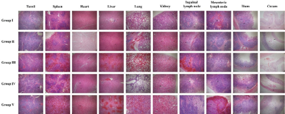
Figure 1 Histopathologic examination of classical swine fever virus-infected piglets, based on H&E stained tonsil section (×40). Congestion of the blood vessels (red arrow) and accumulation of cellular debris including neutrophil and lymphocytes (black arrow) were observed in the tonsil. Diffuse hemorrhages (black arrow) in the red pulp and depletion of the lymph follicles were observed in the spleen. Congestion of capillary (black arrow) was observed in the heart. Congestion of central vein (red arrow) and necrosis with lymphocytic infiltration in periportal areas (black arrow) were observed in the liver. Extensive hemorrhages in alveoli and interstitium, diffuse severe infiltration of mononuclear cells in alveoli, bronchi, and bronchiolar lumen were observed in the lung (black arrow). Congestion of the blood vessels (black arrow) and interstitial nephritis characterized by infiltration of mononuclear cells in the interstitial spaces were observed in the kidney. Hemorrhage was observed in the inguinal lymph node (black arrow). Hemorrhage was observed in the mesenteric lymph node (black arrow). Depletion of lymphocytes (black arrow) and infiltration of eosinophils in the lymphoid follicles and submucosal layer (red arrow) were observed in the ileum. Development of blood vessel was observed in the cecum (black arrow). Control group I; unvaccinated group, experimental group II (challenge at 7 days post vaccination); vaccinated with plant-produced E2 fusion protein, experimental group III (challenge at 11 days post vaccination); vaccinated with plant-produced E2 fusion protein, experimental group IV (challenge at 14 days post vaccination); vaccinated with plant-produced E2 fusion protein, experimental group V (challenge at 14 days post vaccination); vaccinated with LOM strain vaccine.
Reference
-
1. Laevens H, Koenen F, Deluyker H, de Kruif A. Experimental infection of slaughter pigs with classical swine fever virus: transmission of the virus, course of the disease and antibody response. Vet Rec. 1999; 145:243–248.
Article2. Fritzemeier J, Teuffert J, Greiser-Wilke I, Staubach C, Schlüter H, Moennig V. Epidemiology of classical swine fever in Germany in the 1990s. Vet Microbiol. 2000; 77:29–41.
Article3. Moennig V, Floegel-Niesmann G, Greiser-Wilke I. Clinical signs and epidemiology of classical swine fever: a review of new knowledge. Vet J. 2003; 165:11–20.
Article4. Rümenapf T, Unger G, Strauss JH, Thiel HJ. Processing of the envelope glycoproteins of pestiviruses. J Virol. 1993; 67:3288–3294.
Article5. Meyers G, Thiel HJ. Molecular characterization of pestiviruses. Adv Virus Res. 1996; 47:53–118.
Article6. Madera R, Gong W, Wang L, Burakova Y, Lleellish K, Galliher-Beckley A, et al. Pigs immunized with a novel E2 subunit vaccine are protected from subgenotype heterologous classical swine fever virus challenge. BMC Vet Res. 2016; 12:197.
Article7. Huang YL, Deng MC, Wang FI, Huang CC, Chang CY. The challenges of classical swine fever control: modified live and E2 subunit vaccines. Virus Res. 2014; 179:1–11.
Article8. Pauly T, Elbers K, König M, Lengsfeld T, Saalmüller A, Thiel HJ. Classical swine fever virus-specific cytotoxic T lymphocytes and identification of a T cell epitope. J Gen Virol. 1995; 76:3039–3049.
Article9. Sasahara J, Kumagai T, Shimizu Y, Furuuchi S. Field experiments of hog cholera live vaccine prepared in guinea-pig kidney cell culture. Natl Inst Anim Health Q (Tokyo). 1969; 9:83–91.10. Aynaud JM, Lejolly JC, Bibard C, Galicher C. [Studies of the properties of cold induced classical swine fever virus mutants. Application to vaccination]. Bull Off Int Epizoot. 1971; 75:654–659.11. Je SH, Kwon T, Yoo SJ, Lee DU, Lee S, Richt JA, et al. Classical Swine Fever Outbreak after Modified Live LOM Strain Vaccination in Naive Pigs, South Korea. Emerg Infect Dis. 2018; 24:798–800.
Article12. Hulst MM, Westra DF, Wensvoort G, Moormann RJ. Glycoprotein E1 of hog cholera virus expressed in insect cells protects swine from hog cholera. J Virol. 1993; 67:5435–5442.
Article13. Moormann RJ, Bouma A, Kramps JA, Terpstra C, De Smit HJ. Development of a classical swine fever subunit marker vaccine and companion diagnostic test. Vet Microbiol. 2000; 73:209–219.
Article14. van Rijn PA, Bossers A, Wensvoort G, Moormann RJ. Classical swine fever virus (CSFV) envelope glycoprotein E2 containing one structural antigenic unit protects pigs from lethal CSFV challenge. J Gen Virol. 1996; 77:2737–2745.
Article15. Aboul-Ata AA, Vitti A, Nuzzaci M, El-Attar AK, Piazzolla G, Tortorella C, et al. Plant-based vaccines: novel and low-cost possible route for Mediterranean innovative vaccination strategies. Adv Virus Res. 2014; 89:1–37.16. Lai H, Chen Q. Bioprocessing of plant-derived virus-like particles of Norwalk virus capsid protein under current Good Manufacture Practice regulations. Plant Cell Rep. 2012; 31:573–584.
Article17. Guan ZJ, Guo B, Huo YL, Guan ZP, Dai JK, Wei YH. Recent advances and safety issues of transgenic plant-derived vaccines. Appl Microbiol Biotechnol. 2013; 97:2817–2840.
Article18. Lau OS, Sun SSM. Plant seeds as bioreactors for recombinant protein production. Biotechnol Adv. 2009; 27:1015–1022.
Article19. Kumar GB, Ganapathi TR, Bapat VA. Production of hepatitis B surface antigen in recombinant plant systems: an update. Biotechnol Prog. 2007; 23:532–539.
Article20. Kumar GB, Srinivas L, Ganapathi TR, Bapat VA. Hepatitis B surface antigen expression in transgenic tobacco (Nicotiana tabacum) plants using four different expression cassettes. Plant Cell Tissue Organ Cult. 2006; 84:315–323.
Article21. Conley AJ, Zhu H, Le LC, Jevnikar AM, Lee BH, Brandle JE, et al. Recombinant protein production in a variety of Nicotiana hosts: a comparative analysis. Plant Biotechnol J. 2011; 9:434–444.
Article22. Park Y, An DJ, Choe S, Lee Y, Park M, Park S, et al. Development of Recombinant Protein-Based Vaccine Against Classical Swine Fever Virus in Pigs Using Transgenic Nicotiana benthamiana. Front Plant Sci. 2019; 10:624.
Article23. Moennig V, Floegel-Niesmann G, Greiser-Wilke I. Clinical signs and epidemiology of classical swine fever: a review of new knowledge. Vet J. 2003; 165:11–20.
Article24. Straw BE, Zimmerman JJ, D'Allaire S, Taylor DJ. Classical swine fever and other pestiviruses. Diseases of Swine. 9thed. Blackwell Publishing;2006. p. 309–322.25. Dunne HW. Hog cholera (European swine fever). Adv Vet Sci Comp Med. 1973; 17:315–359.26. Cha SH, Choi EJ, Park JH, Yoon SR, Kwon JH, Yoon KJ, et al. Phylogenetic characterization of classical swine fever viruses isolated in Korea between 1988 and 2003. Virus Res. 2007; 126:256–261.
Article27. Yoon SS, Byun JW, Bae YC, Roh IS, Lee KH, Yeo DY, et al. Retrospective pathologic study on the classical swine fever outbreak in Korea. Korean Journal of Veterinary Public Health. 2010; 34:117–123.28. Floegel-Niesmann G, Bunzenthal C, Fischer S, Moennig V. Virulence of recent and former classical swine fever virus isolates evaluated by their clinical and pathological signs. J Vet Med B Infect Dis Vet Public Health. 2003; 50:214–220.
Article29. Kim JS, Shon SG, Lee JH. Hisopathological Study on the Field Cases of Hog Cholera. J Inst Develop of Livestock Prod. 1992; 19:15–31.30. Patchimasiri T, Pinyochon W, Damrongwatanapokin S. Pathological study of chronic swine fever in experimental pigs. 24. Thai Veterinary Medical Association and 4. Bangkok(Thailand): Thailand Veterinary Practitioner Association Meeting;1998. 08. 5–7.31. Narita M, Kawashima K, Kimura K, Mikami O, Shibahara T, Yamada S, et al. Comparative immunohistopathology in pigs infected with highly virulent or less virulent strains of hog cholera virus. Vet Pathol. 2000; 37:402–408.
Article
- Full Text Links
-
- Actions
-
Cited
- CITED
-
- Close
- Share
-
- Similar articles
-
- The potential efficacy of the E2-subunit vaccine to protect pigs against different genotypes of classical swine fever virus circulating in Vietnam
- Toward the development of a one-dose classical swine fever subunit vaccine: antigen titration, immunity onset, and duration of immunity
- Establishment and application of a solid-phase blocking ELISA method for detection of antibodies against classical swine fever virus
- Epidemiological investigation and phylogenetic analysis of Classical Swine Fever virus in Yunnan province from 2015 to 2021
- Establishment and characterization of an infectious cDNA clone of a classical swine fever virus LOM strain
