J Korean Foot Ankle Soc.
2019 Dec;23(4):212-215. 10.14193/jkfas.2019.23.4.212.
Visually Indistinguishable Intractable Neuroma Management after Below Knee Amputation: A Case Report
- Affiliations
-
- 1Department of Orthopedic Surgery, Seoul Medical Center, Seoul, Korea. 711000e@naver.com
- 2Department of Rehabilitation, Seoul Medical Center, Seoul, Korea.
- KMID: 2465947
- DOI: http://doi.org/10.14193/jkfas.2019.23.4.212
Abstract
- Symptomatic neuromas after amputation can be troublesome to treat and make it difficult to properly fit a brace. Surgical management is required when conservative management such as prosthetic socket modification or local injections fail. However, small cutaneous nerves adhere to adjacent soft tissue and they are difficult to locate. The authors suggest that ultrasonography guided tattoo localization using a charcoal suspension is useful to find a visually indistinguishable neuroma.
Keyword
Figure
-
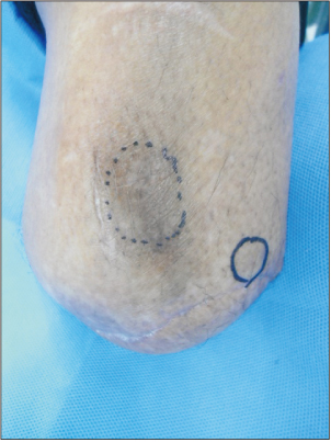
Figure 1 Preoperative photograph shows blue circle marked amputee end marked with Tinel's sign.
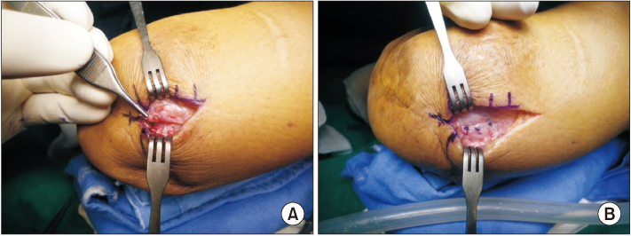
Figure 2 A cutaneous nerve on lateral side of the amputee end was dissected (A) and embedded into muscle layer (B).

Figure 3 A missed cutaneous nerve on anterior and medial side of the amputee end in first exploration (A) was marked with activated charcoal suspension assisted by ultrasonograph (B). (C) A preoperative ultrasonographic image shows tip of needle (arrow) approaching to nerve ending.
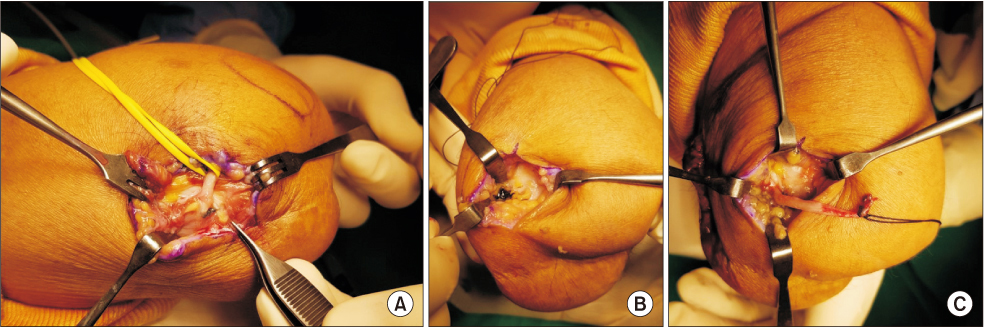
Figure 4 A cutaneous nerve was found near charcoal deposit on medial (A) and anterior side of amputee end (B). (C) This nerve was a L-shaped single branch.
Reference
-
1. Nashold BS Jr, Goldner JL, Mullen JB, Bright DS. Long-term pain control by direct peripheral-nerve stimulation. J Bone Joint Surg Am. 1982; 64:1–10.
Article2. Kim WH, Kim HJ, Kim SH, Jung JH, Park HY, Lee J, et al. Ultrasound-guided dual-localization for axillary nodes before and after neoadjuvant chemotherapy with clip and activated charcoal in breast cancer patients: a feasibility study. BMC Cancer. 2019; 19:859. DOI: 10.1186/s12885-019-6095-1.
Article3. Helvie MA, Ikeda DM, Adler DD. Localization and needle aspiration of breast lesions: complications in 370 cases. AJR Am J Roentgenol. 1991; 157:711–714. DOI: 10.2214/ajr.157.4.1892023.
Article4. Czarnecki DJ, Feider HK, Splittgerber GF. Toluidine blue dye as a breast blocalization marker. AJR Am J Roentgenol. 1989; 153:261–263. DOI: 10.2214/ajr.153.2.261.5. Saarela AO, Kiviniemi HO, Rissanen TJ. Preoperative methylene blue staining of galactographically suspicious breast lesions. Int Surg. 1997; 82:403–405.6. Sewak S, Graham P, Nankervis J. Tattoo allergy in patients receiving adjuvant radiotherapy for breast cancer. Australas Radiol. 1999; 43:558–561. DOI: 10.1046/j.1440-1673.1999.00733.x.
Article7. Soran A, Kanbour-Shakir A, Bas O, Bonaventura M. A tattoo pigmented node and breast cancer. Bratisl Lek Listy. 2014; 115:311–312. DOI: 10.4149/BLL_2014_063.
Article8. Lager DJ, O'Connor JC, Robinson RA, Brown RC, Urdaneta LF. Factitious microcalcifications in breast biopsy material: laboratory-induced error by use of tattoo powder for specimen mammography. J Surg Oncol. 1989; 40:281–282. DOI: 10.1002/jso.2930400415.
Article9. Vilska J. [Indications and contra-indications of activated charcoal in a poison control center. The viewpoint of the Poison Control Center of Finland]. J Toxicol Clin Exp. 1989; 9:295–298. French.10. Sperry K. Tattoos and tattooing. Part i: history and methodology. Am J Forensic Med Pathol. 1991; 12:313–319. DOI: 10.1097/00000433-199112000-00008.
- Full Text Links
-
- Actions
-
Cited
- CITED
-
- Close
- Share
-
- Similar articles
-
- Spinal cord stimulation for the treatment of intractable stump pain after amputation of the both lower limbs 36 years ago: A case report
- Treatment of Recurrent Neuroma after Forearm Amputation: End to End Neurorrhaphy
- Pulsed Radiofrequency Ablation Under Ultrasound Guidance for Huge Neuroma
- Amputation Neuroma Mimicking Common Bile Duct Cancer: A Case Report
- Statistical Survey on the Amputees
