Epithelial-Myoepithelial Carcinoma of the Inferior Turbinate: A Case Report
- Affiliations
-
- 1Department of Otorhinolaryngology-Head and Neck Surgery, Soonchunhyang University College of Medicine, Bucheon Hospital, Bucheon, Korea. handsomemd@naver.com
- 2Department of Pathology, Soonchunhyang University College of Medicine, Bucheon Hospital, Bucheon, Korea. mj@schmc.ac.kr
- KMID: 2465543
- DOI: http://doi.org/10.18787/jr.2019.26.2.113
Abstract
- Epithelial-myoepithelial carcinoma (EMC) is a rare and low-grade malignant salivary gland tumor including epithelial and myoepithelial components. EMC frequently arises in the parotid gland but infrequently originates from the salivary glands of the nasal cavity. Here, we report the case of an EMC arising from the inferior turbinate, one of the most uncommon sites. A 60-year-old female patient presented with left nasal obstruction for several months, and PNS CT showed an about 4×1.4-cm-sized heterogeneously enhancing polypoid mass originating from the inferior turbinate of the left nasal cavity. After surgical treatment, the patient was diagnosed with EMC based on pathologic examinations including histopathological and immunohistochemical tests. We report a case of a patient with EMC in the inferior turbinate who was observed over 18 months without radiation therapy after successful wide excision.
MeSH Terms
Figure
-
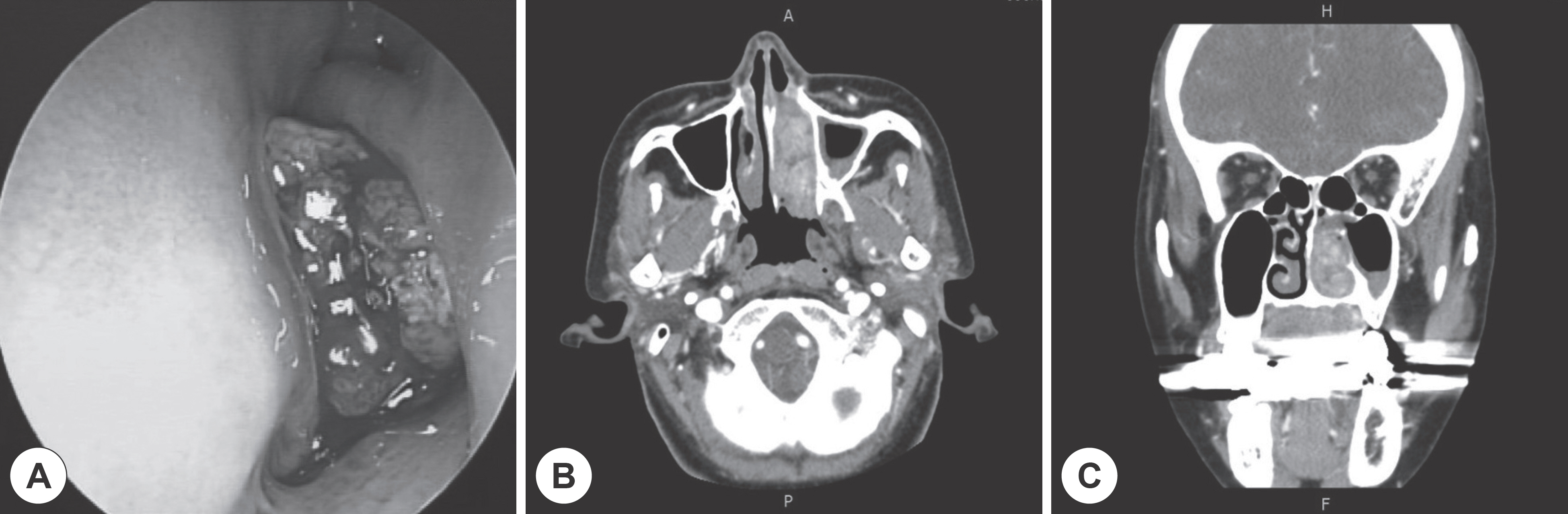
Fig. 1. Preoperative endoscopic and CT findings. (A) Preoperative endoscopic finding presents bloody red-colored, necrotic mass originating from inferior turbinate of left nasal cavity. (B) Axial and (C) coronal paranasal sinus CT images present about 4×1.4 cm sized heterogeneously enhancing polypoid mass originated from left inferior turbinate.

Fig. 2. Microscopic findings. (A) The polypoid tumor shows predominantly biphasic tubular histology, which are characteristic features of epithelial-myoepithelial carcinoma (H&E, ×40). (B) The inner luminal layer is formed by more hyperchromatic ductal cells and the outer layer is formed by myoepithelial cells with indistinct borders. Mitotic features are frequently identified (B, H&E, ×100). (C, D) Cyto-keratin and p63 immunostains highlight luminal-ductal cells and myoepithelial cells, respectively. This biphasic appearance of immu-nohistochemical stain supports the diagnosis of epithelial-myoepithelial carcinoma ([ C], Cytokeratin immunostain, ×40; [ D], p63 im-munostain, ×40).
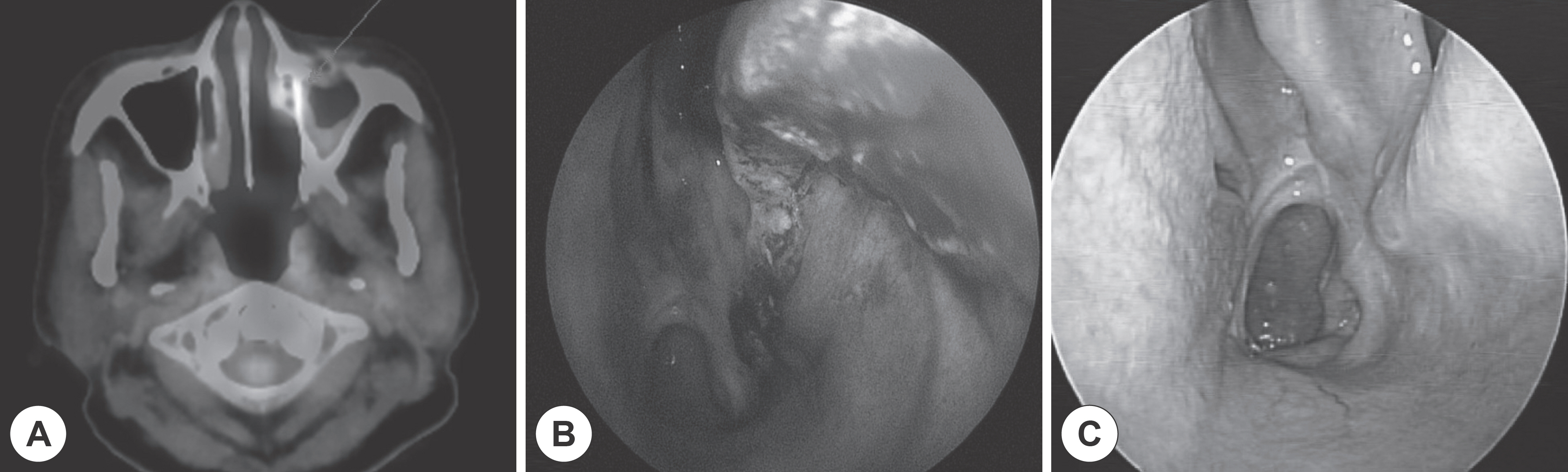
Fig. 3. Postoperative PET-CT and endoscopic findings. (A) Focal hypermetabolism of the left nasal cavity in PET-CT findings after 1st op-eration. (B) 2nd operation was performed due to the possibility of residual tumor. (C) There was no any local recurrence in the follow-up for 18 months.
Reference
-
References
1. Hamper K, Brügmann M, Koppermann R, Caselitz J, Arps H, Asken-sten U, et al. Epithelial-myoepithelial duct carcinoma of salivary glands: a follow-up and cytophotometric study of 21 cases. J Oral Pathol Med. 1989; 18(5):299–304.
Article2. Simpson RH, Clarke TJ, Sarsfield PT, Gluckman PG. Epithelial-myoepithelial carcinoma of salivary glands. J Clin Pathol. 1991; 44(5):419–23.
Article3. Schuman TA, Kimple AJ, Edgerly CH, Ebert CS, Zanation AM, Thorp BD. Sinonasal epithelial-myoepithelial carcinoma: Report of a novel subsite and review of the literature. Allergy Rhinol (Prov-idence). 2018; 9:2152656718764229.
Article4. Savera AT, Sloman A, Huvos AG, Klimstra DS. Myoepithelial carcinoma of the salivary glands: a clinicopathologic study of 25 patients. Am J Surg Pathol. 2000; 24(6):761–74.5. Bong JP, Park JH, Choi HM, Kim JH, Lee KK, Eom MS. A case of malignant myoepithelioma in parotid gland. Korean J Otolaryngol-Head Neck Surg. 2002; 45(6):624–7.6. Magliulo G, Pulice G, Fusconi M, Cuiuli G. Malignant myoepithelioma of the rhinopharynx: case report. Skull Base. 2005; 15(2):113–6. ; discussion 117.
Article7. Lee HM, Choi CS, Kim A, Lee SH. Epithelial-myoepithelial carcinoma arising in the nasal cavity-immunohistochemical and electron microscopic study. Korean J Otolaryngol-Head Neck Surg. 2000; 43(4):383–6.8. Yoo SD, Shim WS, Kim IK, Song HG. Myoepithelial Carcinoma Originated from the Maxillary Sinus. Korean J Otorhinolaryngol-Head Neck Surg. 2008; 51(2):191–3.9. Cho KS, Shin SC, Mun MJ, Roh WJ. A Case of Myoepithelial Car-cinoma Originated from Inferior Turbinate. Korean J Otorhinolaryngol-Head Neck Surg. 2010; 53(12):791–4.
Article10. Flam JO, Brook CD, Sobel R, Lee JC, Platt MP. Nasal epithelial myoepithelial carcinoma: An unusual cause of epiphora, a case report and review of the literature. Allergy Rhinol (Providence). 2015; 6(2):133–7.
Article11. Park JO, Jung CK, Sun DI, Kim MS. An unusual presentation of aggressive epithelial-myoepithelial carcinoma of the nasal cavity with high-grade histology. J Laryngol Otol. 2011; 125(12):1286–9.
Article12. Yamanegi K, Uwa N, Hirokawa M, Ohyama H, Hata M, Yamada N, et al. Epithelial-myoepithelial carcinoma arising in the nasal cavity. Auris Nasus Larynx. 2008; 35(3):408–13.
Article
- Full Text Links
-
- Actions
-
Cited
- CITED
-
- Close
- Share
-
- Similar articles
-
- A Case of Myoepithelial Carcinoma Originated from Inferior Turbinate
- Epithelial-myoepithelial carcinoma arising in pleomorphic adenoma of palate
- Epithelial-Myoepithelial Carcinoma of Intercalated Duct of Parotid Gland
- Fine Needle Aspiration Cytology of Metastatic Epithelial-Myoepithelial Carcinoma of the Scalp: A Case Report
- Epithelial-Myoepithelial Carcinoma of the Lung: one case report
