Salvage Procedure of a Lower Leg in a Contact Burn: Latissimus Dorsi Muscle Free Flap and Scalp Skin Graft
- Affiliations
-
- 1Department of Burn Reconstructive Surgery, Bestian Seoul Hospital, Seoul, Korea. sjoh46@nate.com
- 2Department of Burn Surgery, Bestian Seoul Hospital, Seoul, Korea.
- KMID: 2464475
- DOI: http://doi.org/10.12790/ahm.2019.24.4.381
Abstract
- Intentional carbon monoxide (CO) poisoning has become rapidly increased occurrence, also if the surviving patient is burnt and unconscious, the patient's burn wound is likely to be severely deep and to need flap surgery. A 44-year-old female patient suffered from a deep contact burn to the left posterior lower leg during intentional CO poisoning. The patient underwent escharotomy and treatment with a latissimus dorsi muscle neuro-vascularized free flap and scalp skin grafts. While harvesting 0.008 inches (300 cm²) of the scalp skin, some 25 cm² of the skin was accidentally harvested as a thick split-thickness skin graft (SSG) from the right occipital scalp. Interfollicular epidermal and dermal regeneration was achieved at eleven days postoperatively at the thick SSG donor site. The patient's deep burn wound was reconstructed by two staged surgeries. The patient was able to walk with the help of a supportive shoe one year postoperatively.
Keyword
MeSH Terms
Figure
-
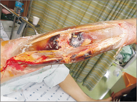
Fig. 1 Fasciotomy of a severe burn wound involving the deep muscles of the posterior aspect of the left lower leg.
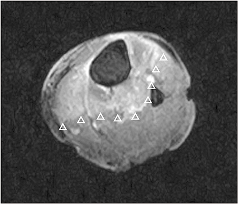
Fig. 2 Water-fat saturated T1-weighted magnetic resonance imaging of the proximal 1/3 of the left lower leg showing the border of the muscle damage marked with arrowheads.
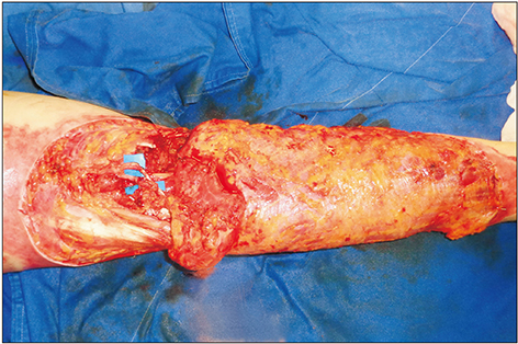
Fig. 3 Neuro-vascular microanastomosis area showing a latissimus dorsi muscle flap.

Fig. 4 Thick skin graft with a histological thickness of 0.035 inches harvested at an incorrect depth setting (H&E, ×40).

Fig. 5 Scalp wound of the 0.035-inch-thick split-thickness skin graft donor site in the right occipital area showing complete epithelization 11 days after surgery.
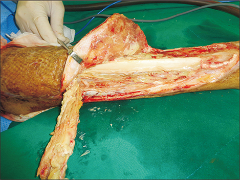
Fig. 6 Re-exploration of the wound showing granulation tissue under the surface of the free flap, residual damaged muscle in the anterior compartment and exposed fibular bone 64 days after the first surgery.
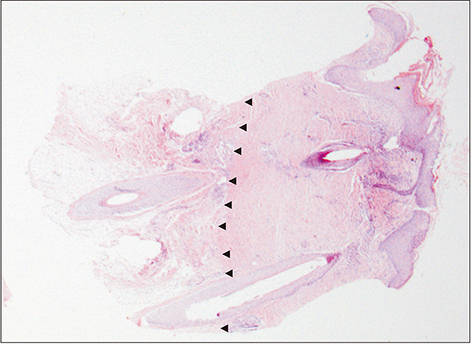
Fig. 7 Regenerated tissue of the deep donor wound 64 days after surgery (H&E, ×12.5); arrowheads mark the border between residual reticular and regenerated fibrotic tissue.

Fig. 8 The inconspicuous scar-less healing of scalp donor sites harvested with thick split-thickness skin one year after surgery.
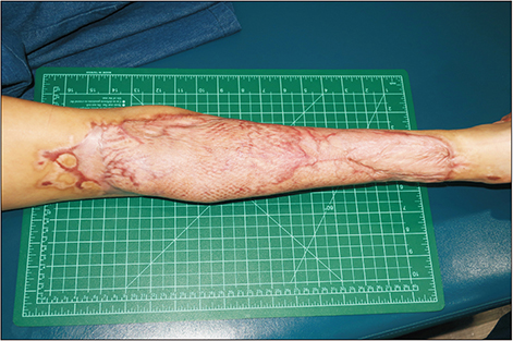
Fig. 9 The reconstructed contour of the posterior aspect of the left leg one year after surgery.
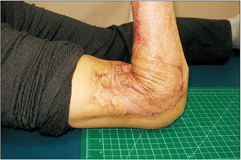
Fig. 10 Full flexion appearance of the lateral aspect of the left knee one year after surgery.
Reference
-
1. Chang SS, Chen YY, Yip PS, Lee WJ, Hagihara A, Gunnell D. Regional changes in charcoal-burning suicide rates in East/Southeast Asia from 1995 to 2011: a time trend analysis. PLoS Med. 2014; 11:e1001622.
Article2. Choi YR, Cha ES, Chang SS, Khang YH, Lee WJ. Suicide from carbon monoxide poisoning in South Korea: 2006-2012. J Affect Disord. 2014; 167:322–325.
Article3. Chai J, Song H, Sheng Z, Chen B, Yang H, Li L. Repair and reconstruction of massively damaged burn wounds. Burns. 2003; 29:726–732.
Article4. Rednam RS, Rinker BD. Reconstruction of posterior compartment of lower extremity using a functional latissimus dorsi free flap: a case report. Microsurgery. 2016; 36:77–80.
Article5. Crawford BS. An unusual skin donor site. Br J Plast Surg. 1964; 17:311–313.
Article6. Gyger D, Genin B, Bugmann P, Lironi A, Coultre CL. Skin harvesting on the scalp in children: utopia or reality. Eur J Pediatr Surg. 1996; 6:166–169.
Article7. Weyandt GH, Bauer B, Berens N, Hamm H, Broecker EB. Split-skin grafting from the scalp: the hidden advantage. Dermatol Surg. 2009; 35:1873–1879.8. Chang LY, Yang JY, Chuang SS, Hsiao CW. Use of the scalp as a donor site for large burn wound coverage: review of 150 patients. World J Surg. 1998; 22:296–299. discussion 299-300.
Article9. Malpass KG, Snelling CF, Tron V. Comparison of donorsite healing under Xeroform and Jelonet dressings: unexpected findings. Plast Reconstr Surg. 2003; 112:430–439.
Article10. McBride CA, Kempf M, Kimble RM, Stockton K. Variability in split-thickness skin graft depth when using an air-powered dermatome: a paediatric cohort study. Burns. 2017; 43:1552–1560.
Article
- Full Text Links
-
- Actions
-
Cited
- CITED
-
- Close
- Share
-
- Similar articles
-
- Neovascularization in the "Cross-Leg Fashioned" Muscular Free Flap
- Reconstruction of Extensive Lower Extermity Soft Tissue Defect Using Free Latissimus Dorsi Muscle Flap with STSG
- Reconstruction of Knee in Long - term Flexion Contracture Using Latissimus Dorsi Flap
- Reconstruction of Midface Defect with Latissimus Dorsi Myocutaneous Free Flap
- A Retrospective Study for Method and Timing of Reconstruction Using Free Latissimus Dorsi Muscle Flap at Foot in Electrical Burn Patients
