Hair Follicle Nevus Located on the Neck: Comparison with Accessory Tragus, Cervical Chondrocutaneous Branchial Remnants and Trichofolliculoma
- Affiliations
-
- 1Department of Dermatology, SMG-SNU Boramae Medical Center, Seoul, Korea. sycho@snu.ac.kr
- KMID: 2461989
- DOI: http://doi.org/10.5021/ad.2019.31.6.662
Abstract
- Hair follicle nevus (HFN) is a rare, benign, follicular hamartoma that most frequently presents as a congenital nodule on the face. We experienced a rare case of HFN on the neck of a 14-year-old boy and performed a pilot immunohistochemical study with cytokeratin 19 (CK19) to compare the staining pattern of hair follicles in HFN and its differential diagnoses, accessory tragus, cervical chondrocutaneous branchial remnants (CCBR) and trichofolliculoma. With hematoxylin and eosin stain, HFN showed numerous tiny hair follicles in the dermis with several sebaceous and eccrine glands, and perifollicular fibrous thickening. With CK19 stain, some hair follicles in HFN and CCBR showed positive expression, a few hair follicles in accessory tragus showed weak expression, and no hair follicles in trichofolliculoma showed expression. The present report supports the view that HFN, accessory tragus and CCBR are within the same spectrum of hamartomas.
Keyword
MeSH Terms
Figure
-
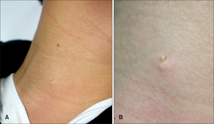
Fig. 1 Two separate papules on the right neck. (A) Asymptomatic and soft pedunculated skin-colored papule on the right lower neck and red-colored papule with bleeding tendency on the right upper neck. (B) Closer view of the lower flesh-colored lesion.
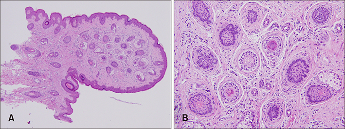
Fig. 2 Histopathology of the lower skin-colored papule. (A) Numerous vellus hair follicles are located in the dermis, and several sebaceous and eccrine glands are connected to the hair follicles (H&E, ×40). (B) Perifollicular stroma shows fibrous thickening (H&E, ×200).
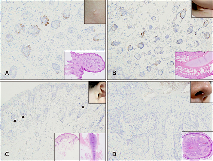
Fig. 3 Cytokeratin 19 (CK19) expression in the hair follicles in hair follicle nevus (HFN) (14-year-old), accessory tragus (8-year-old), cervical chondrocutaneous branchial remnants (CCBR) (24-month-old), and trichofolliculoma (37-year-old) (upper right insets, clinical pictures; lower right insets, hematoxylin and eosin-stained pictures in 40 fold magnification). (A, B) Some hair follicles in HFN and CCBR showed positive expression of CK19 (CK19, ×100). (C) Few hair follicles in accessory tragus showed weak expression of CK19 (triangles) (CK19, ×100). (D) Hair follicles in trichofolliculoma showed no expression of CK19 (CK19, ×100).
Reference
-
1. Larson KN, O'Shea P, Zedek DC, Morrell DS. Hair follicle nevus located on the chin of an infant: case report and review of literature. Pediatr Dermatol. 2016; 33:e106–e108.
Article2. Karabulut YY, Şenel E, Karabulut HH, Dölek Y. Three different clinical faces of the same histopathological entity: hair follicle nevus, trichofolliculoma and accessory tragus. An Bras Dermatol. 2015; 90:519–522.
Article3. Nagase K, Nagase K, Misago N, Narisawa Y. A preauricular hairy papule in an infant: hair follicle nevus closely similar to accessory tragus. Arch Dermatol. 2012; 148:266–268.
Article4. Asahina A, Mitomi H, Sakurai N, Fujita H. Multiple accessory tragi without cartilage: relationship with hair follicle naevi? Acta Derm Venereol. 2009; 89:316–317.
Article5. Jedrych J, Akilov O, Gehris R, Ho J. Hair follicle nevus of the abdominal skin: an unusual extracephalic presentation. Pediatr Dermatol. 2014; 31:e85–e86.
Article6. Davis DA, Cohen PR. Hair follicle nevus: case report and review of the literature. Pediatri Dermatol. 1996; 13:135–138.
Article7. Serra-Guilléna C, Travesb V, Echeverriaa B, Martorell A. Hair follicle nevus: a case report and review of the literature. Actas Dermosifiliogr. 2009; 100:822–824.
Article8. Motegi S, Amano H, Tamura A, Ishikawa O. Hair follicle nevus in a 2-year old. Pediatr Dermatol. 2008; 25:60–62.
Article9. Begovic N, Simic R, Vlahovic A, Kravljanac D, Djuricic S, Mijovic T. Cervical chondrocutaneous branchial remnants--report of 17 cases. Int J Pediatr Otorhinolaryngol. 2014; 78:1961–1964.10. Ackerman AB, Viragh PAD, Chongchitnant N. Neoplasms with follicular differentiation. Philadelphia: Lea & Febiger;1993. p. xviii–703.11. Michel M, Török N, Godbout MJ, Lussier M, Gaudreau P, Royal A, et al. Keratin 19 as a biochemical marker of skin stem cells in vivo and in vitro: keratin 19 expressing cells are differentially localized in function of anatomic sites, and their number varies with donor age and culture stage. J Cell Sci. 1996; 109 (Pt 5):1017–1028.
Article12. Misago N, Kimura T, Toda S, Mori T, Narisawa Y. A revaluation of trichofolliculoma: the histopathological and immunohistochemical features. Am J Dermatopathol. 2010; 32:35–43.
Article
