J Korean Assoc Oral Maxillofac Surg.
2019 Feb;45(1):43-47. 10.5125/jkaoms.2019.45.1.43.
Nasal septum angiofibroma: a rare condition with an unusual onset
- Affiliations
-
- 1ENT Clinic, S.M. alle Scotte University Hospital of Siena, Siena, Italy. mariacarla.spinosi@gmail.com
- 2Institute of Pathologic Anatomy and Histopathology, University of Siena, Siena, Italy.
- KMID: 2461491
- DOI: http://doi.org/10.5125/jkaoms.2019.45.1.43
Abstract
- The characteristics of extra-nasopharyngeal angiofibromas tend to be different from angiofibromas of the nasopharynx according to patient gender, patient age, prevalence, affected site, pathogenesis, and clinical and epidemiological features. We report a case of an extra-nasopharyngeal angiofibroma in a 28-year-old man referred to the ENT Clinic for right-sided epistaxis, airflow impairment and nasal swelling. The right nostril was completely occluded works by a reddish-yellow mass that bled easily. The computed tomography scan revealed an "inhomogeneous solid lesion in the nasal fossa". With the patient under general anesthesia, the formation in the anterior portion of the right side of the nasal septum was removed up to its vascular base. Although electrical cauterization efficiently controlled the bleeding, we abraded the sub-perichondral area to prevent further bleeding as well as recurrence. The histological exam report confirmed the diagnosis of angiofibroma. As in our case, epistaxis is commonly the presenting sign of angiofibroma. Yet its onset was peculiar, given that the bleeding started with a low impact trauma. The nasal swelling was also a relevant feature as well as the breathing impairment. Although uncommon, nasal septal angiofibromas should considered in patients with epistaxis.
Keyword
Figure
-
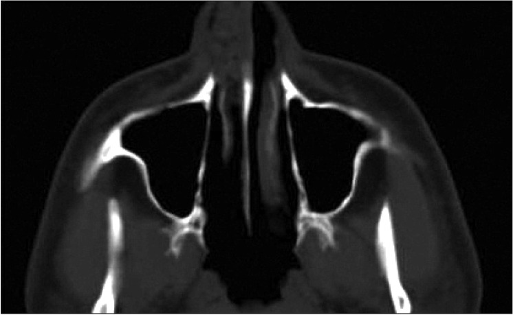
Fig. 1 Computed tomography scan of the nasal angiofibroma.
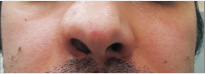
Fig. 2 Angiofibroma; frontal view.
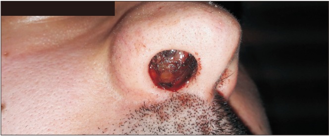
Fig. 3 Angiofibroma; inferolateral view.
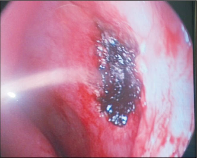
Fig. 4 Endoscopic view of the nasal fossa after surgical removal of the angiofibroma.
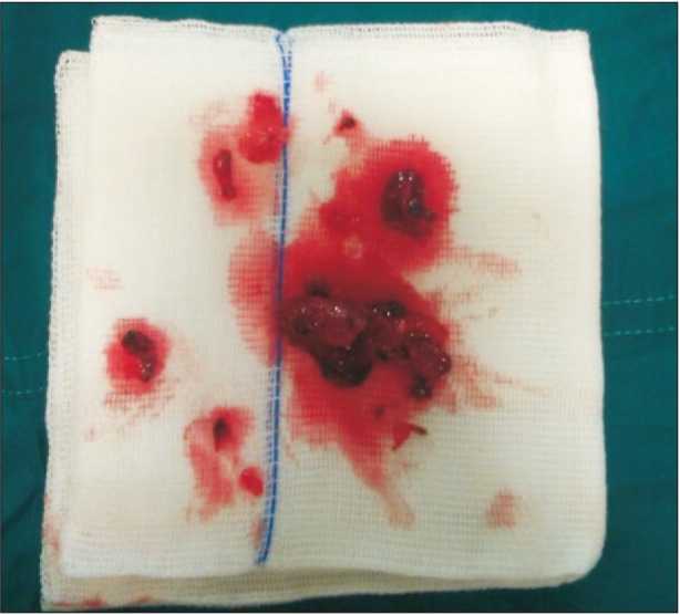
Fig. 5 Specimen collection.

Fig. 6 Histologic sample (H&E staining, ×5).

Fig. 7 Histological sample (CD34 staining, ×5).
Reference
-
1. Barnes L, Brandwein M, Som PM. Diseases of the nasal cavity, paranasal sinuses, and nasopharynx. In : Barnes L, editor. Surgical pathology of the head and neck. New York: Marcel Dekker;2001. p. 440–516.2. Doğan S, Yazici H, Baygit Y, Metin M, Soy FK. Extranasopharyngeal angiofibroma of the nasal septum: a rare clinical entity. J Craniofac Surg. 2013; 24:e390–e393. PMID: 23851880.3. Windfuhr JP, Remmert S. Extranasopharyngeal angiofibroma: etiology, incidence and management. Acta Otolaryngol. 2004; 124:880–889. PMID: 15513521.
Article4. Sarpa JR, Novelly NJ. Extranasopharyngeal angiofibroma. Otolaryngol Head Neck Surg. 1989; 101:693–697. PMID: 2556678.
Article5. Ewe S, Dayana F, Fadzilah FM, Gendeh BS. Nasal septal angiofibroma in a post-menopausal woman: a rare entity. J Clin Diagn Res. 2015; 9:MD03–MD05.
Article6. Windfuhr JP, Remmert S. [Extranasopharyngeal angiofibroma of the nasal cavity and paranasal sinuses]. Laryngorhinootologie. 2004; 83:308–316. German. PMID: 15143448.7. Perić A, Sotirović J, Cerović S, Zivić L. Immunohistochemistry in diagnosis of extranasopharyngeal angiofibroma originating from nasal cavity: case presentation and review of the literature. Acta Medica (Hradec Kralove). 2013; 56:133–141. PMID: 24693794.
Article8. Correia FG, Simões JC, Mendes-Neto JA, Seixas-Alves MT, Gregório LC, Kosugi EM. Extranasopharyngeal angiofibroma of the nasal septum--uncommon presentation of a rare disease. Braz J Otorhinolaryngol. 2013; 79:646. PMID: 24141686.9. Garcia-Rodriguez L, Rudman K, Cogbill CH, Loehrl T, Poetker DM. Nasal septal angiofibroma, a subclass of extranasopharyngeal angiofibroma. Am J Otolaryngol. 2012; 33:473–476. PMID: 21978647.
Article10. Fassih M, Taali L, Abada A, Rouadi S, Roubal M, Mahtar M, et al. [Vascular tumors of the nasal cavities: a retrospective study of 10 cases]. Rev Laryngol Otol Rhinol (Bord). 2012; 133:87–92. French. PMID: 23393743.11. Tasca I, Compadretti GC. Extranasopharyngeal angiofibroma of nasal septum. A controversial entity. Acta Otorhinolaryngol Ital. 2008; 28:312–314. PMID: 19205598.12. Thomas RL. Computed tomography in the assessment of patients with juvenile post-nasal angiofibroma. J Otolaryngol. 1980; 9:334–341. PMID: 6252326.13. Briant TD, Fitzpatrick PJ, Berman J. Nasopharyngeal angiofibroma: a twenty year study. Laryngoscope. 1978; 88:1247–1251. PMID: 209265.14. Hamdan AL, Moukarbel RV, Kattan M, Natout M. Angiofibroma of the nasal septum. Middle East J Anaesthesiol. 2012; 21:653–655. PMID: 23327044.15. Son YH, Baik SK, Kang MS, Kim YD. Recurrent arteriovenous malformation on palate after embolization combined surgical resection: preoperative magnetic resonance features and intraoperative angiographic findings. J Korean Assoc Oral Maxillofac Surg. 2015; 41:346–351. PMID: 26734564.
Article16. Atmaca S, Bayraktar C, Yıldız L. Extranasopharyngeal angiofibroma of the posterior nasal septum: a rare clinical entity. Kulak Burun Bogaz Ihtis Derg. 2013; 23:295–298. PMID: 24010805.
Article17. Hiraide F, Matsubara H. Juvenile nasal angiofibroma: a case report. Arch Otorhinolaryngol. 1984; 239:235–241. PMID: 6329151.
Article18. Uyar M, Turanli M, Pak I, Bakir S, Osma U. Extranasopharyngeal angiofibroma originating from the nasal septum: a case report. Kulak Burun Bogaz Ihtis Derg. 2009; 19:41–44. PMID: 19793047.19. Handa KK, Kumar A, Singh MK, Chhabra AH. Extranasopharyngeal angiofibroma arising from the nasal septum. Int J Pediatr Otorhinolaryngol. 2001; 58:163–166. PMID: 11278025.
Article20. Mohamed AA. [Angiofibroma of the nasal fossae: apropos of 12 cases observed in Mali]. Med Trop (Mars). 1994; 54:247–248. French. PMID: 7885205.
- Full Text Links
-
- Actions
-
Cited
- CITED
-
- Close
- Share
-
- Similar articles
-
- Extranasopharyngeal Angiofibroma of the Nasal Septum: A Case Report
- Mucocele of the Nasal Septum: Case Report and Review of the Literature
- A Case of Squamous Cell Carcinoma of Nasal Septum Removed Through the External Rhinoplasty Approach
- A Case of Extranasopharyngeal Angiofibroma Originating from Superior Turbinate
- Glomus Tumor of the Nasal Septum : A Case Report
