Treatment of Pseudoaneurysm of Internal Maxillary Artery Resulting from Needle Injury
- Affiliations
-
- 1Department of Neurosurgery, Wonju Severance Christian Hospital, Yonsei University Wonju College of Medicine, Wonju, Korea. jjonse@hanmail.net
- KMID: 2461124
- DOI: http://doi.org/10.13004/kjnt.2019.15.e33
Abstract
- Pseudoaneurysm of internal maxillary artery (IMA) after trauma is rare, and most cases reported are caused by maxilla-facial blunt trauma. Pseudoaneurysm is discontinuity in the vascular wall leading to an extravascular hematoma that freely communicates with the intravascular space producing pulsatile hematoma rapidly. A 44-years-old woman presented with a pulsatile swelling and pain in the left parotid region. She underwent the masticatory muscle reduction using needle injection in dentistry 1 month ago. The left facial pulsatile swelling developed after the procedure immediately and uncontrolled bleeding occurred on the day of visit to our institution. We performed emergency angiography and diagnosed pseudoaneurysm of left IMA. We treated by embolization with Histoacryl Glue through left IMA. IMA total occlusion was confirmed and symptoms improved. Pseudoaneurysm following blunt trauma of the face have been reported but are few. Furthermore, there is no report of IMA pseudoaneurysm due to direct injury by needle. Recently, many cosmetic surgery procedures using injection techniques have been performed, and it is necessary to pay attention to the direct vessel injury by the needle. And endovascular therapies can give early recovery with minimal morbidity and avoids injury to the facial nerve and its branches.
MeSH Terms
Figure
-
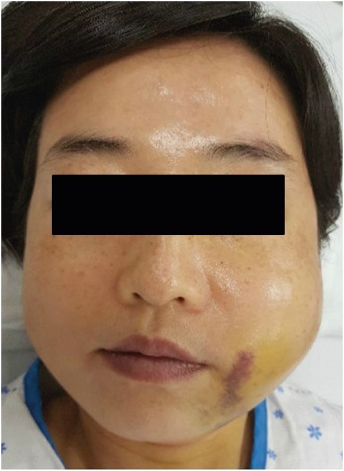
FIGURE 1 Patient's appearance at the emergency room. Left facial swelling and ecchymosis was seen.
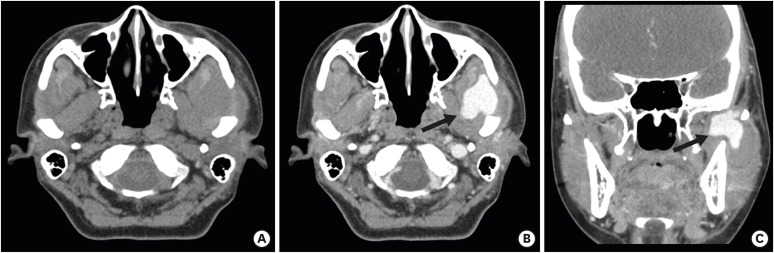
FIGURE 2 Neck contrast CT images. (A) Pre-contrast axial, (B) Post-contrast axial, and (C) Post-contrast coronal. CT shows a low-density mass-like lesion at left masticator space with contrast extravasation (arrow).CT: computed tomography.
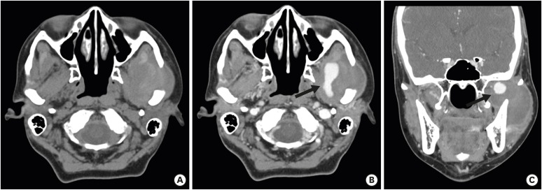
FIGURE 3 Para-nasal sinus CT images. (A) Pre-contrast axial, (B) Post-contrast axial, and (C) Post-contrast coronal. CT shows that Further increased status of previous noted low-density mass-like lesion or loculated fluid at left masticator space but decreased contrast extravasation (arrow).CT: computed tomography.
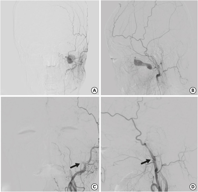
FIGURE 4 Trans-femoral cerebral angiography images. Pre-operative; (A) Antero-posterior view, (B) Lateral view. Contrast filling mass-like pseudoaneurysm was seen in the left IMA proximal area. Post-operative; (C) Antero-posterior view, (D) Lateral view. Left proximal IMA was totally occluded by Histoacryl Glue (arrow).IMA: internal maxillary artery.
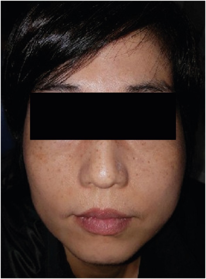
FIGURE 5 Post-operative patient's appearance at the emergency room. There was seen that decreased left mandibular swelling.
Reference
-
1. Chakrabarty S, Majumdar SK, Ghatak A, Bansal A. Management of pseudoaneurysm of internal maxillary artery resulting from trauma. J Maxillofac Oral Surg. 2015; 14:203–208. PMID: 25838698.
Article2. D'Orta JA, Shatney CH. Post-traumatic pseudoaneurysm of the internal maxillary artery. J Trauma. 1982; 22:161–164. PMID: 7062363.3. Kim DY, Hyun DK, Park H, Chung J. Endovascular treatment of life-threatening bleeding of bilateral maxillary arteries in a patient with multiple facial bone fractures- a case report. J Korean Neurotraumatol Soc. 2011; 7:108–111.
Article4. Elden L, Montanera W, Terbrugge K, Willinsky R, Lasjaunias P, Charles D. Angiographic embolization for the treatment of epistaxis: a review of 108 cases. Otolaryngol Head Neck Surg. 1994; 111:44–50. PMID: 8028941.
Article5. Kawano K, Mizuki H, Mori H, Yanagisawa S. Mandibular arteriovenous malformation treated by transvenous coil embolization: a long-term follow-up with special reference to bone regeneration. J Oral Maxillofac Surg. 2001; 59:326–330. PMID: 11243618.
Article6. Kuehne JP, Weaver FA, Papanicolaou G, Yellin AE. Penetrating trauma of the internal carotid artery. Arch Surg. 1996; 131:942–947. PMID: 8790179.
Article7. McDonald PT, Rich NM, Collins GJ Jr, Andersen CA, Kozloff L. Vascular trauma secondary to diagnostic and therapeutic procedures: laparoscopy. Am J Surg. 1978; 135:651–655. PMID: 148217.
Article8. Satoh T, Sakurai M, Yamamoto Y, Asari S. Spontaneous closure of a traumatic middle meningeal arterio-venous fistula. Neuroradiology. 1983; 25:105–109. PMID: 6877587.
Article9. Schwartz HC, Kendrick RW, Pogorel BS. False aneurysm of the maxillary artery. An unusual complication of closed facial trauma. Arch Otolaryngol. 1983; 109:616–618. PMID: 6882272.
Article10. Tsumoto T, Nakakita K, Hayashi S, Terada T. Bone defect associated with middle meningeal arteriovenous fistula treated by embolization--case report. Neurol Med Chir (Tokyo). 2001; 41:42–47. PMID: 11218640.
- Full Text Links
-
- Actions
-
Cited
- CITED
-
- Close
- Share
-
- Similar articles
-
- A Case of Pseudoaneurysm of the Superior Thyroid Artery after Core Needle Biopsy
- Traumatic Internal Maxillary Artery Pseudoaneurysm Caused by Fracture of the Mandible Ramus: A Case Report
- Traumatic Pseudoaneurysm of the External and Internal Carotid Artery Presenting as Epistaxis: Case Report
- Pseudoaneurysm of the Superficial Femoral Artery Following Gamma nail Fixation for Trochanteric Fracture: A Case Report
- Embolization for treating posttraumatic pseudoaneurysm of the sphenopalatine artery
