Ann Pediatr Endocrinol Metab.
2019 Sep;24(3):207-211. 10.6065/apem.2019.24.3.207.
Long-term follow-up on MURCS (Müllerian duct, renal, cervical somite dysplasia) association and a review of the literature
- Affiliations
-
- 1Department of Pediatrics, Inha University Graduate school of Medicine, Inha University Hospital, Incheon, Korea. anicca@inha.ac.kr
- 2Department of Urology, Inha University College of Medicine, Incheon, Korea.
- 3Department of Internal Medicine, Inha University College of Medicine, Incheon, Korea.
- 4Department of Orthopaedic Surgery, Inha University Hospital, Incheon, Korea.
- KMID: 2460770
- DOI: http://doi.org/10.6065/apem.2019.24.3.207
Abstract
- Müllerian duct aplasia-renal aplasia-cervicothoracic somite dysplasia (MURCS) association is a unique development disorder with four common types of malformations that include uterine aplasia or hypoplasia, renal ectopy or agenesis, vertebral anomalies, and short stature. The majority of MURCS patients are diagnosed with primary amenorrhea from late-adolescence. However, a few cases with MURCS association are not well diagnosed during childhood and long-term outcomes are not well reported. We report a case of an 8-year-old girl with MURCS association who presented with recurrent urinary tract infections and multiple congenital malformations, and who was followed for 10 years until adulthood. MURCS association should be considered as one of the differential diagnoses when evaluating prepubertal females with vertebral and renal malformations.
Keyword
MeSH Terms
Figure
-
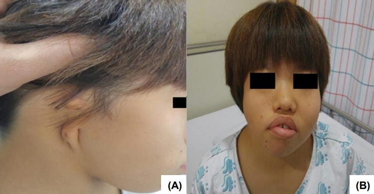
Fig. 1. Image showing external ear atresia (A), short neck and facial asymmetry (B).

Fig. 2. X-ray C-spine showed a bony fusion on C2-3, C4 vertebrae (arrow) (A) and scoliosis of L-spine (arrowhead) (B).

Fig. 3. Abdominal computed tomography showed a single dysplastic kidney (arrow) (A) and uterine aplasia (arrowhead) (B).
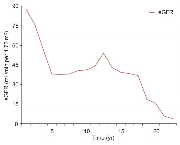
Fig. 4. Estimated glomerular filtration rate (eGFR) of our patient. It declines gradually during follow-up period.
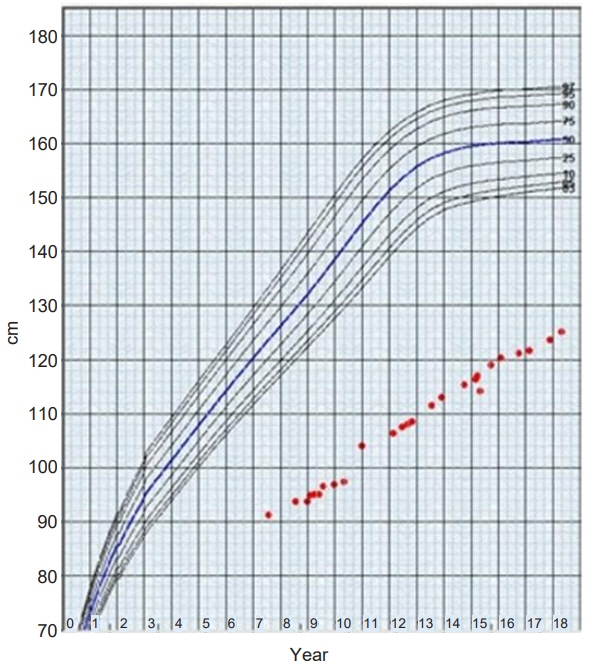
Fig. 5. Growth curve of our patient. It shows extreme short stature with significant catch-down growth during follow-up period.
Reference
-
References
1. Kang Ben, Park SH, Kim DH, Lee BI, Kim MY, Lee JE. Mayer-Rokitansky-Kuster-Hauser (MRKH) syndrome in a child with idiopathic precocious puberty. Ann Pediatr Endocrinol Metab. 2012; 17:126–9.2. Mahajan P, Kher A, Khungar A, Bhat M, Sanklecha M, Bharucha BA. MURCS association--a review of 7 cases. J Postgrad Med. 1992; 38:109–11.3. Duncan PA, Shapiro LR, Stangel JJ, Klein RM, Addonizio JC. The MURCS association: Müllerian duct aplasia, renal aplasia, and cervicothoracic somite dysplasia. J Pediatr. 1979; 95:399–402.4. Guerrier D, Mouchel T, Pasquier L, Pellerin I. The Mayer-Rokitansky-Küster-Hauser syndrome (congenital absence of uterus and vagina)--phenotypic manifestations and genetic approaches. J Negat Results Biomed. 2006; 5:1.5. Park JY, Kim SY, Kim JN, Yang SJ, Park JR, Kwan BS, et al. A case of colon cancer in Mayer-Rokitansky-Küster-Hauser (MRKH) syndrome with gonadal agenesis. J Korean Endocri Soc. 2006; 21:414–8.6. Braun-Quentin C, Billes C, Böwing B, Kotzot D. MURCS association: case report and review. J Med Genet. 1996; 33:618–20.7. Gilliam ML, Shulman LP. Tetralogy of Fallot, imperforate anus, and Müllerian, renal, and cervical spine (MURCS) anomalies in a 15-year-old girl. J Pediatr Adolesc Gynecol. 2002; 15:231–3.8. Kumar S, Sharma S. MURCS (Müllerian duct aplasiarenal agenesis-cervicothoracic somite dysplasia): a rare cause of primary amenorrhoea. Oxf Med Case Reports. 2016; 2016:73–5.9. Al Kaissi A, Ben Chehida F, Ben Ghachem M, Grill F, Klaushofer K. Occipitoatlantoaxial junction malformation and early onset senile ankylosing vertebral hyperostosis in a girl with MURCS association. Am J Med Genet A. 2009; 149A:470–4.10. Källén K, Mastroiacovo P, Castilla EE, Robert E, Källén B. VATER non-random association of congenital malformations: study based on data from four malformation registers. Am J Med Genet. 2001; 101:26–32.11. Miller C, Sassoon DA. Wnt-7a maintains appropriate uterine patterning during the development of the mouse female reproductive tract. Development. 1998; 125:3201–11.12. Gendron RL, Paradis H, Hsieh-Li HM, Lee DW, Potter SS, Markoff E. Abnormal uterine stromal and glandular function associated with maternal reproductive defects in Hoxa-11 null mice. Biol Reprod. 1997; 56:1097–105.13. Burel A, Mouchel T, Odent S, Tiker F, Knebelmann B, Pellerin I, et al. Role of HOXA7 to HOXA13 and PBX1 genes in various forms of MRKH syndrome (congenital absence of uterus and vagina). J Negat Results Biomed. 2006; 5:4.14. Edmonds DK. Congenital malformations of the genital tract and their management. Best Pract Res Clin Obstet Gynaecol. 2003; 17:19–40.
- Full Text Links
-
- Actions
-
Cited
- CITED
-
- Close
- Share
-
- Similar articles
-
- Monostotic Fibrous Dysplasia of the Cervical Spine: A Case Report
- A Case of Cervical Thoracic Duct Cyst
- Pancreaticoduodenectomy for recurrence of intraductal papillary neoplasm of bile duct at seven years after curative resection
- Renal Dysplasia: A Clinicopathologic Review of Six Cases
- Promotion of Cervical Screening among Long-term Nonattendees by Human Papillomavirus Self-sampling
