Position and size of the sphenoid door jamb in the lateral orbital wall for the orbital decompression
- Affiliations
-
- 1Department of Anatomy and Cell Biology, Dong-A University College of Medicine, Busan, Korea.
- 2Department of Anatomy, Research Institute of Medical Science, Konkuk University School of Medicine, Seoul, Korea.
- 3Department of Ophthalmology, Konkuk University Medical Center, Konkuk University School of Medicine, Seoul, Korea.
- 4Department of Medical Education, Hanyang University College of Medicine, Seoul, Korea. paikdj@hanyang.ac.kr
- KMID: 2459538
- DOI: http://doi.org/10.5115/acb.19.101
Abstract
- The aim of this study was to identify the three-dimensional topography of the sphenoid door jamb (SDJ) in the lateral orbital wall and to propose navigational guidelines for safe deep lateral decompression using surgical landmarks. The 120 orbits and SDJs of 60 subjects were three-dimensionally reconstructed using Mimics software. The mean volumes of the orbit and SDJ were 24.3 mm³ and 2.0 mm³, respectively. The mean distances from the lateral orbital margin (LOM) to the anterior and posterior margins of the SDJ were 13.2 and 36.3 mm, respectively. The mean distances from the superior orbital fissure to the LOM and to the posterior margin of the SDJ were 40.2 mm and 4.6 mm, respectively. The mean distances from the inferior orbital fissure (IOF) to the anterior and posterior margins of the SDJ were 3.8 mm and 20.5 mm, respectively. In the superior approach of the orbit, it can be predicted that the area up to 3 cm posterior from the LOM is safe, while 1 cm posterior from the safe zone could be a dangerous zone. In the inferior approach of the orbit, the safe area will be about 1 cm posterior from the anterior tip of the IOF, and the area up to 1 cm posterior from the safe zone should be approached with extreme care.
MeSH Terms
Figure
-
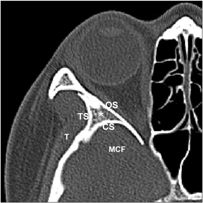
Fig. 1 Transverse plane of the sphenoid door jamb (SDJ). CS, the cranial surface of the SDJ; MCF, the middle cranial fossa; OS, the orbital surface of the SDJ; T, the temporalis muscle; TS, the temporal surface of the SDJ.
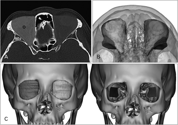
Fig. 2 Segmentation and 3D volume rendering of the orbit and sphenoid door jamb (SDJ). (A) the outlines of the orbit and SDJ by manual segmentation. (B) The superior view of reconstructed orbit and SDJ. (C) Frontal view reconstructed orbit and SDJ. O, orbit.
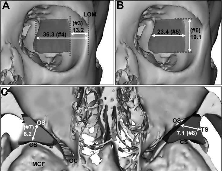
Fig. 3 (A–C) Mean distances of the sphenoid door jamb (SDJ ) at the intermediated level (mm). CS, the cranial surface; LOM, the lateral orbital margin; MCF, the middle cranial fossa; OC, the optic canal; OS, the orbital surface; TS, the temporal surface.

Fig. 4 Mean distances of the sphenoid door jamb at the superior orbital fissure (SOF) and inferior orbital fissure (IOF) level (mm). LOM, lateral orbital margin.
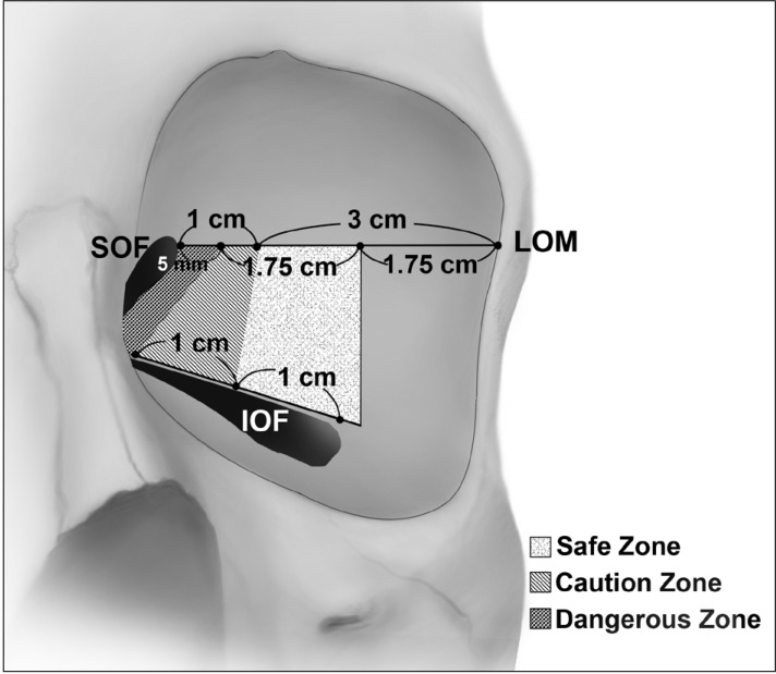
Fig. 5 The navigational guidelines for the effective and safe deep lateral orbital decompression. IOF, inferior orbital fissure; LOM, lateral orbital margin; SOF, superior orbital fissure.
Reference
-
1. Bahn RS. Graves' ophthalmopathy. N Engl J Med. 2010; 362:726–738. PMID: 20181974.2. Choi SW, Lee JY, Lew H. Customized orbital decompression surgery combined with eyelid surgery or strabismus surgery in mild to moderate thyroid-associated ophthalmopathy. Korean J Ophthalmol. 2016; 30:1–9. PMID: 26865797.3. Khoo TK, Bahn RS. Pathogenesis of Graves’ ophthalmopathy: the role of autoantibodies. Thyroid. 2007; 17:1013–1018. PMID: 17935483.4. Leong SC, White PS. Outcomes following surgical decompression for dysthyroid orbitopathy (Graves' disease). Curr Opin Otolaryngol Head Neck Surg. 2010; 18:37–43. PMID: 19940769.5. Baldeschi L. Small versus coronal incision orbital decompression in Graves' orbitopathy. Orbit. 2010; 29:177–182. PMID: 20812832.6. Cascone P, Rinna C, Reale G, Calvani F, Iannetti G. Compression and stretching in Graves orbitopathy: emergency orbital decompression techniques. J Craniofac Surg. 2012; 23:1430–1433. PMID: 22948632.7. Verity DH, Rose GE. Acute thyroid eye disease (TED): principles of medical and surgical management. Eye (Lond). 2013; 27:308–319. PMID: 23412559.8. Baldeschi L, MacAndie K, Hintschich C, Wakelkamp IM, Prummel MF, Wiersinga WM. The removal of the deep lateral wall in orbital decompression: its contribution to exophthalmos reduction and influence on consecutive diplopia. Am J Ophthalmol. 2005; 140:642–647. PMID: 16140250.9. Goldberg RA. Old wine and new wine. Arch Ophthalmol. 2000; 118:1006–1007.10. Abràmoff MD, Kalmann R, de Graaf ME, Stilma JS, Mourits MP. Rectus extraocular muscle paths and decompression surgery for Graves orbitopathy: mechanism of motility disturbances. Invest Ophthalmol Vis Sci. 2002; 43:300–307. PMID: 11818370.11. McCord CD Jr. Current trends in orbital decompression. Ophthalmology. 1985; 92:21–33. PMID: 3838377.12. Goldberg RA, Kim AJ, Kerivan KM. The lacrimal keyhole, orbital door jamb, and basin of the inferior orbital fissure: three areas of deep bone in the lateral orbit. Arch Ophthalmol. 1998; 116:1618–1624. PMID: 9869791.13. Beden U, Edizer M, Elmali M, Icten N, Gungor I, Sullu Y, Erkan D. Surgical anatomy of the deep lateral orbital wall. Eur J Ophthalmol. 2007; 17:281–286. PMID: 17534804.14. Feneis H, Dauber W. Anatomisches Bildwörterbuch der internationalen Nomenklatur. Stuttgart: Thieme;1998.15. Unal M, Ileri F, Konuk O, Hasanreisoğlu B. Balanced orbital decompression in Graves’ orbitopathy: Upper eyelid crease incision for extended lateral wall decompression. Orbit. 2000; 19:109–117. PMID: 12045955.16. Mehta P, Durrani OM. Outcome of deep lateral wall rim-sparing orbital decompression in thyroid-associated orbitopathy: a new technique and results of a case series. Orbit. 2011; 30:265–268. PMID: 22132843.17. Kakizaki H. Advantageous surgeon's position in deep lateral orbital wall decompression. Orbit. 2011; 30:131. PMID: 21574801.18. Takahashi Y, Miyazaki H, Ichinose A, Nakano T, Asamoto K, Kakizaki H. Anatomy of deep lateral and medial orbital walls: implications in orbital decompression surgery. Orbit. 2013; 32:409–412. PMID: 24063541.19. Oh DH, Lee JK. Surgical anatomy of deep lateral wall by adults cadavers and computed tomography. J Korean Ophthalmol Soc. 2011; 52:964–969.20. Kim KW, Byun JS, Lee JK. Surgical effects of various orbital decompression methods in thyroid-associated orbitopathy: computed tomography-based comparative analysis. J Craniomaxillofac Surg. 2014; 42:1286–1291. PMID: 24793198.21. Kamer L, Noser H, Kirsch E, Hammer B. Anatomy-based surgical concepts for individualized orbital decompression surgery in graves orbitopathy. II. Orbital rim position and angulation. Ophthalmic Plast Reconstr Surg. 2012; 28:251–255. PMID: 22785582.22. Kakizaki H, Nakano T, Asamoto K, Iwaki M. Posterior border of the deep lateral orbital wall: appearance, width, and distance from the orbital rim. Ophthalmic Plast Reconstr Surg. 2008; 24:262–265. PMID: 18645427.23. Lee H, Lee Y, Ha S, Park M, Baek S. Measurement of width and distance of the posterior border of the deep lateral orbital wall using computed tomography. J Craniomaxillofac Surg. 2011; 39:606–609. PMID: 21875811.24. Rootman DB. Orbital decompression for thyroid eye disease. Surv Ophthalmol. 2018; 63:86–104. PMID: 28343872.
- Full Text Links
-
- Actions
-
Cited
- CITED
-
- Close
- Share
-
- Similar articles
-
- Evaluation of Stereotactic Navigation During Orbital Decompression in Thyroid-Associated Orbitopathy Patients
- Surgical Anatomy of Deep Lateral Wall by Adults Cadavers and Computed Tomography
- Orbital DEcompression Usiing Caruncular Approach
- Effect of Posterior Strut Removal during Orbital Decompression for Graves’ Orbitopathy
- Orbital wall restoring surgery with primary orbital wall fragments in blowout fracture
