Primary Epithelioid Hemangioendothelioma of the Central Nervous System: A Case Report
- Affiliations
-
- 1Department of Radiology, Uijeongbu St. Mary's Hospital, College of Medicine, The Catholic University of Korea, Uijeongbu, Korea. violet2@catholic.ac.kr
- 2Department of Pathology, Uijeongbu St. Mary's Hospital, College of Medicine, The Catholic University of Korea, Uijeongbu, Korea.
- KMID: 2459025
- DOI: http://doi.org/10.3348/jksr.2019.80.5.981
Abstract
- Primary epithelioid hemangioendothelioma (EHE) of the central nervous system is an extremely rare sarcoma of vascular origin. Imaging findings have been reported for few cases. Herein, we present a case of intracranial EHE manifesting as spontaneous intracranial hemorrhage. The tumor presented as a well-demarcated hemorrhagic lesion. It had a peripheral location, and showed signs of two-layered target-like mild enhancement in the early phase and gradual fill-in delayed enhancement on MRI.
MeSH Terms
Figure
-

Fig. 1. A 72-year-old man with primary epithelioid hemangioendothelioma. A. Unenhanced axial CT scan shows a well-defined, hyperattenuating lesion in the right frontal lobe abutting the pial surface of the brain. Laterally, a band-like heterogeneous hypoattenuating area can be seen with vasogenic edema surrounding the lesion. B. The mass exhibits heterogeneous signal intensity, mainly hypointensity, on the axial T2-weighted image (upper left panel) with peripheral edema, heterogeneously isointense with the gray matter on T1-weighted images (upper central panel), and strongly hypointense “blooming” on susceptibility-weighted images (upper right panel). Axial T1-weighted image immediately after contrast injection (lower left panel) demonstrates two-layered target-like, mild peripheral enhancements. Five-minutes delayed postcontrast axial T1-weighted image shows a greater increase of contrast enhancement area and intensity that is pronounced at the periphery (lower right panel).
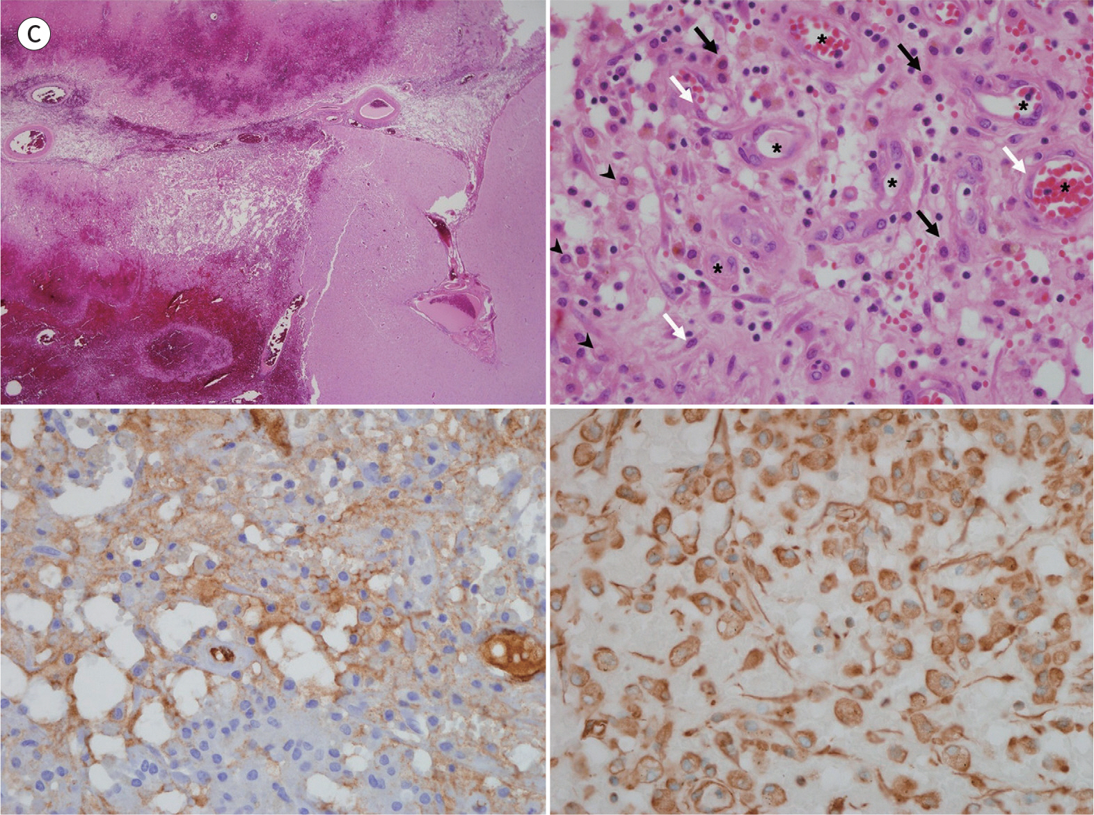
Fig. 1. A 72-year-old man with primary epithelioid hemangioendothelioma. C. Photomicrograph of hematoxylin and eosin-stained slide (left upper panel, × 12) reveals a relatively well demarcated and hemorrhagic tumor mass. The tumor was composed of epithelioid (black arrows) or spin-dle shaped cells (white arrows) in nests or cords (asterisks) within a myxoid stoma. Some tumor cells show small intracytoplasmic lumens (“blister cells”) that contain red cells (arrowheads) (right upper panel, × 400). Immunohistochemistry reveals positive factor VIII-related antigen staining (left lower panel, × 400) indicating an endothelial origin. Vimentin stain (right lower panel, × 400) reveals positive staining of part of the tumor, indicating a mesenchymal component.
Reference
-
References
1. Weiss SW, Enzinger FM. Epithelioid hemangioendothelioma: a vascular tumor often mistaken for a carcinoma. Cancer. 1982; 50:970–981.
Article2. Taratuto AL, Zurbriggen G, Sevlever G, Saccoliti M. Epithelioid hemangioendothelioma of the central nervous system. Immunohistochemical and ultrastructural observations of a pediatric case.Pediatr Neurosci. 1988; 14:11–14.
Article3. Batista KP, Gómez GL, Quintana EM, Astudillo A, Fernandez-Vega I, Fernandez BA, et al. Giant cranionasal epithelioid haemangioendothelioma with invasive growth pattern mimicking a skull base chondrosarcoma. Contemp Oncol (Pozn). 2018; 22:118–123.4. Parajón A, Vaquero J. Meningel intracranial epithelioid hemangioendothelioma: case report and literature review.J Neurooncol. 2008; 88:169–173.5. Zheng J, Liu L, Wang J, Wang S, Cao Y, Zhao J. Primary intracranial epithelioid hemangioendothelioma: a low-proliferation tumor exhibiting clinically malignant behavior.J Neurooncol. 2012; 110:119–127.6. Chan YL, Ng HK, Poon WS, Cheung HS. Epithelioid haemangioendothelioma of the brain: a case report. Neuroradiology. 2001; 43:848–850.
Article7. Paolantonio P, Laghi A, Vanzulli A, Grazioli L, Morana G, Ragozzino A, et al. MRI of hepatic epithelioid hemangioendothelioma (HEH).J Magn Reson Imaging. 2014; 40:552–558.8. Brat DJ, Van Meir EG. Vaso-occlusive and prothrombotic mechanisms associated with tumor hypoxia, necrosis, and accelerated growth in glioblastoma.Lab Invest. 2004; 84:397–405.9. Rapacki TF, Brantley MJ, Furlow TW Jr, Geyer CA, Toro VE, George ED. Heterogeneity of cerebral cavernous hemangiomas diagnosed by MR imaging.J Comput Assist Tomogr. 1990; 14:18–25.
- Full Text Links
-
- Actions
-
Cited
- CITED
-
- Close
- Share
-
- Similar articles
-
- Epithelioid Hemangioendothelioma of the Spinal Cord
- A Case Pulmonary Epithelioid Hemangioendothelioma that Underwent Unusual Malignant Course
- Primary epithelioid hemangioendothelioma arising at vulva: A case report
- A Case of Epithelioid Hemangioendothelioma on the Choana
- Intracranial Epithelioid Hemangioendothelioma
