Maxillary ameloblastoma in an 8-year-old child: A case report with a review of the literature
- Affiliations
-
- 1Division of Oral and Maxillofacial Radiology, Department of Diagnostic Sciences, Rutgers School of Dental Medicine, NJ, USA. sangeetha7455@gmail.com
- 2Department of Oral and Maxillofacial Surgery, Rutgers School of Dental Medicine, NJ, USA.
- 3Department of Pathology, Immunology and Laboratory Medicine, Rutgers New Jersey Medical School, NJ, USA.
- KMID: 2458374
- DOI: http://doi.org/10.5624/isd.2019.49.3.241
Abstract
- Ameloblastoma is a benign locally invasive tumor with a high tendency to recur. It is considered rare in the pediatric population, with most cases diagnosed in the third to fifth decades of life. Approximately 80% of ameloblastomas occur in the molar and ramus region of the mandible, while 20% of cases occur in the maxillary posterior region. This report presents a case of plexiform ameloblastoma in an uncommon location in an 8-year-old child. The lesion was initially thought to be a dentigerous cyst, based on its location and radiographic appearance. The clinical and radiographic features, histopathology, and treatment of solid, plexiform, maxillary ameloblastoma are reviewed, with an added emphasis on a literature review of ameloblastoma in children. This report emphasize the importance of long-term follow-up, since recurrence may occur many years after initial tumor removal.
Keyword
MeSH Terms
Figure
-
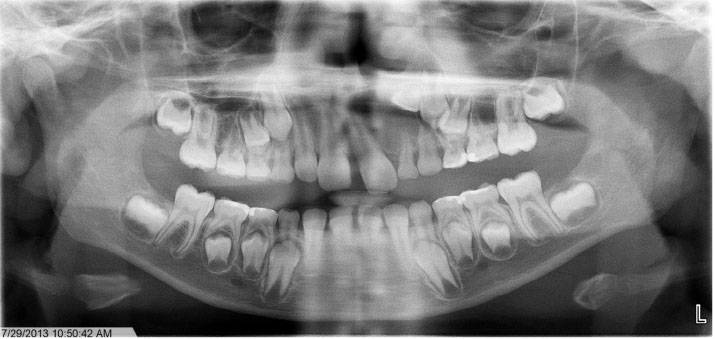
Fig. 1 Panoramic radiograph taken on the first visit. A small oval radiolucency can be observed around the impacted maxillary left lateral incisor.
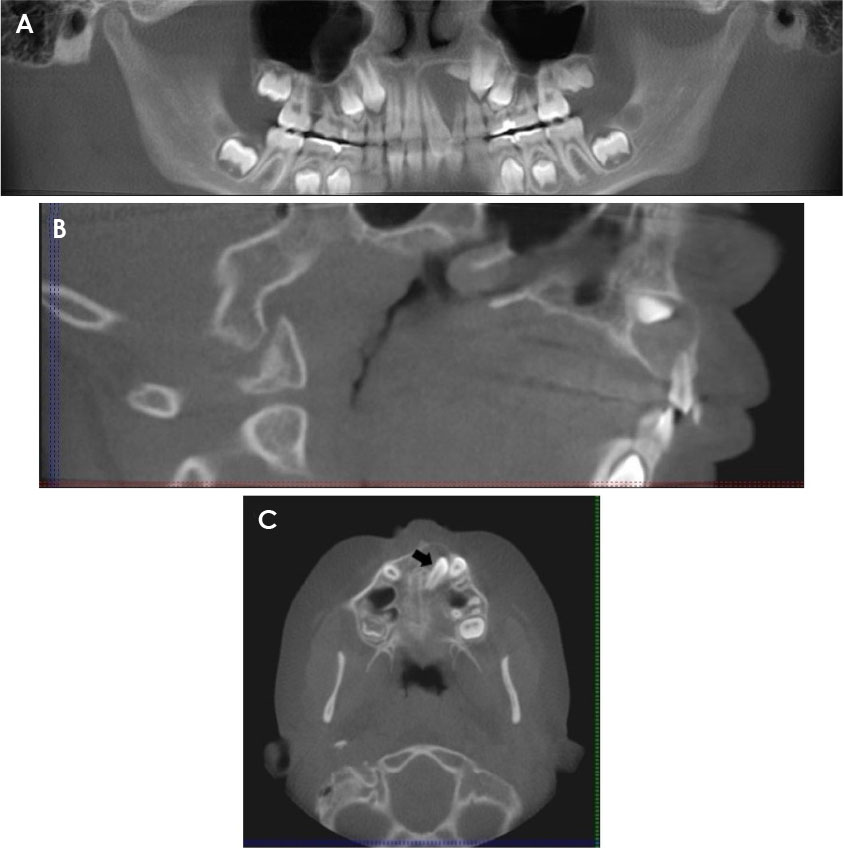
Fig. 2 Cone-beam computed tomography (CBCT) images taken on the first visit. A. CBCT panoramic reconstruction shows a low-density area surrounding the permanent maxillary left central and lateral incisor and causing displacement of the teeth. B. Sagittal CBCT view shows expansion of the buccal and palatal cortices in the anterior maxilla. C. Axial CBCT view at the level of the maxilla. The arrow indicates the impacted maxillary left lateral incisor.

Fig. 3 Clinical photographs taken at the time of surgical enucleation. A and B. Impacted maxillary left lateral incisor exposed, bonded, and ligated at the time of the procedure. C. Fragments of the lesion and extracted primary maxillary left lateral incisor and canine.
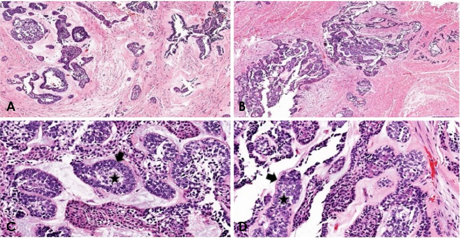
Fig. 4 Hematoxylin and eosin stained sections of the lesion demonstrating cords and sheets of anastomosing odontogenic epithelial cells consistent in appearance with the plexiform variant of ameloblastoma. Note the epithelial cells, which show reverse polarization away from the basement membrane (arrowheads), and the stellate reticulum-like cells and suprabasal cells, which compose loosely arranged angular cells (star). A and B are ×4 magnification fields, C and D are ×10 magnification fields.
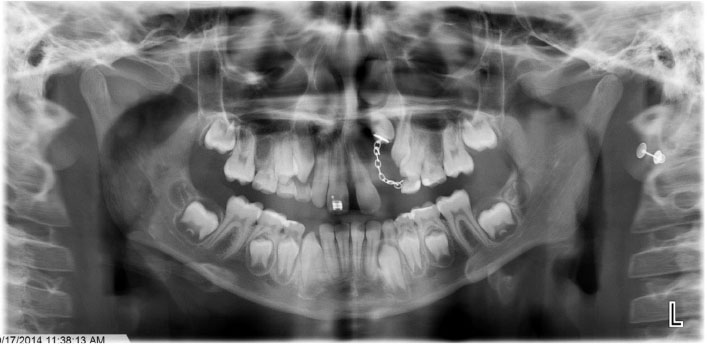
Fig. 5 Postoperative panoramic radiograph (2014) taken after 1 year shows the maxillary left lateral incisor protruding through the mucosa.

Fig. 6 Panoramic radiograph (2015) shows a radiolucency distal to the maxillary left lateral incisor.

Fig. 7 Cone-beam computed tomography cross-sectional images show an enlarged periodontal ligament space, missing buccal plate, and bone loss around the maxillary left lateral incisor.
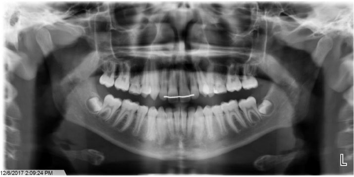
Fig. 8 Panoramic radiograph (2017) shows no distinct radiographic evidence of recurrence.
Reference
-
1. Neville BW, Damm DD, Allen CM, Bouquot JE. Oral and maxillofacial pathology. 3rd ed. St. Louis: Saunders/Elsevier;2009.2. Zhang J, Gu Z, Jiang L, Zhao J, Tian M, Zhou J, et al. Ameloblastoma in children and adolescents. Br J Oral Maxillofac Surg. 2010; 48:549–554.
Article3. Kreppel M, Zöller J. Ameloblastoma - clinical, radiological, and therapeutic findings. Oral Dis. 2018; 24:63–66.4. Bansal S, Desai RS, Shirsat P, Prasad P, Karjodkar F, Andrade N. The occurrence and pattern of ameloblastoma in children and adolescents: an Indian institutional study of 41 years and review of the literature. Int J Oral Maxillofac Surg. 2015; 44:725–731.
Article5. Arotiba GT, Ladeinde AL, Arotiba JT, Ajike SO, Ugboko VI, Ajayi O. Ameloblastoma in Nigerian children and adolescents: a review of 79 cases. J Oral Maxillofac Surg. 2005; 63:747–751.
Article6. Butt FM, Guthua SW, Awange DA, Dimba EA, Macigo FG. The pattern and occurrence of ameloblastoma in adolescents treated at a university teaching hospital, in Kenya: a 13-year study. J Craniomaxillofac Surg. 2012; 40:e39–e45.
Article7. Giraddi GB, Arora K, Saifi AM. Ameloblastoma: a retrospective analysis of 31 cases. J Oral Biol Craniofac Res. 2017; 7:206–211.
Article8. Kashyap B, Reddy PS, Desai RS. Plexiform ameloblastoma mimicking a periapical lesion: a diagnostic dilemma. J Conserv Dent. 2012; 15:84–86.
Article9. Singer SR, Mupparapu M, Philipone E. Cone beam computed tomography findings in a case of plexiform ameloblastoma. Quintessence Int. 2009; 40:627–630.10. Castro-Silva II, Israel MS, Lima GS, de Queiroz Chaves Lourenço S. Difficulties in the diagnosis of plexiform ameloblastoma. Oral Maxillofac Surg. 2012; 16:115–118.
Article11. Payne SJ, Albert T, Lighthall JG. Management of ameloblastoma in the pediatric population. Oper Tech Otolaryngol Head Neck Surg. 2015; 26:168–174.
Article12. Takahashi K, Miyauchi K, Sato K. Treatment of ameloblastoma in children. Br J Oral Maxillofac Surg. 1998; 36:453–456.
Article13. Al-Khateeb T, Ababneh KT. Ameloblastoma in young Jordanians: a review of the clinicopathologic features and treatment of 10 cases. J Oral Maxillofac Surg. 2003; 61:13–18.
Article14. Chukwuneke FN, Anyanechi CE, Akpeh JO, Chukwuka A, Ekwueme OC. Clinical characteristics and presentation of ameloblastomas: an 8-year retrospective study of 240 cases in Eastern Nigeria. Br J Oral Maxillofac Surg. 2016; 54:384–387.
Article15. Huang IY, Lai ST, Chen CH, Chen CM, Wu CW, Shen YH. Surgical management of ameloblastoma in children. Oral Surg Oral Med Oral Pathol Oral Radiol Endod. 2007; 104:478–485.
Article16. Pogrel MA, Montes DM. Is there a role for enucleation in the management of ameloblastoma? Int J Oral Maxillofac Surg. 2009; 38:807–812.
Article17. Iordanidis S, Makos C, Dimitrakopoulos J, Kariki H. Ameloblastoma of the maxilla. Case report. Aust Dent J. 1999; 44:51–55.
Article18. McClary AC, West RB, McClary AC, Pollack JR, Fischbein NJ, Holsinger CF, et al. Ameloblastoma: a clinical review and trends in management. Eur Arch Otorhinolaryngol. 2016; 273:1649–1661.
Article
