J Korean Foot Ankle Soc.
2019 Sep;23(3):116-120. 10.14193/jkfas.2019.23.3.116.
Surgical Treatment of Calcaneal Fractures of Sanders Type II and III by A Minimally Invasive Technique with 6.5 mm Cancellous Screw
- Affiliations
-
- 1Department of Orthopedic Surgery, Dong-A University College of Medicine, Busan, Korea. tynitus@dau.ac.kr
- 2Department of Orthopedic Surgery, Good Samsun Hospital, Busan, Korea.
- KMID: 2458013
- DOI: http://doi.org/10.14193/jkfas.2019.23.3.116
Abstract
- PURPOSE
This study evaluated the clinical and radiological results of 6.5 mm full threaded cancellous bone screw fixation of calcaneal fractures.
MATERIALS AND METHODS
Thirty seven patients diagnosed with Sanders type II or III calcaneal fractures, who underwent open reduction and internal fixation with a 6.5 mm full threaded cancellous bone screw between August 2014 and August 2017, were analyzed. Both the preoperative and postoperative Böhler angle and Gissane angle were measured radiographically. American Orthopaedic Foot and Ankle Society (AOFAS) ankle-hindfoot scale on the final follow-up were also assessed.
RESULTS
The mean age of the patients was 52.7 years and the mean follow-up period was 29.5 months. In the Sanders classification, type II and III were 16 and 24 cases, respectively. The Böhler and Gissane angles improved from 21.2° and 122.6° preoperatively to 21.6° and 120.3°, respectively, in the postoperative radiographs. All cases achieved bony union, and the AOFAS ankle-hindfoot scale was 90.7 and 91.3 in Sanders type II and III, respectively, at the final follow-up.
CONCLUSION
The treatment of calcaneal fractures using a 6.5 mm full threaded cancellous bone screw can reduce the complications with minimally invasive surgery and achieve firm fixation.
MeSH Terms
Figure
-
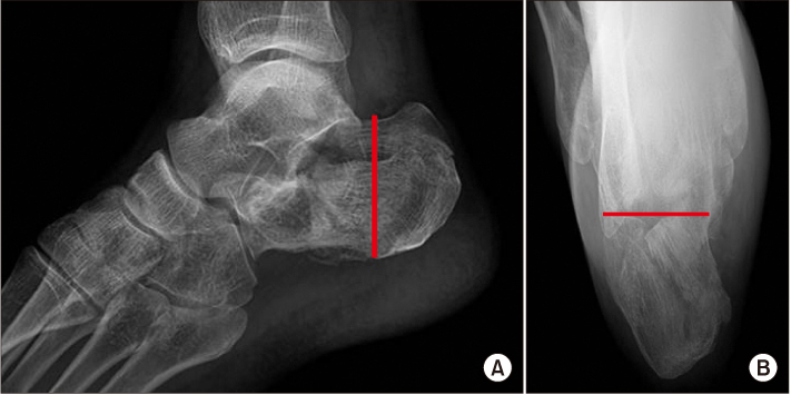
Figure 1 Measurement of calcaneal height and width in X-ray. (A) Calcaneal height is measured by the line drawn from the superior point of the posterior facet to anterior point of lateral process of tuberosity in lateral view. (B) Calcaneal width is measured by the line drawn from calcaneus medial groove to peroneus tubercle in axial view.
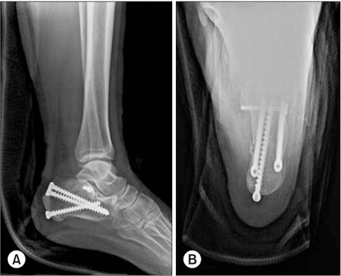
Figure 2 Postoperative images show that 4.0 mm cortical screws were used to fix the posterior articular surface in the transverse direction. After the reduction of the calcaneal fracture, a 6.5 mm cancellous bone full threaded screw was used to fix from the superior and inferior calcaneus tubercle to articular facet. (A) Lateral view. (B) Axial view.
Reference
-
1. Cave EF. Fracture of the os calcis--the problem in general. Clin Orthop Relat Res. 1963; 30:64–66.2. Fitzgibbons TC, Mcmullen ST, Mormino MA. Fractures and dislocations of the calcaneus. In : Bucholz RW, Heckman JD, editors. Rockwood and Green's fractures in adults vol 2. 5th ed. Philadelphia: Lippincott Williams & Wilkins;2001. p. 2133–2179.3. Murphy GA. Fractures and dislocations of foot. In : Campbell WC, Canale ST, editors. Campbell's operative orthopaedics. 10th ed. St. Louis (MO): Mosby;2003. p. 4231–4283.4. Susan N. Ishikawa. Fractures and dislocations of the foot. In : Canale ST, Azar FM, Beaty JH, Campbell WC, editors. Campbell's operative orthopaedics volume 1. 13th ed.Philadelphia: Elsevier;2017. p. 4278.5. Abidi NA, Dhawan S, Gruen GS, Vogt MT, Conti SF. Woundhealing risk factors after open reduction and internal fixation of calcaneal fractures. Foot Ankle Int. 1998; 19:856–861. DOI: 10.1177/107110079801901211.
Article6. Benirschke SK, Sangeorzan BJ. Extensive intraarticular fractures of the foot. Surgical management of calcaneal fractures. Clin Orthop Relat Res. 1993; (292):128–134.7. Wang T, Boone C, Behn AW, Ledesma JB, Bishop JA. Cancellous screws are biomechanically superior to cortical screws in metaphyseal bone. Orthopedics. 2016; 39:e828–e832. DOI: 10.3928/01477447-20160509-01.
Article8. Patel PS, Shepherd DE, Hukins DW. The effect of screw insertion angle and thread type on the pullout strength of bone screws in normal and osteoporotic cancellous bone models. Med Eng Phys. 2010; 32:822–828. DOI: 10.1016/j.medengphy.2010.05.005.
Article9. Barei DP, Bellabarba C, Sangeorzan BJ, Benirschke SK. Fractures of the calcaneus. Orthop Clin North Am. 2002; 33:263–285. x
Article10. Sanders R, Fortin P, DiPasquale T, Walling A. Operative treatment in 120 displaced intraarticular calcaneal fractures. Results using a prognostic computed tomography scan classification. Clin Orthop Relat Res. 1993; (290):87–95.11. Ross SD, Sowerby MR. The operative treatment of fractures of the os calcis. Clin Orthop Relat Res. 1985; (199):132–143.
Article12. Letournel E. Open reduction and internal fixation of calcaneus fractures. In : Spiegel PG, editor. Topics in orthopedic trauma. Baltimore: University Parkpress;1984. p. 173–192.13. Thordarson DB, Krieger LE. Operative vs. nonoperative treatment of intra-articular fractures of the calcaneus: a prospective randomized trial. Foot Ankle Int. 1996; 17:2–9. DOI: 10.1177/107110079601700102.
Article14. Basile A. Operative versus nonoperative treatment of displaced intra-articular calcaneal fractures in elderly patients. J Foot Ankle Surg. 2010; 49:25–32. DOI: 10.1053/j.jfas.2009.08.001.
Article15. Klaue K, Perren SM, Kowalski M. Internal fixation with a selfcompressing plate and lag screw: improvements of the plate hole and screw design. 1. Mechanical investigation. J Orthop Trauma. 1991; 5:280–288.16. Abdelgaid SM. Closed reduction and percutaneous cannulated screws fixation of displaced intra-articular calcaneus fractures. Foot Ankle Surg. 2012; 18:164–179. DOI: 10.1016/j.fas.2011.07.005.
Article17. Park JW, Park CH. Outcomes of arthroscopic assisted reduction and percutaneous fixation for tongue-type sanders type II calcaneal fractures. J Korean Foot Ankle Soc. 2017; 21:144–150. DOI: 10.14193/jkfas.2017.21.4.144.
Article18. Lee MJ, Sohn SK, Lee KY, Kim SS, Kang MS, Kim HJ, et al. Open reduction and internal fixation with AO calcaneal plate for displaced intra-articular calcaneal fracture. J Korean Fract Soc. 2010; 23:303–309. DOI: 10.12671/jkfs.2010.23.3.303.
Article19. Moon JS, Lee WC. A comparison of extensile lateral approach and sinus tarsi approach for the sanders type II calcaneal fracture. J Korean Fract Soc. 2009; 22:13–18. DOI: 10.12671/jkfs.2009.22.1.13.
Article20. Lee YT, Oh HC, Yoon HK, Jang JW, Jang KJ. Comparison of F calcaneal plate and locking calcaneal plate fixation using an lateral extensile approach to intra-articular calcaneal fractures. J Korean Foot Ankle Soc. 2012; 16:175–180.
- Full Text Links
-
- Actions
-
Cited
- CITED
-
- Close
- Share
-
- Similar articles
-
- Outcomes of Minimally Invasive Surgery in Intra-Articular Calcaneal Fractures: Sanders Type III, Joint Depressive Type Calcaneal Fracture
- Minimally-invasive Percutaneous Screw Fixation of Displaced Intra-articular Calcaneal Fractures
- Usefulness of Treatment with 6.5 mm Cancellous Screw and Steinmann Pin Fixation for Calcaneal Joint Depression Fracture
- The Result Treated by Open Reduction and Internal Fixation with Minimally Invasive Technique in Joint Depressive Calcaneal Fracture
- Clinical Results of Surgical Treatment with Minimally Invasive Percutaneous Plate Osteosynthesis for Displaced Intra-articular Fractures of Calcaneus
