Hip Pelvis.
2019 Sep;31(3):129-135. 10.5371/hp.2019.31.3.129.
Understanding Painful Hip in Young Adults: A Review Article
- Affiliations
-
- 1Department of Orthopedic Surgery, Khoula Hospital, Muscat, Sultanate of Oman. Drsalimalhabsi@hotmail.com
- 2Department of Radiology, Khoula Hospital, Muscat, Sultanate of Oman.
- KMID: 2455985
- DOI: http://doi.org/10.5371/hp.2019.31.3.129
Abstract
- A wide number of disorders, including pathologies outside the hip, can cause and refer pain to hip. However, determining the cause of a painful hip can be a major challenge to orthopedic surgeons. Failure to diagnose and appropriately investigate pathologies of the hip in adults may result in delayed management and prolonged patient morbidity. A systematic approach to investigating the etiology of hip pain in adults (e.g., history, careful clinical and radiographic examination), will help identify the majority of clinically important pathologies which can cause hip pain. Conservative treatment and selective use of injection therapies has proven quite successful for the treatment of most causes of hip pain.
Keyword
Figure
-
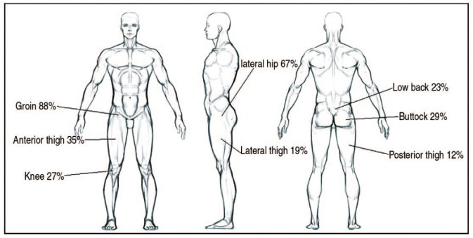
Fig. 1 Location of pain generated from hip pathology.
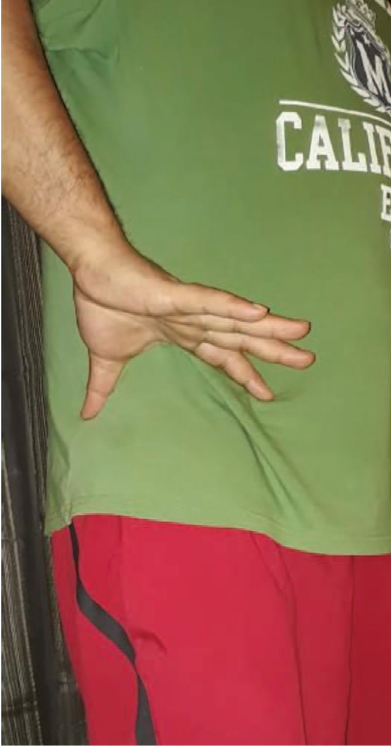
Fig. 2 C-sign in patients with hip pain. Patient with hip pathology usually point to the site of pain by cupping the thumb and index finger in the shape of the letter C.
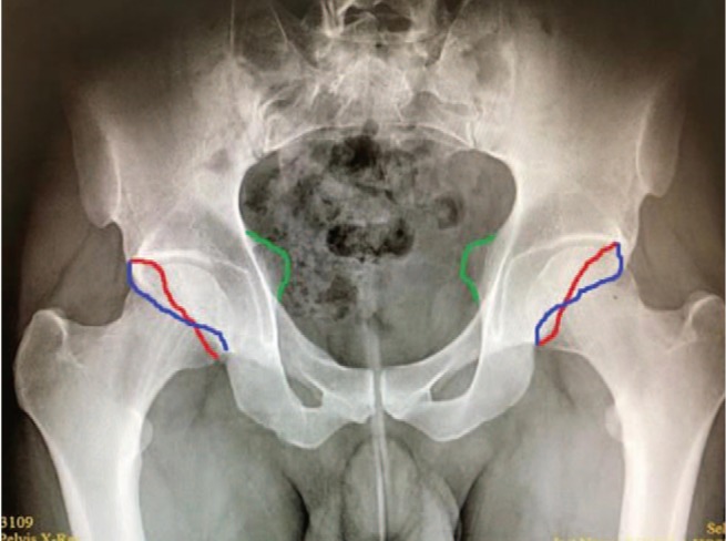
Fig. 3 Anteroposterior radiograph of the pelvis and both hips revealing cross over sign and ischial spine sign.
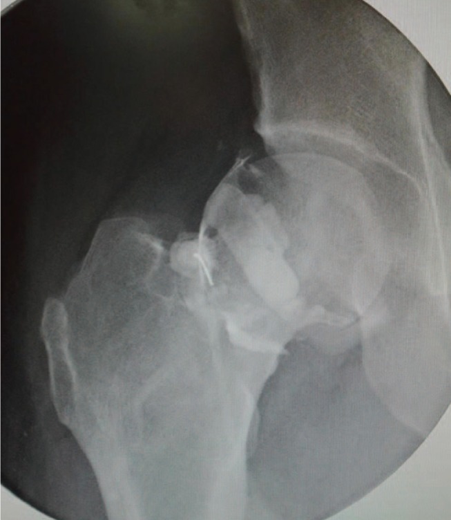
Fig. 4 Intra-articular injection of right hip. The hip was approached anteriorly with spinal needle. Contrast was injected to confirm intra-articular position.
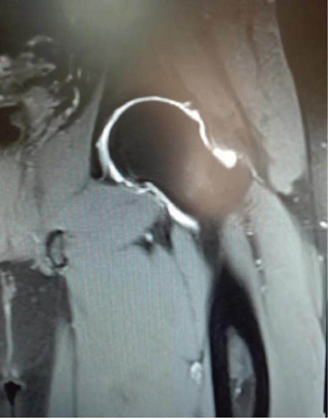
Fig. 5 Magnetic resonance arthrogram of left hip showing thickening of superior labrum with focal area of high signal intensity indicating superior labrum tear.
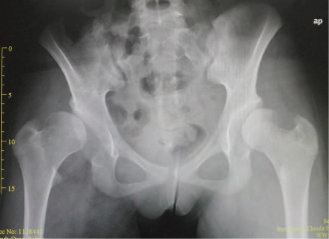
Fig. 6 Anteroposterior view of pelvis with hips showing bilateral hip dysplasia.
Reference
-
1. Dick AG, Houghton JM, Bankes MJK. An approach to hip pain in a young adult. BMJ. 2018; 361:k1086. PMID: 29674488.
Article2. Clohisy JC, Beaulé PE, O’Malley A, Safran MR, Schoenecker P. AOA symposium. Hip disease in the young adult: current concepts of etiology and surgical treatment. J Bone Joint Surg Am. 2008; 90:2267–2281. PMID: 18829926.3. Ganz R, Parvizi J, Beck M, Leunig M, Nötzli H, Siebenrock KA. Femoroacetabular impingement: a cause for osteoarthritis of the hip. Clin Orthop Relat Res. 2003; (417):112–120.4. Röling MA, Mathijssen NM, Bloem RM. Incidence of symptomatic femoroacetabular impingement in the general population: a prospective registration study. J Hip Preserv Surg. 2016; 3(3):203–207. PMID: 27583159.
Article5. Byrd JWT. Operative hip arthroscopy. New York: Springer;2013.6. Wilson JJ, Furukawa M. Evaluation of the patient with hip pain. Am Fam Physician. 2014; 89:27–34. PMID: 24444505.7. Ali F. Clinical examination of the knee. Orthop Trauma. 2013; 27:50–55.
Article8. Ward D, Parvizi J. Management of hip pain in young adults. Orthop Clin North Am. 2016; 47:485–496. PMID: 27241373.
Article9. Clohisy JC, Nunley RM, Otto RJ, Schoenecker PL. The frog-leg lateral radiograph accurately visualized hip cam impingement abnormalities. Clin Orthop Relat Res. 2007; 462:115–121. PMID: 17589367.
Article10. Meyer DC, Beck M, Ellis T, Ganz R, Leunig M. Comparison of six radiographic projections to assess femoral head/neck asphericity. Clin Orthop Relat Res. 2006; 445:181–185. PMID: 16456309.
Article11. Lequesne M, de Seze. [False profile of the pelvis. A new radiographic incidence for the study of the hip. Its use in dysplasias and different coxopathies]. Rev Rhum Mal Osteoartic. 1961; 28:643–652. French. PMID: 14464207.12. Ito K, Minka MA 2nd, Leunig M, Werlen S, Ganz R. Femoroacetabular impingement and the cam-effect. A MRI-based quantitative anatomical study of the femoral head-neck offset. J Bone Joint Surg Br. 2001; 83:171–176. PMID: 11284559.13. Klaue K, Wallin A, Ganz R. CT evaluation of coverage and congruency of the hip prior to osteotomy. Clin Orthop Relat Res. 1988; (232):15–25. PMID: 3383480.
Article14. Nishii T, Tanaka H, Sugano N, Miki H, Takao M, Yoshikawa H. Disorders of acetabular labrum and articular cartilage in hip dysplasia: evaluation using isotropic high-resolutional CT arthrography with sequential radial reformation. Osteoarthritis Cartilage. 2007; 15:251–257. PMID: 16990027.
Article15. Keeney JA, Peelle MW, Jackson J, Rubin D, Maloney WJ, Clohisy JC. Magnetic resonance arthrography versus arthroscopy in the evaluation of articular hip pathology. Clin Orthop Relat Res. 2004; (429):163–169.
Article16. Pateder DB, Hungerford MW. Use of fluoroscopically guided intra-articular hip injection in differentiating the pain source in concomitant hip and lumbar spine arthritis. Am J Orthop (Belle Mead NJ). 2007; 36:591–593. PMID: 18075606.17. Wenger DE, Kendell KR, Miner MR, Trousdale RT. Acetabular labral tears rarely occur in the absence of bony abnormalities. Clin Orthop Relat Res. 2004; (426):145–150.
Article18. Byrd JWT, Phillips JC. Hip tears of the acetabular labrum. In : Volpi P, editor. Arthroscopy and sport injuries. Cham: Springer;2016.19. Briggs K, Philippon M, Ho C, McNamara S. Prevalence of acetabular labral tears in asymptomatic young athletes. Br J Sports Med. 2017; 51:303.
Article20. Dangin A, Tardy N, Wettstein M, May O, Bonin N. Microinstability of the hip: a review. Orthop Traumatol Surg Res. 2016; 102(8S):S301–S309. PMID: 27744000.
Article21. Safran MR. Microinstability of the hip-gaining acceptance. J Am Acad Orthop Surg. 2019; 27:12–22. PMID: 30475277.
Article22. Beck M, Kalhor M, Leunig M, Ganz R. Hip morphology influences the pattern of damage to the acetabular cartilage: femoroacetabular impingement as a cause of early osteoarthritis of the hip. J Bone Joint Surg Br. 2005; 87:1012–1018. PMID: 15972923.23. Robertson WJ, Kadrmas WR, Kelly BT. Arthroscopic management of labral tears in the hip: a systematic review of the literature. Clin Orthop Relat Res. 2007; 455:88–92. PMID: 17119461.24. Reurink G, Jansen SP, Bisselink JM, Vincken PW, Weir A, Moen MH. Reliability and validity of diagnosing acetabular labral lesions with magnetic resonance arthrography. J Bone Joint Surg Am. 2012; 94:1643–1648. PMID: 22878728.
Article25. Narveson JR, Haberl MD, Nathan Vannatta C, Rhon DI. Conservative treatment continuum for managing femoroacetabular impingement syndrome and acetabular labral tears in surgical candidates: a case series. Int J Sports Phys Ther. 2018; 13:1032–1048. PMID: 30534469.
Article26. Potter BK, Freedman BA, Andersen RC, Bojescul JA, Kuklo TR, Murphy KP. Correlation of Short Form-36 and disability status with outcomes of arthroscopic acetabular labral debridement. Am J Sports Med. 2005; 33:864–870. PMID: 15827367.
Article27. Ayeni OR, Adamich J, Farrokhyar F, et al. Surgical management of labral tears during femoroacetabular impingement surgery: a systematic review. Knee Surg Sports Traumatol Arthrosc. 2014; 22:756–762. PMID: 24519616.
Article28. Sankar WN, Nevitt M, Parvizi J, Felson DT, Agricola R, Leunig M. Femoroacetabular impingement: defining the condition and its role in the pathophysiology of osteoarthritis. J Am Acad Orthop Surg. 2013; 21 Suppl 1:S7–S15. PMID: 23818194.
Article29. Thier S, Gerisch D, Weiss C, Fickert S, Brunner A. Prevalence of cam and pincer deformities in the X-rays of asymptomatic individuals. Biomed Res Int. 2017; 2017:856232.
Article30. Beall DP, Sweet CF, Martin HD, et al. Imaging findings of femoroacetabular impingement syndrome. Skeletal Radiol. 2005; 34:691–701. PMID: 16172860.
Article31. Griffin DR, Dickenson EJ, O'Donnell J, et al. The Warwick Agreement on femoroacetabular impingement syndrome (FAI syndrome): an international consensus statement. Br J Sports Med. 2016; 50:1169–1176. PMID: 27629403.32. Nwachukwu BU, Rebolledo BJ, McCormick F, Rosas S, Harris JD, Kelly BT. Arthroscopic versus open treatment of femoroacetabular impingement: a systematic review of medium- to long-term outcomes. Am J Sports Med. 2016; 44:1062–1068. PMID: 26059179.33. Wall PD, Fernandez M, Griffin DR, Foster NE. Nonoperative treatment for femoroacetabular impingement: a systematic review of the literature. PM R. 2013; 5:418–426. PMID: 23419746.
Article34. Bracken J, Tran T, Ditchfield M. Developmental dysplasia of the hip: controversies and current concepts. J Paediatr Child Health. 2012; 48:963–972. quiz 972-3. PMID: 23126391.35. Klaue K, Durnin CW, Ganz R. The acetabular rim syndrome. A clinical presentation of dysplasia of the hip. J Bone Joint Surg Br. 1991; 73:423–429. PMID: 1670443.
Article36. Harris WH. Etiology of osteoarthritis of the hip. Clin Orthop Relat Res. 1986; (213):20–33.37. Solomon L. Patterns of osteoarthritis of the hip. J Bone Joint Surg Br. 1976; 58:176–183. PMID: 932079.
Article38. Gala L, Clohisy JC, Beaulé PE. Hip dysplasia in the young adult. J Bone Joint Surg Am. 2016; 98:63–73. PMID: 26738905.
Article39. Nötzli HP, Müller SM, Ganz R. [The relationship between fovea capitis femoris and weight bearing area in the normal and dysplastic hip in adults: a radiologic study]. Z Orthop Ihre Grenzgeb. 2001; 139:502–506. German. PMID: 11753770.40. James S, Miocevic M, Malara F, Pike J, Young D, Connell D. MR imaging findings of acetabular dysplasia in adults. Skeletal Radiol. 2006; 35:378–384. PMID: 16570172.
Article41. Ganz R, Klaue K, Vinh TS, Mast JW. A new periacetabular osteotomy for the treatment of hip dysplasias. Technique and preliminary results. Clin Orthop Relat Res. 1988; (232):26–36.
Article42. Koulouvaris P, Stafylas K, Aznaoutoglou C, Zacharis K, Xenakis T. Isolated varus intertrochanteric osteotomy for hip dysplasia in 52 patients: long-term results. Int Orthop. 2007; 31:193–198. PMID: 16783549.
Article43. Hospital for Special Surgery. Hip conditions: chondral lesions or injuries. New York: HSS;cited 2019 Feb 23. Available from: https://www.hss.edu/hip-pain-center-hip-conditions.asp#4.44. Yen YM, Kocher MS. Chondral lesions of the hip: microfracture and chondroplasty. Sports Med Arthrosc Rev. 2010; 18:83–89. PMID: 20473126.45. Meyers MH. Resurfacing of the femoral head with fresh osteochondral allografts. Long-term results. Clin Orthop Relat Res. 1985; (197):111–114.
Article
- Full Text Links
-
- Actions
-
Cited
- CITED
-
- Close
- Share
-
- Similar articles
-
- Total Hip Replacement in Painful Stiff and Deformed Hips
- Plain Radiography of the Hip: A Review of Radiographic Techniques and Image Features
- Acute Isolated Tear of Gluteus Medius in Young Male
- Hematogenous Septic Arthritis of the Hip in Extensive Burn
- Clinical Application of the Ultrasound in Painful Pediatric Hip
