Sequential traction of a labio-palatal horizontally impacted maxillary canine with a custom three-directional force device in the space of a missing ipsilateral first premolar
- Affiliations
-
- 1Center of Craniofacial Orthodontics, Department of Oral and Craniomaxillofacial Surgery, Ninth People's Hospital, Shanghai Jiaotong University School of Medicine, Shanghai Key Laboratory of Stomatology & Shanghai Research Institute of Stomatology, National Clinical Research Center of Stomatology, Shanghai, China. jly117@sina.com
- 2Polyclinic Department, Zhejiang Stomatology Hospital, Stomatology Hospital Affiliated to Zhejiang University of Medicine, Hangzhou, China.
- 3The 2nd Dental Center, Ninth People's Hospital, Shanghai Jiaotong University School of Medicine, Shanghai Key Laboratory of Stomatology & Shanghai Research Institute of Stomatology, National Clinical Research center of Stomatology, Shanghai, China.
- KMID: 2453976
- DOI: http://doi.org/10.4041/kjod.2019.49.2.124
Abstract
- Orthodontic treatment is more complicated when both soft and hard tissues must be considered because an impacted maxillary canine has important effects on function and esthetics. Compared with extraction of impacted maxillary canines, exposure followed by orthodontic traction can improve esthetics and better protect the patient's teeth and alveolar bone. Therefore, in order to achieve desirable tooth movement with minimal unexpected complications, a precise diagnosis is indispensable to establish an effective and efficient force system. In this report, we describe the case of a 31-year-old patient who had a labio-palatal horizontally impacted maxillary left canine with a severe occlusal alveolar bone defect and a missing maxillary left first premolar. Herein, with the aid of three-dimensional imaging, sequential traction was performed with a three-directional force device that finally achieved acceptable occlusion by bringing the horizontally impacted maxillary left canine into alignment. The maxillary left canine had normal gingival contours and was surrounded by a substantial amount of regenerated alveolar bone. The 1-year follow-up stability assessment demonstrated that the esthetic and functional outcomes were successful.
MeSH Terms
Figure
-
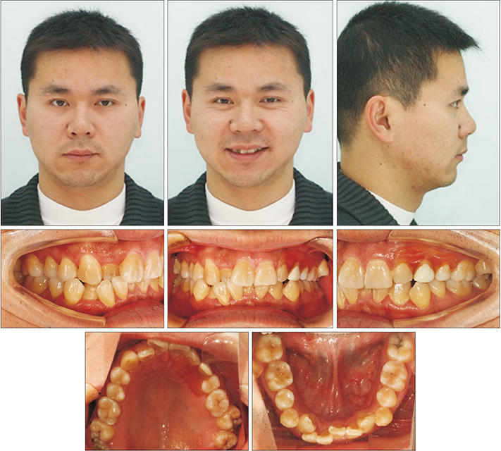
Figure 1 Pretreatment extraoral and intraoral photographs. A removable partial denture is in the left maxillary edentulous region.
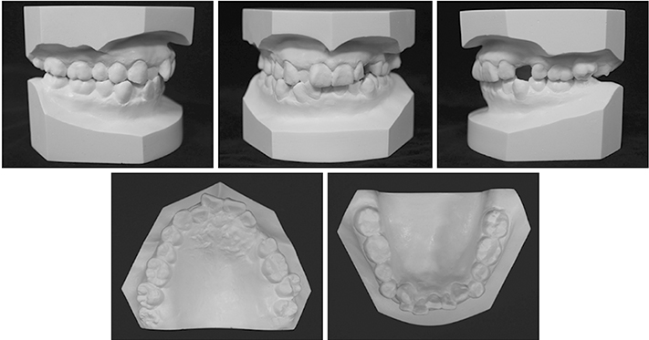
Figure 2 Pretreatment dental casts.

Figure 3 A pretreatment panoramic radiograph.
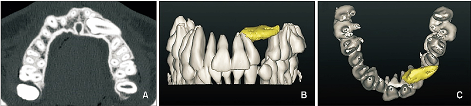
Figure 4 A, Pretreatment axial computed tomography images of the impacted left maxillary canine. B and C, Pretreatment three-dimensional reconstructed computed tomography images of the impacted left maxillary canine are shown in yellow.

Figure 5 A pretreatment cephalometric radiograph (A) and cephalometric tracing (B).
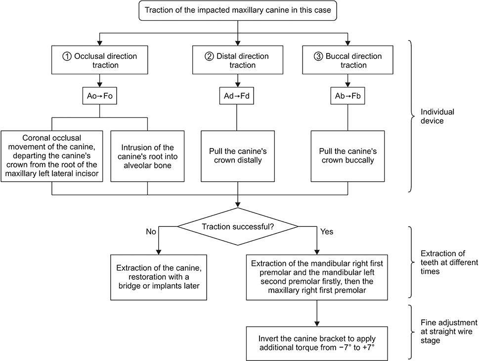
Figure 6 The proposed treatment procedure for the impacted maxillary canine in this case. Ao, Occlusal arm; Fo, occlusal force; Ad, distal arm; Fd, distal force; Ab, buccal arm; Fb, buccal force.
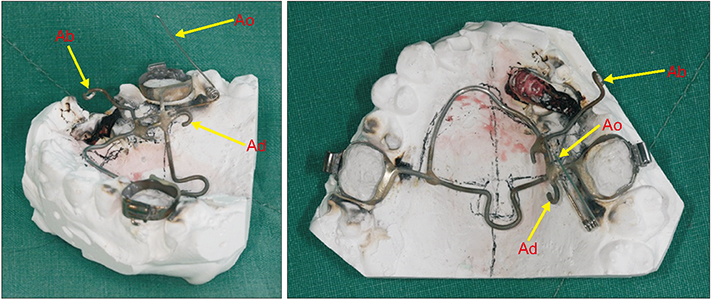
Figure 7 The custom-designed device with three arms (Ao, Ad, and Ab) was implemented on the cast model. Ao, Occlusal arm; Ad, distal arm; Ab, buccal arm.
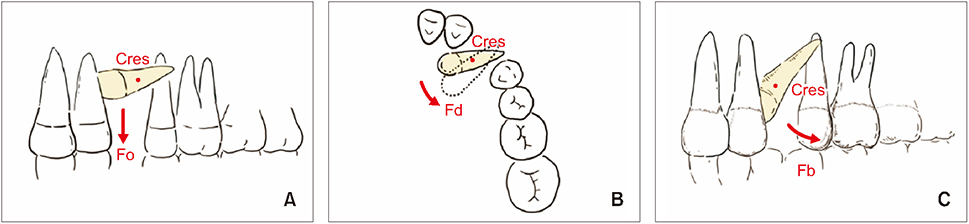
Figure 8 Biomechanics of the custom traction device. A, Occlusal direction; B, distal direction; C, buccal direction. Cres, Center of resistance; Fo, occlusal force; Fd, distal force; Fb, buccal force.
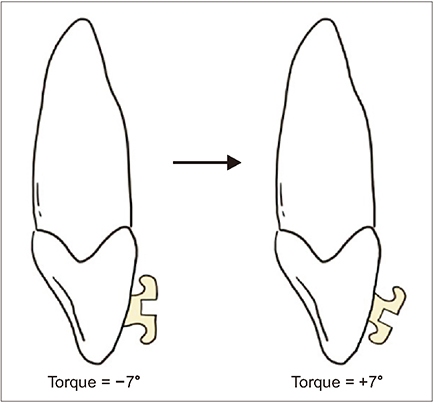
Figure 9 The bracket was inverted and bonded to the maxillary left canine to apply additional torque.
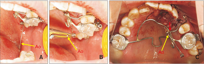
Figure 10 Traction of the maxillary left canine in the occlusal direction. The occlusal arm (Ao) applies an occlusal force (Fo). A, Before activation; B, activation procedure using weingart plier; C, activation by connecting the Ao and the impacted tooth.
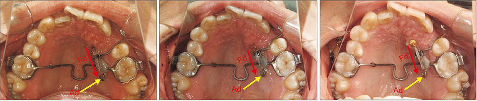
Figure 11 Traction of the maxillary canine in the distal direction. The distal arm (Ad) applies a distal force (Fd).

Figure 12 Traction of the maxillary canine in the buccal direction. The buccal arm (Ab) applies a buccal force (Fb).
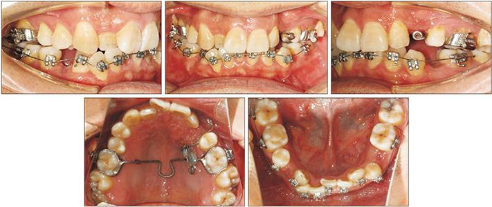
Figure 13 Intraoral photographs obtained at 10 months. The alignment and leveling on the mandibualr arch have commenced.
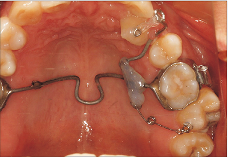
Figure 14 A helix was added to correct the scissor bite of the maxillary left second molar and mandibular left second molar.
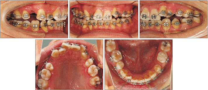
Figure 15 Intraoral photographs obtained at 22 months. The alignment and leveling on the maxillary arch have commenced.

Figure 16 Post-treatment extraoral and intraoral photographs.
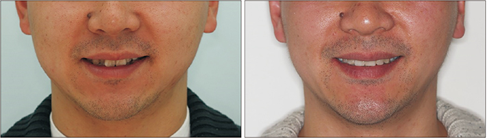
Figure 17 Pretreatment and post-treatment smile photographs.
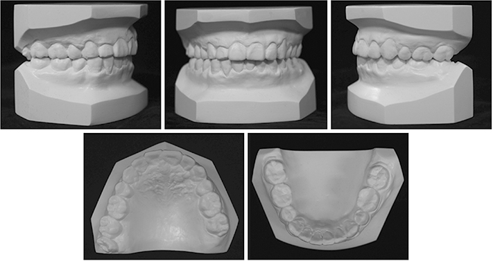
Figure 18 Post-treatment dental casts.
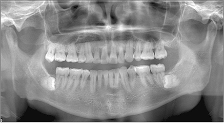
Figure 19 A post-treatment panoramic radiograph.
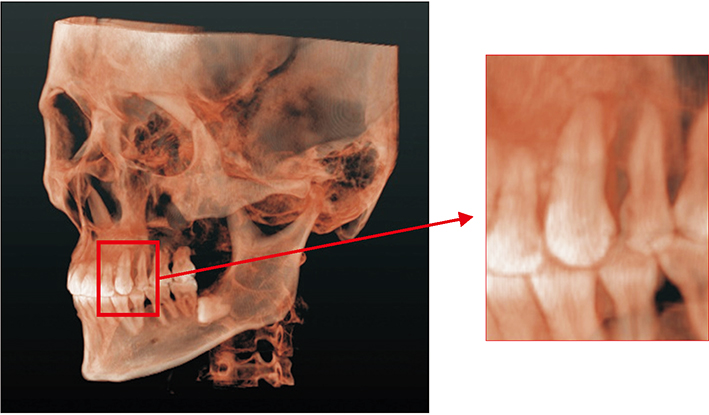
Figure 20 Three-dimensional cone-beam computed tomography images.

Figure 21 Pretreatment (white) and post-treatment (red) superimposed three-dimensional images of the dentition. The impacted left maxillary canine is shown in yellow.
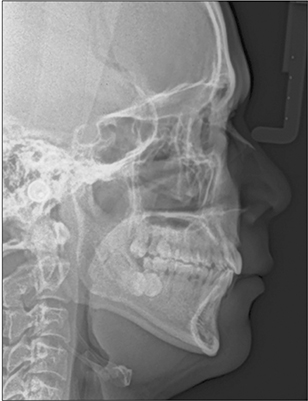
Figure 22 A post-treatment cephalometric radiograph.

Figure 23 A post-treatment cephalometric tracing.

Figure 24 A superimposition of pretreatment (black lines) and post-treatment (red lines) cephalometric tracings.

Figure 25 One-year retention is shown in extraoral and intraoral photographs.
Reference
-
1. Grover PS, Lorton L. The incidence of unerupted permanent teeth and related clinical cases. Oral Surg Oral Med Oral Pathol. 1985; 59:420–425.
Article2. Aydin U, Yilmaz HH, Yildirim D. Incidence of canine impaction and transmigration in a patient population. Dentomaxillofac Radiol. 2004; 33:164–169.
Article3. Hitchin AD. The impacted maxillary canine. Br Dent J. 1956; 100:1–14.4. Counihan K, Al-Awadhi EA, Butler J. Guidelines for the assessment of the impacted maxillary canine. Dent Update. 2013; 40:770–772. 775–777.
Article5. Bishara SE. Impacted maxillary canines: a review. Am J Orthod Dentofacial Orthop. 1992; 101:159–171.
Article6. Gonnissen H, Politis C, Schepers S, Lambrichts I, Vrielinck L, Sun Y, et al. Long-term success and survival rates of autogenously transplanted canines. Oral Surg Oral Med Oral Pathol Oral Radiol Endod. 2010; 110:570–578.
Article7. Kim E, Jung JY, Cha IH, Kum KY, Lee SJ. Evaluation of the prognosis and causes of failure in 182 cases of autogenous tooth transplantation. Oral Surg Oral Med Oral Pathol Oral Radiol Endod. 2005; 100:112–119.
Article8. Kokich VG. Surgical and orthodontic management of impacted maxillary canines. Am J Orthod Dentofacial Orthop. 2004; 126:278–283.
Article9. Kaczor-Urbanowicz K, Zadurska M, Czochrowska E. Impacted teeth: an interdisciplinary perspective. Adv Clin Exp Med. 2016; 25:575–585.
Article10. Dindaroğlu F, Doğan S. Root resorption in orthodontics. Turk J Orthod. 2016; 29:103–108.
Article11. Mirabella AD, Artun J. Risk factors for apical root resorption of maxillary anterior teeth in adult orthodontic patients. Am J Orthod Dentofacial Orthop. 1995; 108:48–55.
Article12. Krishnan V. Root resorption with orthodontic mechanics: pertinent areas revisited. Aust Dent J. 2017; 62:Suppl 1. 71–77.
Article13. Nieto-Nieto N, Solano JE, Yañez-Vico R. External apical root resorption concurrent with orthodontic forces: the genetic influence. Acta Odontol Scand. 2017; 75:280–287.
Article14. Al-Qawasmi RA, Hartsfield JK Jr, Everett ET, Flury L, Liu L, Foroud TM, et al. Genetic predisposition to external apical root resorption in orthodontic patients: linkage of chromosome-18 marker. J Dent Res. 2003; 82:356–360.
Article15. Dewel BF. The upper cuspid: its development and impaction. Angle Orthod. 1949; 19:79–90.16. Botticelli S, Verna C, Cattaneo PM, Heidmann J, Melsen B. Two-versus three-dimensional imaging in subjects with unerupted maxillary canines. Eur J Orthod. 2011; 33:344–349.
Article17. Khojasteh A, Morad G, Behnia H. Clinical importance of recipient site characteristics for vertical ridge augmentation: a systematic review of literature and proposal of a classification. J Oral Implantol. 2013; 39:386–398.
Article18. Lindskog-Stokland B, Wennström JL, Nyman S, Thilander B. Orthodontic tooth movement into edentulous areas with reduced bone height. An experimental study in the dog. Eur J Orthod. 1993; 15:89–96.
Article
- Full Text Links
-
- Actions
-
Cited
- CITED
-
- Close
- Share
-
- Similar articles
-
- Evaluation of Impacted Maxillary Canine Position Using Panoramic Radiographs and Cone-beam Computed Tomography
- Modified Mandibular Lingual Arch for Orthodontic Traction of Impacted Mandibular Canine and Premolar: Case Reports
- Orthodontic traction of a horizontally impacted mandibular second premolar
- Treatment of a Horizontally Impacted and Dilacerated Maxillary Central Incisor and an Impacted Canine
- Photoelastic evaluation of maxillary posterior crossbite appliance
