Serous Cystadenoma of the Pancreas with Dilatation of Bile and Pancreatic Duct
- Affiliations
-
- 1Department of Internal Medicine, Yeungnam University College of Medicine, Daegu, Korea. tnkim@yu.ac.kr
- KMID: 2453261
- DOI: http://doi.org/10.4166/kjg.2019.74.1.63
Abstract
- No abstract available.
Figure
-
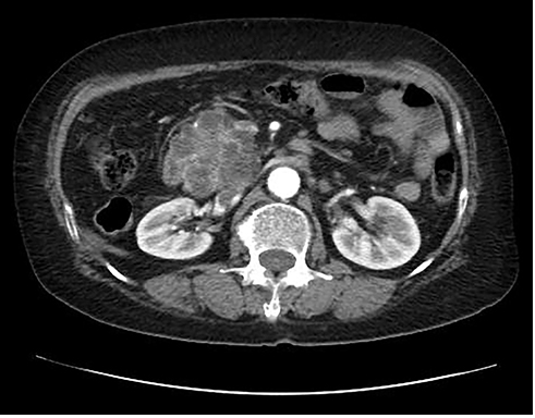
Fig. 1 Abdominal computed tomography (CT). CT image shows about 6 cm sized multilobulated cystic lesion and low-density lesion at center in the head of pancreas.
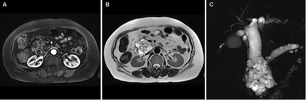
Fig. 2 Pancreatic magnetic resonance images (MRI). (A) MRI shows multilocular cystic lesion with low-signal intensity lesion at center in head of pancreas on T1-weighted axial image (B), multilobular cystic lesion in head of pancreas on T2-weighted axial image. (C) Multilobular cystic lesion in head of pancreas with dilatation of bile and pancreatic duct on T2-weighted magnetic resonance cholangiopancreatography.
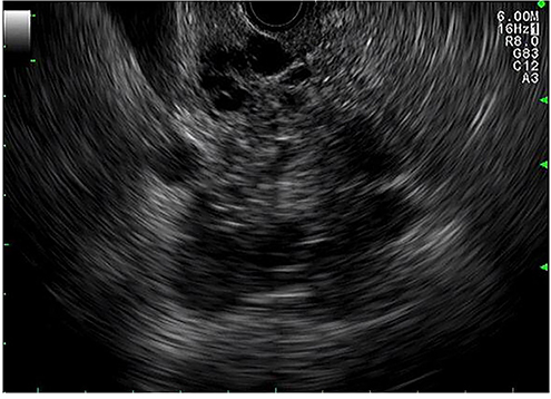
Fig. 3 Endoscopic ultrasound (EUS). EUS image shows multilobulated cystic lesion with hypoechoic lesion at center in head of pancreas.
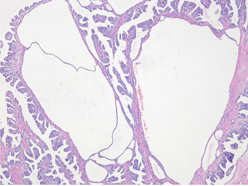
Fig. 4 Pathologic finding of surgical specimen. Microscopic finding of the tumor shows a cystic neoplasm composed of uniform glycogen-rich cuboidal cells lining numerous variable sized cysts (H&E, ×40).
Reference
-
1. Correa-Gallego C, Ferrone CR, Thayer SP, Wargo JA, Warshaw AL, Fernández-Del Castillo C. Incidental pancreatic cysts: do we really know what we are watching. Pancreatology. 2010; 10:144–150.
Article2. Farrell JJ, Fernández-del Castillo C. Pancreatic cystic neoplasms: management and unanswered questions. Gastroenterology. 2013; 144:1303–1315.
Article3. Chu LC, Singhi AD, Haroun RR, Hruban RH, Fishman EK. The many faces of pancreatic serous cystadenoma: radiologic and pathologic correlation. Diagn Interv Imaging. 2017; 98:191–202.
Article4. Federle MP, McGrath KM. Cystic neoplasms of the pancreas. Gastroenterol Clin North Am. 2007; 36:365–376.
Article5. Choi JY, Kim MJ, Lee JY, et al. Typical and atypical manifestations of serous cystadenoma of the pancreas: imaging findings with pathologic correlation. AJR Am J Roentgenol. 2009; 193:136–142.
Article6. Woo YS, Lee KT. Clinical approach to incidental pancreatic cystic neoplasm in outpatient clinics. Korean J Gastroenterol. 2017; 70:13–20.
Article7. Lombardo C, Iacopi S, Menonna F, et al. Incidence and reasons of pancreatic resection in patients with asymptomatic serous cystadenoma. Pancreatology. 2018; 18:577–584.
Article8. Khalpey Z, Rajab TK, Ashley SW. Serous cystadenoma causing the double duct sign. J Gastrointest Surg. 2012; 16:1282–1283.
Article9. Scheiman JM, Hwang JH, Moayyedi P. American gastroenterological association technical review on the diagnosis and management of asymptomatic neoplastic pancreatic cysts. Gastroenterology. 2015; 148:824–848.
Article10. Huh J, Byun JH, Hong SM, et al. Malignant pancreatic serous cystic neoplasms: systematic review with a new case. BMC Gastroenterol. 2016; 16:97.
Article
- Full Text Links
-
- Actions
-
Cited
- CITED
-
- Close
- Share
-
- Similar articles
-
- A Case of Serous Cystadenoma of the Pancreas Communicating with the Pancreatic Duct
- Serous Cystadenoma of the Pancreas
- Simultaneous Pancreatic Serous Microcystic Adenoma and Intraductal Papillary Mucinous Tumor of the Pancreas: A Case Report
- Pancreatic serous cystadenoma mimicking pseudocyst
- Intrahepatic Bile Duct Dilatation Caused by Pancreatic Pseudocyst: A Case Report
